Immune-Regulatory and Molecular Effects of Antidepressants on the Inflamed Human Keratinocyte HaCaT Cell Line
- PMID: 33945102
- PMCID: PMC8275564
- DOI: 10.1007/s12640-021-00367-5
Immune-Regulatory and Molecular Effects of Antidepressants on the Inflamed Human Keratinocyte HaCaT Cell Line
Abstract
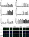
Allergic contact dermatitis (ACD) is a T cell-mediated type of skin inflammation resulting from contact hypersensitivity (CHS) to antigens. There is strong comorbidity between ACD and major depression. Keratinocytes release immunomodulatory mediators including pro-inflammatory cytokines and chemokines, which modulate skin inflammation and are crucial cell type for the development of CHS. Our previous studies showed that fluoxetine and desipramine were effective in suppressing CHS in different mouse strains. However, the immune and molecular mechanisms underlying this effect remain to be explored. The aim of the current study was to determine the immune and molecular mechanisms of action of antidepressant drugs engaged in the inhibition of CHS response in the stimulated keratinocyte HaCaT cell line. The results show that LPS, TNF-α/IFN-γ, and DNFB stimulate HaCaT cells to produce large amounts of pro-inflammatory factors including IL-1β, IL-6, CCL2, and CXCL8. HaCaT stimulation was associated with increased expression of ICAM-1, a cell adhesion molecule, and decreased expression of E-cadherin. Imipramine, desipramine, and fluoxetine suppress the production of IL-1β, CCL2, as well as the expression of ICAM-1. LPS and TNF-α/IFN-γ activate p-38 kinase, but antidepressants do not regulate this pathway. LPS decreases E-cadherin protein expression and fluoxetine normalizes these effects. In summary, the antidepressant drugs examined in this study attenuate the stimulated secretion of pro-inflammatory cytokines, chemokines, and modulate adhesion molecule expression by the HaCaT cell line. Therefore, antidepressants may have some clinical efficacy in patients with ACD and patients with comorbid depression and contact allergy.
Keywords: Adhesion molecules; Antidepressant drugs; Contact hypersensitivity; Cytokines; Depression.
© 2021. The Author(s).
Figures


Similar articles
-
Icariin inhibits TNF-α/IFN-γ induced inflammatory response via inhibition of the substance P and p38-MAPK signaling pathway in human keratinocytes.Int Immunopharmacol. 2015 Dec;29(2):401-407. doi: 10.1016/j.intimp.2015.10.023. Epub 2015 Oct 24. Int Immunopharmacol. 2015. PMID: 26507164
-
Illicium verum extract suppresses IFN-γ-induced ICAM-1 expression via blockade of JAK/STAT pathway in HaCaT human keratinocytes.J Ethnopharmacol. 2013 Oct 7;149(3):626-32. doi: 10.1016/j.jep.2013.07.013. Epub 2013 Jul 18. J Ethnopharmacol. 2013. PMID: 23872327
-
Xanthone suppresses allergic contact dermatitis in vitro and in vivo.Int Immunopharmacol. 2020 Jan;78:106061. doi: 10.1016/j.intimp.2019.106061. Epub 2019 Dec 9. Int Immunopharmacol. 2020. PMID: 31821937
-
Anti-Inflammatory and Anti-Allergic Effects of Saponarin and Its Impact on Signaling Pathways of RAW 264.7, RBL-2H3, and HaCaT Cells.Int J Mol Sci. 2021 Aug 5;22(16):8431. doi: 10.3390/ijms22168431. Int J Mol Sci. 2021. PMID: 34445132 Free PMC article.
-
3-Bromo-4,5-dihydroxybenzaldehyde Isolated from Polysiphonia morrowii Suppresses TNF-α/IFN-γ-Stimulated Inflammation and Deterioration of Skin Barrier in HaCaT Keratinocytes.Mar Drugs. 2022 Aug 31;20(9):563. doi: 10.3390/md20090563. Mar Drugs. 2022. PMID: 36135752 Free PMC article.
Cited by
-
The Anti-Inflammatory Potential of Tricyclic Antidepressants (TCAs): A Novel Therapeutic Approach to Atherosclerosis Pathophysiology.Pharmaceuticals (Basel). 2025 Jan 31;18(2):197. doi: 10.3390/ph18020197. Pharmaceuticals (Basel). 2025. PMID: 40006011 Free PMC article. Review.
References
-
- Aye A, Song YJ, Jeon YD, Jin JS (2020) Xanthone suppresses allergic contact dermatitis in vitro and in vivo. Int Immunopharmacol 78 (September 2019): 106061. 10.1016/j.intimp.2019.106061 - PubMed
-
- Barton WW, Wilcoxen S, Christensen PJ, Paine R (1995) Disparate cytokine regulation of ICAM-1 in rat alveolar epithelial cells and pulmonary endothelial cells in vitro. Am J Physiol Lung Cell Mol Physiol 269 (1 13–1). 10.1152/ajplung.1995.269.1.l127 - PubMed
MeSH terms
Substances
Grants and funding
LinkOut - more resources
Full Text Sources
Other Literature Sources
Medical
Miscellaneous

