. 2021 Sep 30;138(13):1182-1193.
doi: 10.1182/blood.2020009428.

A PSGL-1 glycomimetic reduces thrombus burden without affecting hemostasis

Affiliations

A PSGL-1 glycomimetic reduces thrombus burden without affecting hemostasis

Daniel J Wong et al. Blood. .

Abstract

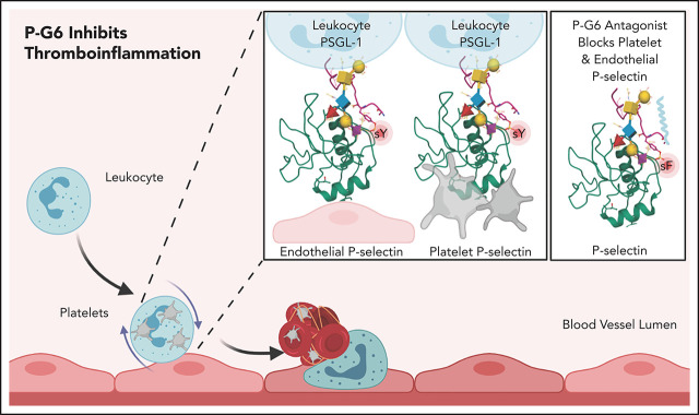
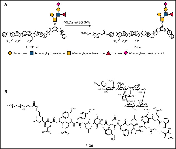
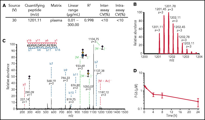
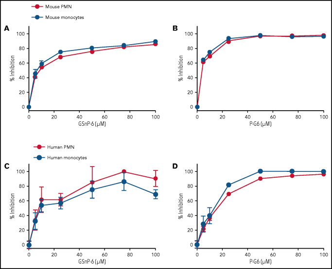
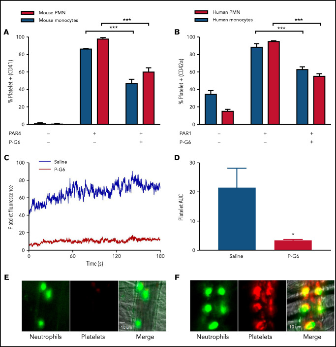
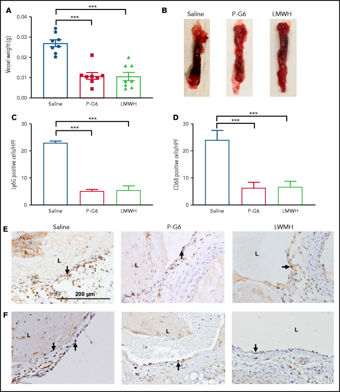
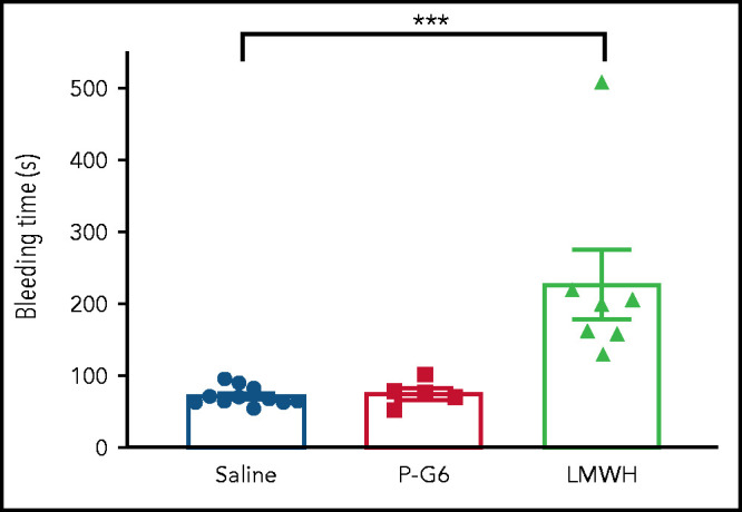
Events mediated by the P-selectin/PSGL-1 pathway play a critical role in the initiation and propagation of venous thrombosis by facilitating the accumulation of leukocytes and platelets within the growing thrombus. Activated platelets and endothelium express P-selectin, which binds P-selectin glycoprotein ligand-1 (PSGL-1) that is expressed on the surface of all leukocytes. We developed a pegylated glycomimetic of the N terminus of PSGL-1, PEG40-GSnP-6 (P-G6), which proved to be a highly potent P-selectin inhibitor with a favorable pharmacokinetic profile for clinical translation. P-G6 inhibits human and mouse platelet-monocyte and platelet-neutrophil aggregation in vitro and blocks microcirculatory platelet-leukocyte interactions in vivo. Administration of P-G6 reduces thrombus formation in a nonocclusive model of deep vein thrombosis with a commensurate reduction in leukocyte accumulation, but without disruption of hemostasis. P-G6 potently inhibits the P-selectin/PSGL-1 pathway and represents a promising drug candidate for the prevention of venous thrombosis without increased bleeding risk.


Figures

None
Graphical abstract
Figure 1.
Figure 1.
Synthesis of PEG40-GSnP-6 (P-G6). P-selectin antagonist GSnP-6 is modeled after the N terminus of human PSGL-1, including sialyl LewisX (N-acetylneuraminic acid)–containing hexasaccharide and sulfopeptide epitopes responsible for high-affinity binding to P-selectin. A linear 40-kDa mPEG-SVA was conjugated to the ε-amino residue of the N terminal lysine of GSnP-6 affording P-G6. The glycan short form (A) and full chemical structure (B) of P-G6 are shown.
Figure 2.
Figure 2.
Quantification of P-G6 in plasma. (A-C) LC-MS/MS was used to generate a standard curve of P-G6 concentration in blood plasma. (A) LC-MS/MS parameters for in-source fragmentation and rigor of the calibration curve. (B) Generation of the quantifying peptide. (C) A stepped higher-energy collisional dissociation approach effectively fragmented P-G6 in a single MS/MS spectrum. Fragment ions: blue, b-ions; red, y-ions; green, glycan loss. Modified amino acid are in bold type. (D) Plasma concentration of P-G6 over time after IV administration of a single weight-based (8 µmol/kg) dose.
Figure 3.
Figure 3.
GSnP-6 and P-G6 inhibit P-selectin binding to leukocytes. GSnP-6 and P-G6 (0-100 μM) were incubated with mouse (A-B) or human (C-D) neutrophils and monocytes. Flow cytometry was used to evaluate the percentage of binding inhibition of species-appropriate P-selectin chimera to neutrophils or monocytes produced by GSnP-6 and P-G6 compared with phosphate-buffered saline control. GSnP-6 and P-G6 inhibit P-selectin leukocyte interactions in a dose-dependent manner. Data are mean ± SEM, n = 3 per agent per study. PMN, polymorphonuclear cells.
Figure 4.
Figure 4.
P-G6 inhibition of platelet-leukocyte aggregation in vitro and in vivo. (A-B) Inhibition of platelet-leukocyte aggregation in vitro. Anticoagulated mouse or human blood was dosed with 120 µM P-G6 or saline control. Platelet-leukocyte aggregation was induced by adding species-specific PAR peptide, and samples were analyzed using flow cytometry to quantify platelet-positive monocytes or neutrophils. P-G6 significantly reduced platelet-leukocyte aggregation in mouse (A) and human (B) blood. Unstimulated control blood is included for reference. (C-F) Inhibition of platelet-leukocyte aggregation in vivo. Intravital microscopy was used to characterize TNF-α–induced venular inflammation. Immediately prior to cremaster exteriorization, mice were infused with P-G6 or saline vehicle control and antiplatelet CD42b-DyLight 649 (red)/antineutrophil Alexa Fluor 488 (green). Platelet fluorescent signal was normalized to vessel area and reported as median integrated fluorescence over time (C) and AUC (D). P-G6 significantly reduced platelet accumulation to adherent neutrophils (E) compared with mice administered saline vehicle (F). Data are mean ± SEM. ***P < .001, *P < .05, Student t test. PMN, polymorphonuclear cells.
Figure 5.
Figure 5.
Treatment with P-G6 reduces venous thrombus formation. A nonocclusive thrombus was induced be electrolytic injury of the inferior vena cava, and vessel thrombus weight was measured 48 hours after injury to determine treatment efficacy. (A) Prophylactic administration of P-G6 (8 µmol/kg IV) and low molecular weight heparin (LMWH; 6 mg/kg, subcutaneously) resulted in a significant reduction in thrombus weight compared with mice administered saline vehicle (8 mice per group). (B) Representative images of excised infrarenal vena cava 48 hours after electrolytic injury. (C-F) Neutrophil and macrophage infiltration within the vein wall 48 hours after thrombus induction. Quantification of Ly6G+ neutrophils (C,E) and CD68+ macrophages (D,F) reveals significantly less wall inflammation compared with mice administered saline vehicle. Arrows indicate positive immunostaining scale bar displayed in saline group applies to all images. Data are mean ± SEM. ***P < .001, ANOVA with Tukey’s multiple-comparison test. L, lumen.
Figure 6.
Figure 6.
Treatment with P-G6 does not affect hemostasis. The effect of P-G6 on hemostasis was assessed using a tail vein bleeding assay. Mice were subjected to IV administration of saline vehicle, P-G6 (8 μmol/kg), or low molecular weight heparin (LMWH; 6 mg/kg) 5 minutes prior to transection of the lateral tail vein. LMWH resulted in a significant increase in bleeding time compared with mice receiving saline vehicle, whereas no increase in bleeding time was observed after administration of P-G6. Data are mean ± SEM. Group comparisons were conducted using Welch’s ANOVA with Dunnett’s multiple-comparison test. ***P < .001.

Comment in

References

    1. Benjamin EJ, Muntner P, Alonso A, et al. ; American Heart Association Council on Epidemiology and Prevention Statistics Committee and Stroke Statistics Subcommittee . Heart disease and stroke statistics-2019 update: a report from the American Heart Association. Circulation. 2019;139(10):e56-e528. - PubMed
    1. Beckman MG, Hooper WC, Critchley SE, Ortel TL. Venous thromboembolism: a public health concern. Am J Prev Med. 2010;38(4 suppl):S495-S501. - PubMed
    1. Kearon C, Akl EA, Ornelas J, et al. . Antithrombotic therapy for VTE disease: Chest guideline and expert panel report [published correction appears in Chest. 2016;150(4):988]. Chest. 2016;149(2):315-352. - PubMed
    1. Carrier M, Abou-Nassar K, Mallick R, et al. ; AVERT Investigators . Apixaban to prevent venous thromboembolism in patients with cancer. N Engl J Med. 2019;380(8):711-719. - PubMed
    1. Coulis AA, Mackey WC. A review of the efficacy and safety profiles of the novel oral anticoagulants in the treatment and prevention of venous thromboembolism. Clin Ther. 2018;40(12):2140-2167. - PubMed

Publication types

MeSH terms