

State-of-Art of Standard and Innovative Materials Used in Cranioplasty
- PMID: 33946170
- PMCID: PMC8124570
- DOI: 10.3390/polym13091452
State-of-Art of Standard and Innovative Materials Used in Cranioplasty
Abstract
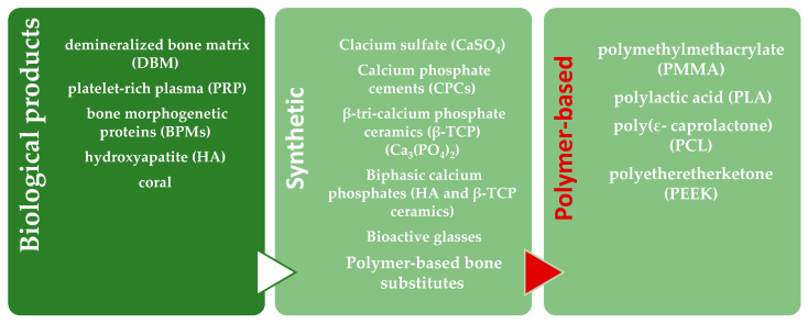
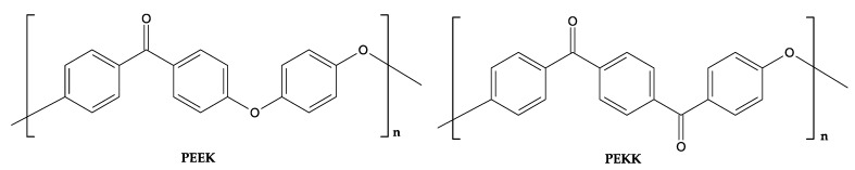
Cranioplasty is the surgical technology employed to repair a traumatic head injury, cerebrovascular disease, oncology resection and congenital anomalies. Actually, different bone substitutes are used, either derived from biological products such as hydroxyapatite and demineralized bone matrix or synthetic ones such as sulfate or phosphate ceramics and polymer-based substitutes. Considering that the choice of the best material for cranioplasty is controversial, linked to the best operation procedure, the intent of this review was to report the outcome of research conducted on materials used for such applications, comparing the most used materials. The most interesting challenge is to preserve the mechanical properties while improving the bioactivity, porosity, biocompatibility, antibacterial properties, lowering thickness and costs. Among polymer materials, polymethylmethacrylate and polyetheretherketone are the most motivating, due to their biocompatibility, rigidity and toughness. Other biomaterials, with ecofriendly attributes, such as polycaprolactone and polylactic acid have been investigated, due to their microstructure that mimic the trabecular bone, encouraging vascularization and cell-cell communications. Taking into consideration that each material must be selected for specific clinical use, the main limitation remains the defects and the lack of vascularization, consequently porous synthetic substitutes could be an interesting way to support a faster and wider vascularization, with the aim to improve patient prognosis.
Keywords: biomaterials; cranial defect; cranioplasty; neurosurgery; polycaprolactone (PCL); polyethereketoneketone (PEKK); polyetheretherketone (PEEK); polyglycolide (PGA); polylactic acid (PLA); polymers; polymethylmethacrylate (PMMA); skull reconstruction; synthetic cranioplasty.
Conflict of interest statement
The authors declare no conflict of interest.
Figures


References
-
- Grant G.A., Jolley M., Ellenbogen R.G., Roberts T.S., Gruss J.R., Loeser J.D. Failure of autologous bone-assisted cranio-plasty following decompressive craniectomy in children and adolescents. J. Neurosurg. 2004;100:163–168. - PubMed
Publication types
LinkOut - more resources
Full Text Sources
