. 2021 Apr 30;10(5):1067.
doi: 10.3390/cells10051067.

Metformin Dysregulates the Unfolded Protein Response and the WNT/β-Catenin Pathway in Endometrial Cancer Cells through an AMPK-Independent Mechanism

Affiliations

Metformin Dysregulates the Unfolded Protein Response and the WNT/β-Catenin Pathway in Endometrial Cancer Cells through an AMPK-Independent Mechanism

Domenico Conza et al. Cells. .

Abstract

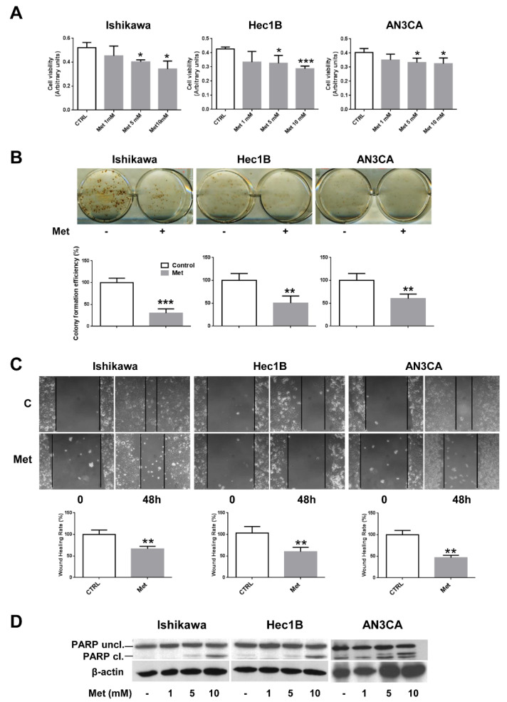
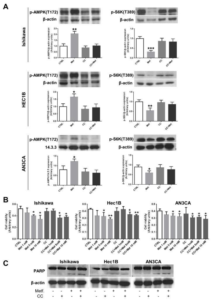
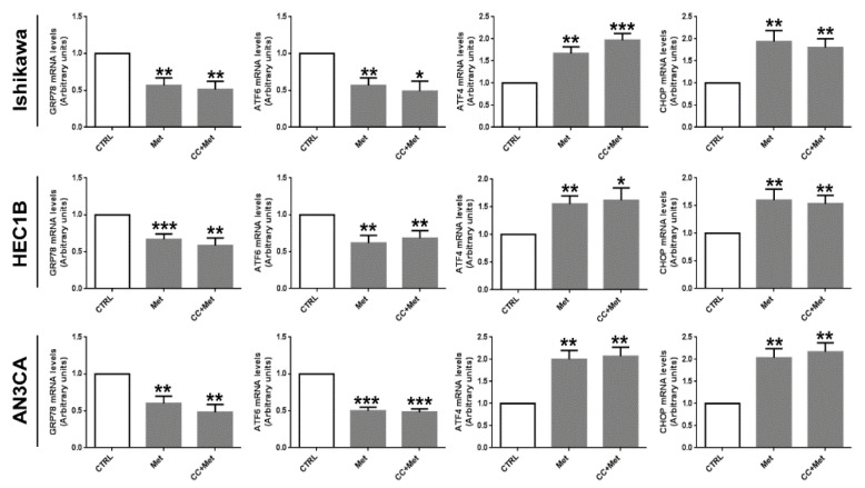
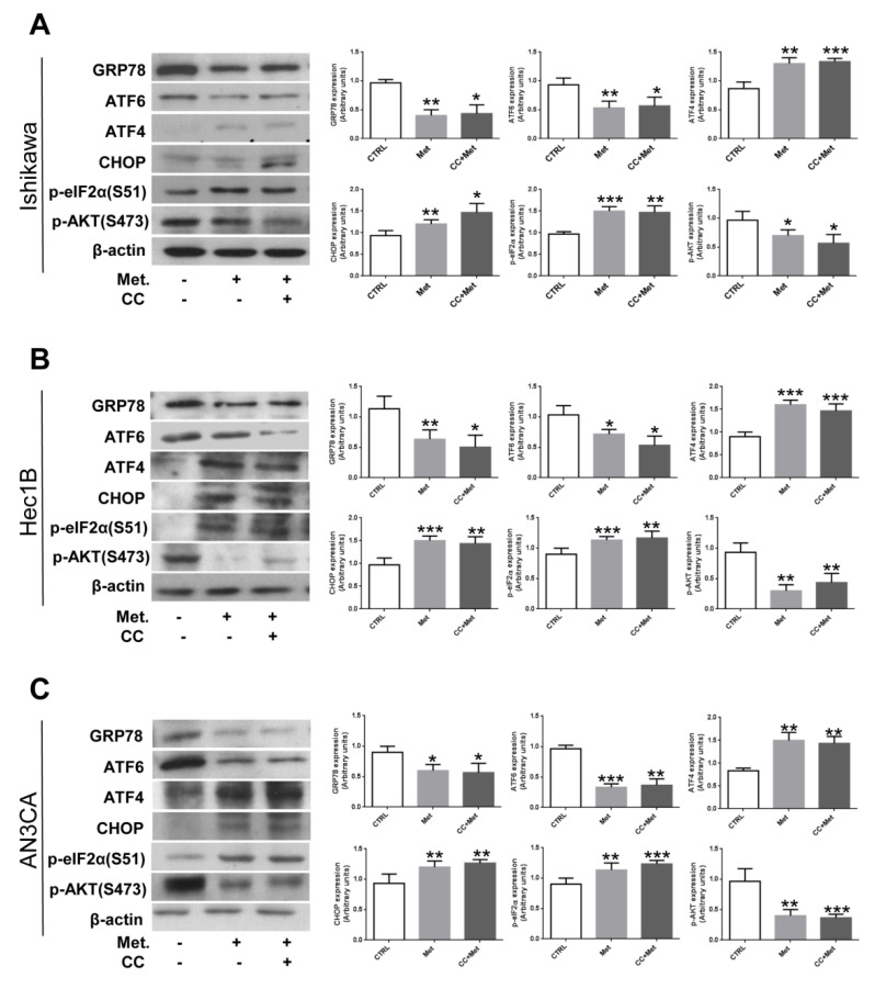
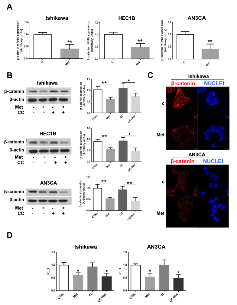
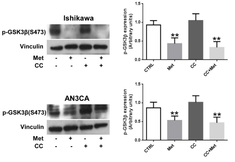
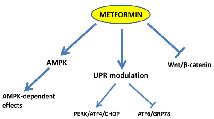
Multiple lines of evidence suggest that metformin, an antidiabetic drug, exerts anti-tumorigenic effects in different types of cancer. Metformin has been reported to affect cancer cells' metabolism and proliferation mainly through the activation of AMP-activated protein kinase (AMPK). Here, we show that metformin inhibits, indeed, endometrial cancer cells' growth and induces apoptosis. More importantly, we report that metformin affects two important pro-survival pathways, such as the Unfolded Protein Response (UPR), following endoplasmic reticulum stress, and the WNT/β-catenin pathway. GRP78, a key protein in the pro-survival arm of the UPR, was indeed downregulated, while GADD153/CHOP, a transcription factor that mediates the pro-apoptotic response of the UPR, was upregulated at both the mRNA and protein level. Furthermore, metformin dramatically inhibited β-catenin mRNA and protein expression. This was paralleled by a reduction in β-catenin transcriptional activity, since metformin inhibited the activity of a TCF/LEF-luciferase promoter. Intriguingly, compound C, a well-known inhibitor of AMPK, was unable to prevent all these effects, suggesting that metformin might inhibit endometrial cancer cells' growth and survival through the modulation of specific branches of the UPR and the inhibition of the Wnt/β-catenin pathway in an AMPK-independent manner. Our findings may provide new insights on the mechanisms of action of metformin and refine the use of this drug in the treatment of endometrial cancer.

Keywords: AMPK; UPR; Wnt/β-catenin; endometrial cancer; metformin.


Conflict of interest statement

The authors declare no conflict of interest.

Figures

Figure 1
Figure 1
Metformin inhibits cell growth and viability in endometrial cancer cells. (A) Ishikawa, HEC1B, or AN3CA cells were seeded at a density of 5 × 10³ cells in a 96-well plate. After 16 h, cells were treated or not with 1, 5, or 10 mM metformin. Cell viability was measured after 48 h using the MTT assay. Values represent the mean absorbance at 570 nm ± SD of triplicates of three independent experiments. * indicates a p-value < 0.05; *** indicates a p-value < 0.001. (B) Ishikawa, HEC1B, or AN3CA cells were seeded onto six-well plates at a density of 1 × 10³ cells per well. After cell attachment, 5 mM metformin was added or not to the wells for 4 h. The cells were then cultured with fresh medium. After 2 weeks, the resultant colonies were fixed with 4% paraformaldehyde and stained with hematoxylin. The colonies were then photographed, counted under a microscope, and colony efficiency formation was calculated. ** indicates a p-value < 0.01; *** indicates a p-value < 0.001. (C) Ishikawa, HEC1B, or AN3CA cells (1 × 10⁶ per well) were seeded in six-well plates and allowed to form a cell monolayer for 24 h. Cell layers were wounded with a micropipette tip and then incubated in fresh culture medium containing or not 5 mM metformin for 48 h. Cell migration toward the wounded area was observed, photographed, and measured. Experiments were performed three times in triplicate. Graphs show the percentage of wound healing rate. ** indicates a p-value < 0.01. (D) Ishikawa, HEC1B, or AN3CA cells were treated or not with 1, 5, or 10 mM metformin for 48 h. Total cellular proteins were extracted and Western blot experiments were performed with antibodies against PARP and β-actin.
Figure 2
Figure 2
Inhibition of AMPK by CC does not alter metformin effects on endometrial cancer cells. (A) Ishikawa, HEC1B, or AN3CA cells were treated or not for 24 h with 5 mM metformin or 10 µM CC or pretreated for 1 h with 10 µM CC followed by treatment with 5 mM metformin. Total cellular proteins were extracted and Western blot experiments were performed, as described in the Section 2.3, using antibodies against p-AMPK (left panels) or p-S6 kinase (right panels). Data represent the mean ± SD of three independent experiments. * p < 0.1; ** p < 0.05; *** p < 0.01. (B) Ishikawa, HEC1B, or AN3CA cells were seeded at a density of 5 × 10³ cells in a 96-well plate. After 16 h, cells were treated or not with increasing concentration of metformin in the presence or absence of 1 h pretreatment with 10 µM CC. Cell viability was measured after 48 h using the MTT assay. Values represent the mean absorbance at 570 nm ±SD of triplicates of three independent experiments. * indicates a p-value < 0.05; ** indicates a p-value < 0.01. (C) Ishikawa, HEC1B, or AN3CA cells were treated or not for 48 h with 5 mM metformin or 10 µM CC or pretreated for 1 h with 10 µM CC followed by treatment with 5 mM metformin. Total cellular proteins were extracted and Western blot experiments were performed, as described in the Section 2.3, with antibodies against PARP or β-actin (loading control).
Figure 3
Figure 3
Metformin modulates the mRNA expression of UPR genes in an AMPK-independent manner in endometrial cancer cells. Ishikawa, HEC1B, or AN3CA cells were treated or not for 24 h with 5 mM metformin in the presence or absence of 1 h pretreatment with 10 µM CC. Total RNA was extracted and real-time RT-PCR experiments were performed using oligonucleotides specific for GRP78, ATF6, ATF4, CHOP, and GAPDH as described in the Section 2.4. Values shown represent the mean (± s.d.) of triplicate samples of three independent experiments. * p < 0.1; ** p < 0.05; *** p < 0.01.
Figure 4
Figure 4
Metformin modulates the expression/phosphorylation of UPR proteins and AKT in an AMPK-independent manner in endometrial cancer cells. Ishikawa (A), HEC1B (B), or AN3CA (C) cells were treated or not for 24 h with 5 mM metformin in the presence or absence of 1 h pretreatment with 10 µM CC. Total cellular proteins were extracted and Western blot experiments were performed with antibodies against GRP78, ATF6, ATF4, p-eIF2α, p-AKT, or β-actin (loading control), as described in the Section 2.3. Values shown represent the mean (± s.d.) of three independent experiments. * p < 0.1; ** p < 0.05; *** p < 0.01.
Figure 5
Figure 5
Metformin inhibits β-catenin expression independently from AMPK activation in endometrial cancer cells. (A) Ishikawa, HEC1B, or AN3CA cells were treated or not for 24 h with 5 mM metformin. Total RNA was extracted and real-time RT-PCR experiments were performed using oligonucleotides specific to β-catenin and GAPDH as described in the Section 2.4. Values shown represent the mean (± s.d.) of triplicate samples of three independent experiments. ** p < 0.05. (B) Ishikawa, HEC1B, or AN3CA cells were treated or not for 24 h with 5 mM metformin in the presence or absence of 1 h pretreatment with 10 µM CC. Total cellular proteins were extracted and Western blot experiments were performed with antibodies against β-catenin or β-actin (loading control), as described in the Section 2.3. Values shown represent the mean (± s.d.) of three independent experiments. * p < 0.1; ** p < 0.05. (C) Ishikawa and AN3CA cells were grown on glass coverslips for 48 h, then were treated or not for 24 h with 5 mM metformin. Cells were fixed in 4% paraformaldehyde in PBS for 20 min, washed twice in 50 mm NH4Cl in PBS, and permeabilized for 5 min in 0.1% Triton X-100 in PBS. Cells were double-stained with anti-β-catenin antibodies and HOECHST 33258 (Nuclei). Bars, 10 μm. (D) Ishikawa and AN3CA cells were plated in six-well plates to approximately 80% confluence 24 h before transfection. Cells were then transfected with 1.0 μg of the reporter vector BAT-LUX TCF/LEF and 50 ng of pRL-TK vector with Lipofectamine 3000. After 24 h, transfection medium was replaced with fresh medium and cells were treated or not for 24 h with 5 mM metformin, 10 µM CC, or pretreated for 1 h with 10 µM CC followed by treatment with 5 mM metformin. Firefly and renilla activities were determined in cell lysates using the Dual-Luciferase Reporter Assay System and a luminometer. Results were expressed as the ratio of firefly to renilla activity. Values shown represent the mean (± s.d.) of triplicate samples of three independent experiments. * p < 0.1.
Figure 6
Figure 6
Metformin inhibits GSK3β phosphorylation in endometrial cancer cells. Ishikawa or AN3CA cells were treated or not for 24 h with 5 mM metformin or 10 µM CC or pretreated for 1 h with 10 µM CC followed by treatment with 5 mM metformin. Total cellular proteins were extracted and Western blot experiments were performed with antibodies against p-GSK3β (Ser9) or vinculin (loading control), as described in the Section 2.3. Values shown represent the mean (± s.d.) of three independent experiments. ** p < 0.05.
Figure 7
Figure 7
Scheme summarizing the observed mechanisms affected by metformin in endometrial cancer cells. Besides AMPK activation, metformin modulates the UPR by activating the PERK/ATF4/CHOP axis and inhibiting the ATF6/GRP78 axis. Furthermore, metformin inhibits the Wnt/β-catenin signaling pathway by reducing β-catenin expression.

References

    1. Pollak M.N. Investigating metformin for cancer prevention and treatment: The end of the beginning. Cancer Discov. 2012;2:2778–2790. doi: 10.1158/2159-8290.CD-12-0263. - DOI - PubMed
    1. Kourelis T.V., Siegel R.D. Metformin and cancer: New applications for an old drug. Med. Oncol. 2012;29:1314–1327. doi: 10.1007/s12032-011-9846-7. - DOI - PubMed
    1. Evans J.M., Donnelly L.A., Emslie-Smith A.M., Alessi D.R., Morris A.D. Metformin and reduced risk of cancer in diabetic patients. BMJ. 2005;330:1304–1305. doi: 10.1136/bmj.38415.708634.F7. - DOI - PMC - PubMed
    1. Bowker S.L., Majumdar S.R., Veugelers P., Johnson J.A. Increased cancer-related mortality for patients with type 2 diabetes who use sulfonylureas or insulin. Diabetes Care. 2006;29:254–258. doi: 10.2337/diacare.29.02.06.dc05-1558. - DOI - PubMed
    1. Ezewuiro O., Grushko T.A., Kocherginsky M., Habis M., Hurteau J.A., Mills K.A., Hunn J., Olopade O.I., Fleming G.F., Romero I.L. Association of Metformin Use with Outcomes in Advanced Endometrial Cancer Treated with Chemotherapy. PLoS ONE. 2016;11:e0147145. doi: 10.1371/journal.pone.0147145. - DOI - PMC - PubMed

Publication types

MeSH terms