. 2021 Apr 30;11(5):676.
doi: 10.3390/biom11050676.

Chitosan Functionalized Magnetic Nanoparticles to Provide Neural Regeneration and Recovery after Experimental Model Induced Peripheral Nerve Injury

Affiliations

Chitosan Functionalized Magnetic Nanoparticles to Provide Neural Regeneration and Recovery after Experimental Model Induced Peripheral Nerve Injury

Nadina Liana Pop et al. Biomolecules. .

Abstract

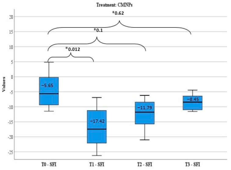
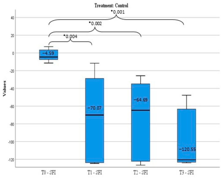
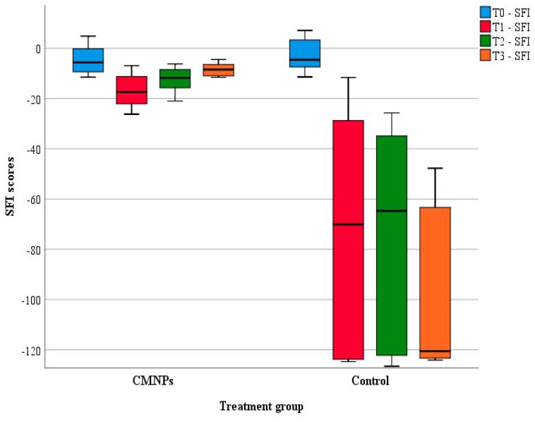
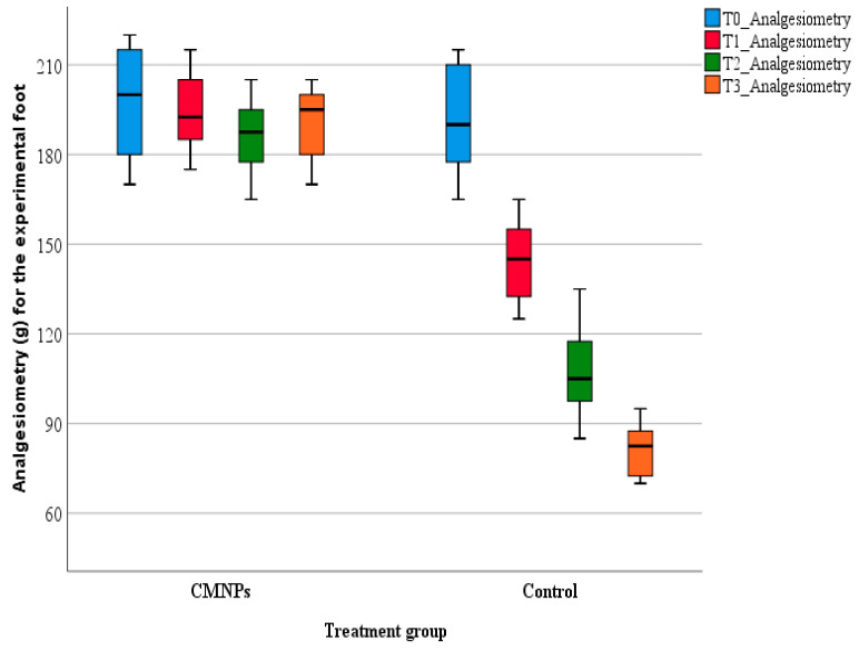
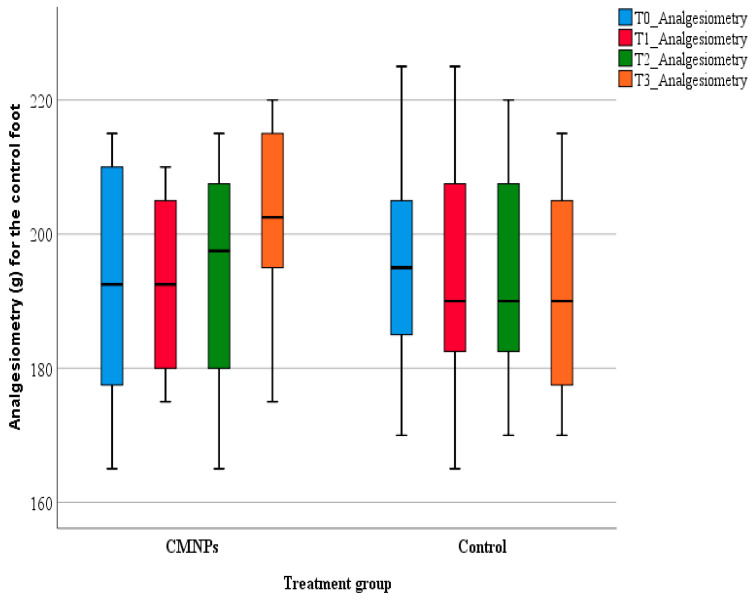
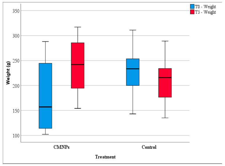
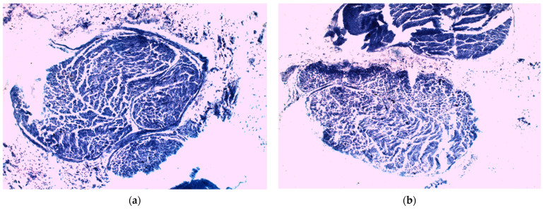
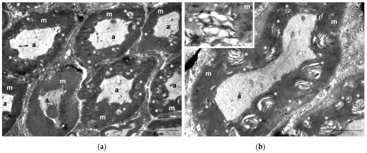
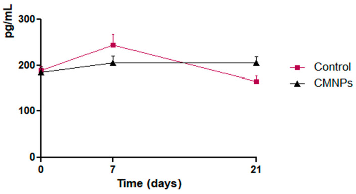
(1) Background: Peripheral nerve injuries have a great impact on a patient's quality of life and a generally poor outcome regarding functional recovery. Lately, studies have focused on different types of nanoparticles and various natural substances for the treatment of peripheral nerve injuries. This is the case of chitosan, a natural compound from the crustaceans' exoskeleton. The present study proposes to combine chitosan benefic properties to the nanoparticles' ability to transport different substances to specific locations and evaluate the effects of magnetic nanoparticles functionalized with chitosan (CMNPs) on peripheral nerve injuries' rehabilitation by using an in vivo experimental model. (2) Methods: CMNPs treatment was administrated daily, orally, for 21 days to rats subjected to right sciatic nerve lesion and compared to the control group (no treatment) by analyzing the sciatic functional index, pain level, body weight, serum nerve growth factor levels and histology, TEM and EDX analysis at different times during the study. (3) Results: Animals treated with CMNPs had a statistically significant functional outcome compared to the control group regarding: sciatic functional index, pain-like behavior, total body weight, which were confirmed by the histological and TEM images. (4) Conclusions: The results of the study suggest that CMNPs appear to be a promising treatment method for peripheral nerve injuries.

Keywords: chitosan magnetic nanoparticles; functional rehabilitation; peripheral nerve injury; sciatic functional index.


Conflict of interest statement

The authors declare no conflict of interest. The funders had no role in the design of the study; in the collection, analyses, or interpretation of data; in the writing of the manuscript, or in the decision to publish the results.

Figures

Scheme 1
Scheme 1
Coating of magnetic nanoparticles with chitosan (MNP—magnetic nanoparticles, CHIT—chitosan, CMNPs—chitosan magnetic nanoparticles, rt—room temperature).
Figure 1
Figure 1
FTIR spectrum of uncovered MNP (black) and MNP covered with chitosan CMNPs (blue).
Figure 2
Figure 2
TEM images of (a) uncovered MNP and (b) chitosan functionalized magnetic nanoparticles CMNPs.
Figure 3
Figure 3
Magnetization curve of uncovered MNPs (black) and of CMNPs (red); M-magnetization saturation; B(T)-magnetic field measured at room temperature.
Figure 4
Figure 4
SFI score distribution (median) and evolution in time (* Wilcoxon signed-rank tests: p-values) for the CMNPs treatment group.
Figure 5
Figure 5
SFI score distribution (median) and evolution in time (* Wilcoxon signed-rank tests: p-values) for the control treatment group.
Figure 6
Figure 6
Comparative analysis of the SFI score between control group and CMNPs treatment group (dynamic evaluation).
Figure 7
Figure 7
Comparative analysis of pain level between CMNPs group and control group for the experimental foot.
Figure 8
Figure 8
Comparative analysis of pain level between CMNPs group and control group for the normal foot.
Figure 9
Figure 9
Comparative analysis of animal body weight evolution for CMNPs group and control group.
Figure 10
Figure 10
Right sciatic nerve samples—histological studies. (a) Control group nerve sample; (b) CMNPs treatment group nerve sample.
Figure 11
Figure 11
Right sciatic nerve samples—TEM studies. (a,b) Control group nerve sample; (c,d) CMNPs treatment group nerve sample. a, axon; m, myelin; n, nucleus; Sc, Schwann cell; *, region of disorganization of myelin layers.
Figure 11
Figure 11
Right sciatic nerve samples—TEM studies. (a,b) Control group nerve sample; (c,d) CMNPs treatment group nerve sample. a, axon; m, myelin; n, nucleus; Sc, Schwann cell; *, region of disorganization of myelin layers.
Figure 12
Figure 12
TEM-EDX analysis of right sciatic nerve sample from CMNPs group. (a) TEM image of the treated sciatic nerve; (b) Point-probe analysis using TEM equipped with EDX, showing the presence of iron (Fe) in the sciatic nerve.
Figure 13
Figure 13
Comparative analysis of serum NGF values’ evolution for CMNPs group and control group.

References

    1. Jung Y., Ng J., Keating C., Senthil-Kumar P., Zhao J., Randolph M., Winograd J.M., Evans C.L. Comprehensive evaluation of peripheral nerve regeneration in the acute healing phase using tissue clearing and optical microscopy in a rodent model. PLoS ONE. 2014;9:e94054. doi: 10.1371/journal.pone.0094054. - DOI - PMC - PubMed
    1. Chen Z.L., Yu W.M., Strickland S. Peripheral regeneration. Annu. Rev. Neurosci. 2007;30:209–233. doi: 10.1146/annurev.neuro.30.051606.094337. - DOI - PubMed
    1. Rotshenker S. Wallerian degeneration: The innate-immune response to traumatic nerve injury. J. Neuroinflamm. 2011;8:109. doi: 10.1186/1742-2094-8-109. - DOI - PMC - PubMed
    1. Vargas M.E., Barres B.A. Why is wallerian degeneration in the CNS so slow? Annu. Rev. Neurosci. 2007;30:153–179. doi: 10.1146/annurev.neuro.30.051606.094354. - DOI - PubMed
    1. Menorca R.M.G., Fussell T.S., Elfar J.C. Peripheral nerve trauma: Mechanisms of injury and recovery. Hand Clin. 2013;29:317–330. doi: 10.1016/j.hcl.2013.04.002. - DOI - PMC - PubMed

Publication types