. 2021 Apr 30;22(9):4796.
doi: 10.3390/ijms22094796.

CHIR99021 Augmented the Function of Late Endothelial Progenitor Cells by Preventing Replicative Senescence

Affiliations

CHIR99021 Augmented the Function of Late Endothelial Progenitor Cells by Preventing Replicative Senescence

Vinoth Kumar Rethineswaran et al. Int J Mol Sci. .

Erratum in

Abstract

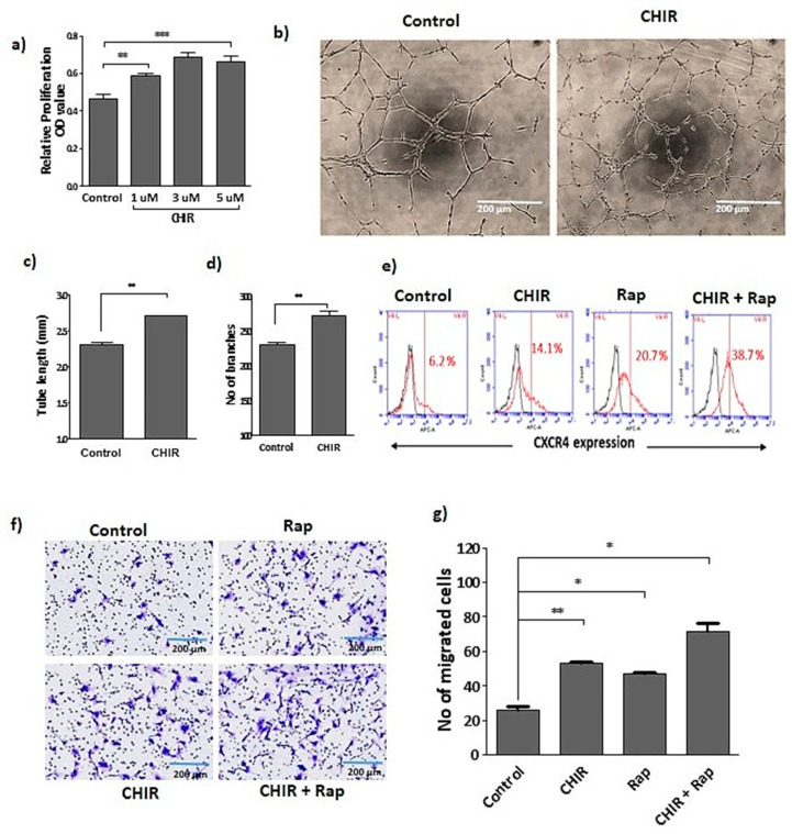
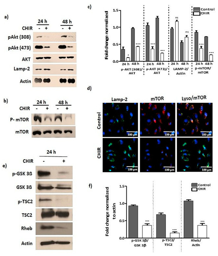
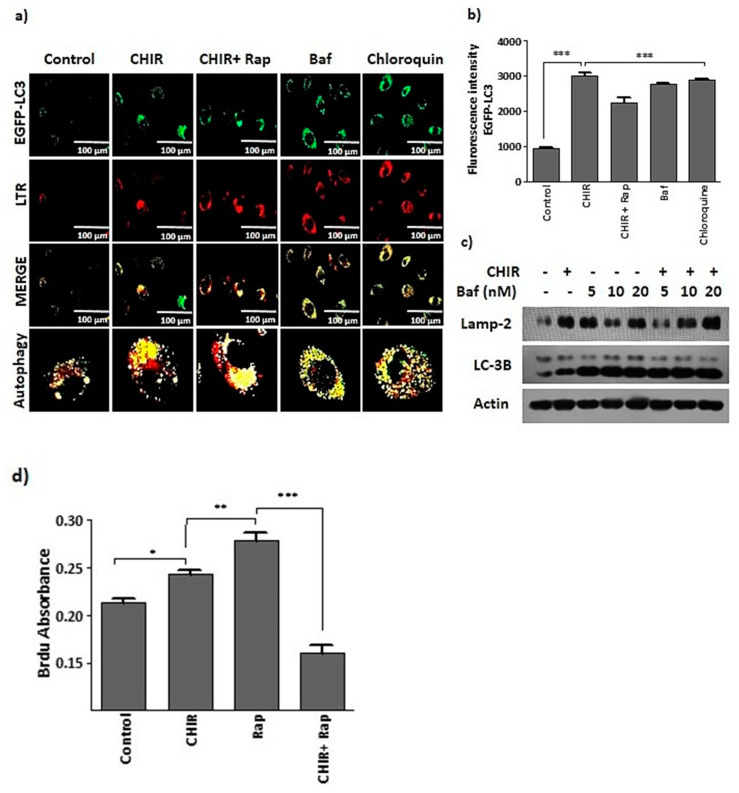
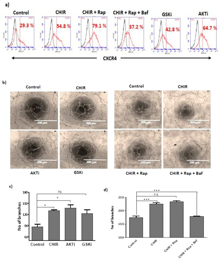
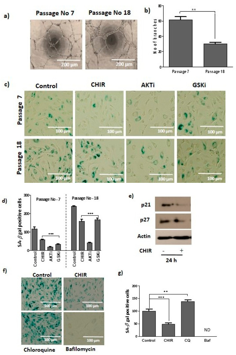
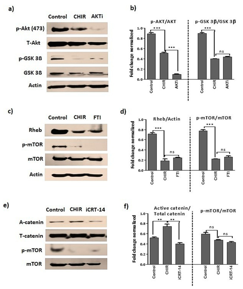
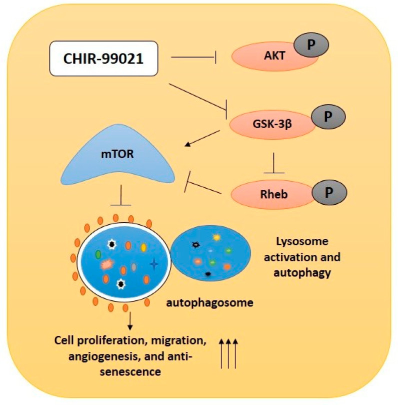
Endothelial progenitor cells (EPCs) are specialized cells in circulating blood, well known for their ability to form new vascular structures. Aging and various ailments such as diabetes, atherosclerosis and cardiovascular disease make EPCs vulnerable to decreasing in number, which affects their migration, proliferation and angiogenesis. Myocardial ischemia is also linked to a reduced number of EPCs and their endothelial functional role, which hinders proper blood circulation to the myocardium. The current study shows that an aminopyrimidine derivative compound (CHIR99021) induces the inhibition of GSK-3β in cultured late EPCs. GSK-3β inhibition subsequently inhibits mTOR by blocking the phosphorylation of TSC2 and lysosomal localization of mTOR. Furthermore, suppression of GSK-3β activity considerably increased lysosomal activation and autophagy. The activation of lysosomes and autophagy by GSK-3β inhibition not only prevented replicative senescence of the late EPCs but also directed their migration, proliferation and angiogenesis. To conclude, our results demonstrate that lysosome activation and autophagy play a crucial role in blocking the replicative senescence of EPCs and in increasing their endothelial function. Thus, the findings provide an insight towards the treatment of ischemia-associated cardiovascular diseases based on the role of late EPCs.

Keywords: CHIR99021; EPC; GSK-3β; autophagy; lysosome; mTOR; senescence.


Conflict of interest statement

The author declares no conflict of interest.

Figures

Figure 1
Figure 1
Stimulation of CHIR99021 established the late EPCs functional activity. (a) Cells were treated with CHIR99021 at different doses (1, 3, 5 uM) for 24 h, and proliferation was assessed by using wst-1. (b) Growth factor reduced Matrigel used for tube formation assay. The vessel structure was visualized using a light microscopy. Tube length and branches were quantified using ImageJ. (c,d) Quantification of total tube length and number of branches. (e) The treatment with CHIR99021 (3 uM) alone or co-treated with rapamycin (20 nM) for 24 h in late EPCs. Fluorescence activated cell sorting was performed by using non-stained cells as a negative control. The fraction of positively stained cells was determined by comparison with non-stained cells. The fraction of positively stained cells is indicated by the positive peaks (red lines indicate cells stained with each antibody, and black lines indicate the negative control). (f) Transwell migration assay was performed by seeding cells in the upper inserts of the transwell chambers, whereas medium containing the drug was loaded into lower chambers of the plate. The cells were incubated for up to 6 h, and the number of migrated cells was counted in three random fields for each membrane filter (20× magnification) under microscope. (g) Quantification of migrated cells. Data are presented as mean ± standard error of the mean (SEM). The results are considered statistically significant at * p< 0.05; ** p< 0.01; *** p < 0.001 when compared to untreated groups. (Rap-Rapamycin).
Figure 2
Figure 2
GSK-3β inactivation by CHIR99021 treatment deregulates AKT and mTOR signaling. (ac) Cells were treated with CHIR99021 (3 uM) for 24, 48 h, then Western blot was performed to detect the phosphorylation and total form of AKT, mTOR, Rheb, LAMP-2 and actin (taken as loading control). (d) The cells were selectively treated with CHIR99021 (3 uM) for 24 h, then subsequently immunostaining was performed to evaluate the lysosomal localization of mTOR. (e,f) Followed by 24 h of CHIR99021 (3 uM) treatment, whole cell lysate fractions were isolated, then Western blot was performed to detect phosphorylation and total form of GSK-3β, TSC2, Rheb, and actin taken as a loading control. Data are presented as mean ± standard error of the mean (SEM). The results are considered statistically significant at * p< 0.05; *** p < 0.001 when compared to untreated group, and ns (no significant).
Figure 3
Figure 3
The suppression of GSK-3β using CHIR99021 upregulates lysosome activation and autophagy. (a,b) EPCs were transfected with GFP-LC3 plasmid using Lipofectamine 3000 reagent. Then, cells were selectively treated with CHIR99021 (3 uM), rapamycin (20 nM), lysosomal blocker bafilomycin A1 (5 nM) and autophagy blocker chloroquine (20 uM) for 4 h. Next, cells were co-stained with lysotracker (200 nM) for 1 h. GFP-LC3 expression was captured using a 40× objective lens on a Lion Heart FX automated microscope. Scale bar = 100 μM. (c) The cells were treated with CHIR99021 (3 uM) alone or co-treated with bafilomycin A1 (5, 10, 20 nM) for 4 h, then Western blots were generated to detect the expression of lysosomal marker proteins LAMP2 and autophagy marker protein LC-3B. Actin was taken as a loading control. (d) The cells were selectively treated with CHIR99021 (3 uM) for 24 h, rapamycin (20 nM) and bafilomycin A1 (5 nM) for 1 h. BrdU incorporation was absorbed at 450 nm to assess the proliferation. Data are presented as mean ± standard error of the mean (SEM). The results are considered statistically significant at * p< 0.05; ** p< 0.01; *** p < 0.001 when compared to untreated groups, and ns (no significant). (Baf—bafilomycin A1, Rap—Rapamycin).
Figure 4
Figure 4
Lysosome activation and autophagy by CHIR99021 expands endothelial functional activity of late EPCs: (a) Cells were single treated with CHIR99021 (3 uM), GSK-3β-specific inhibitor (3 uM) for 24 h and AKT 1/2 inhibitor (3 uM) for 1 h, then subsequently co-treated with rapamycin (20 nM) and bafilomycin A1 (5 nM) for 1 h. FACS was performed for gating the population of non-stained cells as a negative control. The fraction of positively stained cells was determined by comparison with non-stained cells. The fraction of positively stained cells is indicated by the positive peaks (red line indicates cells stained with each antibody, and black lines indicate the negative control). (b) Endothelial functional activity of EPCs was established using vascular network formation. Cells were treated with CHIR99021 (3 uM), AKT 1/2 inhibitor (3 μM) and GSK-3β inhibitor (3 uM) either in the presence or absence of rapamycin (20 nM) and bafilomycin A1 (5 nM) for 6 h. The vascular structure was visualized using a light microscopy; the branches were quantified using ImageJ. (c,d) The number of branches were also quantified. Data are presented as mean ± standard error of the mean (SEM). The results are considered statistically significant at * p < 0.05; *** p < 0.001 when compared to untreated groups and ns (no significant. (Baf—bafilomycin A1, Rap—Rapamycin, AKTi—AKT inhibitor, GSKi—GSK inhibitor).
Figure 5
Figure 5
Late EPCs senescence repressed by lysosome activation and autophagy. (a) The formation of vascular structure was compared with young (p-7) and old (p-18) passage numbers of late EPCs. (b) The number of branches was quantified by young (p-7) and old (p-18) cells. (c) The cells were treated with CHIR99021 (3 uM) and GSK-3β inhibitor (3 uM) for 24 h, and AKT 1/2 inhibitor (3 uM) for 1h, at different passages (p-7, p-18). Following this, the cells were stained to detect senescence by β-galactosidase according to the manufacturers’ instructions. (d) The senescence-positive cells were quantified. (e) Cells were stimulated with CHIR99021 (3 uM) for 24 h, and senescence marker proteins p21 and p27 and loading control actin expression was observed using Western blots. (f) After selective treatment with CHIR99021 (3 uM), chloroquine (20 uM) and bafilomycin A1 (5 nM), cells were stained with β-galactosidase. (g) The senescence-positive cells were captured and quantified. Data are presented as mean ± standard error of the mean (SEM). The results are considered statistically significant at ** p< 0.01; *** p < 0.001 when compared to untreated groups. (Baf—bafilomycin A1, CQ—chloroquine).
Figure 6
Figure 6
GSK-3β inactivation deregulates mTOR through Rheb signaling. (af) Late EPCs stimulated with CHIR99021 (3 μM), AKT 1/2 inhibitor (3 μM), Rheb inhibitor (10μM) and beta-catenin inhibitor (10μM) for 24 h. Then, Western blot was performed to evaluate the protein level expression of p-AKT, AKT, active catenin, total catenin, p-GSK-3β, GSK-3β, Rheb, LAMP2, p-mTOR, mTOR and loading control actin. Data are presented as mean ± standard error of the mean (SEM). The results are considered statistically significant at ** p< 0.01; *** p < 0.001 when compared to untreated groups, and ns (non-significant). (AKTi—AKT inhibitor, FTI—Farnesyltransferase, a Rheb inhibitor, iCRT-14, beta-catenin inhibitor).
Figure 7
Figure 7
Graphical representation. Schematic representation of GSK-3β inhibition in late EPCs using CHIR99021. The CHIR99021-induced GSK-3β inactivation nonspecifically inhibited the phosphorylated form of AKT. Inhibition of GSK-3β subsequently inhibited Rheb activity and suppressed mTOR activity. In addition, CHIR99021 treatment upregulated lysosome activation and autophagy, which further prevents cellular senescence and thereby increases bioactivity such as cell proliferation, migration and angiogenesis in late EPCs.

References

    1. Urbich C., Dimmeler S. Endothelial progenitor cells: Characterization and role in vascular biology. Circ. Res. 2004;95:343–353. doi: 10.1161/01.RES.0000137877.89448.78. - DOI - PubMed
    1. Dai X., Yan X., Zeng J., Chen J., Wang Y., Chen J., Li Y., Barati M.T., Wintergerst K.A., Pan K., et al. Elevating CXCR7 Improves Angiogenic Function of EPCs via Akt/GSK-3β/Fyn-Mediated Nrf2 Activation in Diabetic Limb Ischemia. Circ. Res. 2017;120:e7–e23. doi: 10.1161/CIRCRESAHA.117.310619. - DOI - PMC - PubMed
    1. Zhang X.Y., Su C., Cao Z., Xu S.Y., Xia W.H., Xie W.L., Chen L., Yu B.B., Zhang B., Wang Y., et al. CXCR7 upregulation is required for early endothelial progenitor cell-mediated endothelial repair in patients with hypertension. Hypertension. 2014;63:383–389. doi: 10.1161/HYPERTENSIONAHA.113.02273. - DOI - PubMed
    1. Xu Q. The impact of progenitor cells in atherosclerosis. Nat. Clin. Pract. Cardiovasc. Med. 2006;3:94–101. doi: 10.1038/ncpcardio0396. - DOI - PubMed
    1. Ii M., Takenaka H., Asai J., Ibusuki K., Mizukami Y., Maruyama K., Yoon Y.S., Wecker A., Luedemann C., Eaton E., et al. Endothelial progenitor thrombospondin-1 mediates diabetes-induced delay in reendothelialization following arterial injury. Circ. Res. 2006;98:697–704. doi: 10.1161/01.RES.0000209948.50943.ea. - DOI - PubMed

MeSH terms

LinkOut - more resources