Review
. 2021 Apr 30;22(9):4806.
doi: 10.3390/ijms22094806.

Multidrug Resistance in Mammals and Fungi-From MDR to PDR: A Rocky Road from Atomic Structures to Transport Mechanisms

Affiliations
Review

Multidrug Resistance in Mammals and Fungi-From MDR to PDR: A Rocky Road from Atomic Structures to Transport Mechanisms

Narakorn Khunweeraphong et al. Int J Mol Sci. .

Abstract

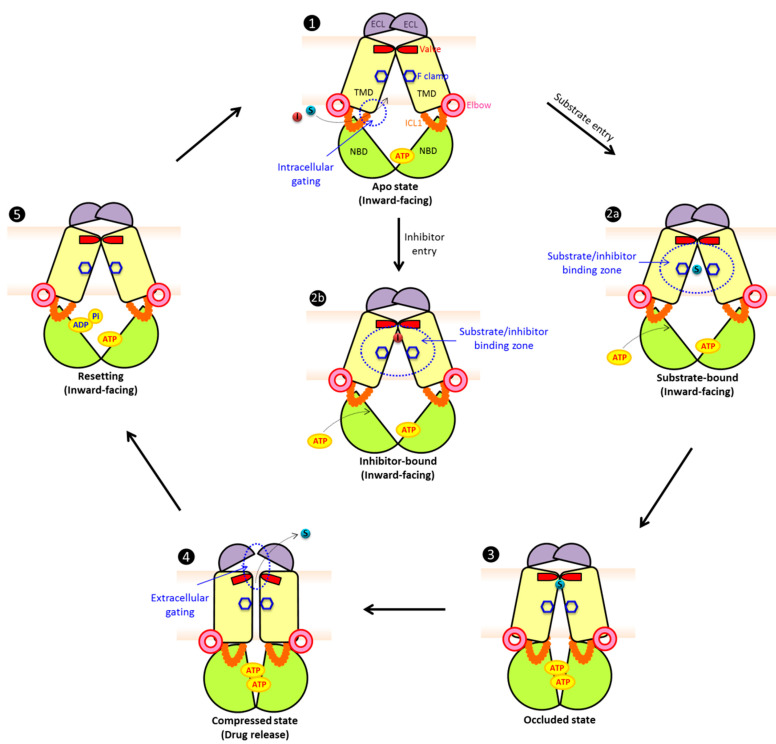
Multidrug resistance (MDR) can be a serious complication for the treatment of cancer as well as for microbial and parasitic infections. Dysregulated overexpression of several members of the ATP-binding cassette transporter families have been intimately linked to MDR phenomena. Three paradigm ABC transporter members, ABCB1 (P-gp), ABCC1 (MRP1) and ABCG2 (BCRP) appear to act as brothers in arms in promoting or causing MDR in a variety of therapeutic cancer settings. However, their molecular mechanisms of action, the basis for their broad and overlapping substrate selectivity, remains ill-posed. The rapidly increasing numbers of high-resolution atomic structures from X-ray crystallography or cryo-EM of mammalian ABC multidrug transporters initiated a new era towards a better understanding of structure-function relationships, and for the dynamics and mechanisms driving their transport cycles. In addition, the atomic structures offered new evolutionary perspectives in cases where transport systems have been structurally conserved from bacteria to humans, including the pleiotropic drug resistance (PDR) family in fungal pathogens for which high resolution structures are as yet unavailable. In this review, we will focus the discussion on comparative mechanisms of mammalian ABCG and fungal PDR transporters, owing to their close evolutionary relationships. In fact, the atomic structures of ABCG2 offer excellent models for a better understanding of fungal PDR transporters. Based on comparative structural models of ABCG transporters and fungal PDRs, we propose closely related or even conserved catalytic cycles, thus offering new therapeutic perspectives for preventing MDR in infectious disease settings.

Keywords: ABC transporters; anticancer; antifungal resistance; mechanism; multidrug transporter; yeast.


Conflict of interest statement

The authors have no competing financial interests.

Figures

Figure 1
Figure 1
Phylogeny and structural organization of ABC transporters in mammals and yeast. (A) The phylogenetic tree shows the evolutionary relationships of ABC transporter subfamilies in yeast and mammals. Some 29 ABC transporters were subjected to amino acid sequence alignments. Branch length was analyzed using MEGA-X, and represents the evolutionary distance in the units of the number of amino acid substitutions per site. Names are given in the UniProt code, protein name and organism, respectively. The analysis reveals two major exporters subfamilies referred to as type I and II. Type I was sub-classified into ABCB/MDR (blue) and ABCC/MRP (green) subgroups. The type II family represents ABCG/PDR subgroups with the fungal (red) and mammalian transporters (orange). Ca: Candida albicans, Cg: Candida glabrata, Caur: Candida auris, Cd: Candida dubliniensis, Ct: Candida tropicalis, Cn: Cryptococcus neoformans, Sc: Saccharomyces cerevisiae, Sp: Schizosaccharomyces pombe. (B) Predicted membrane topologies of three MDR ABC exporter families. The transporters hold several diagnostic hallmark domains, including two NBDs (NBD1: light green; NBD2: green), two TMD regions usually with 6 putative membrane-spanning helices each (TMD1: light yellow; TMD2: bright orange), elbow helix (pink), re-entry helix (blue) and TMD0 (purple), respectively.
Figure 2
Figure 2
Atomic structures, folds and evolutionary conservation of MDR exporters. (A) Crystal or cryo-EM particle structures of multidrug resistance-related ABC exporters from the type I (ABCB and ABCC) and type II families (ABCG subfamily) using the color codes as shown in Figure 1B. (B) Conservation analysis of ABC exporters from panel A. Multiple sequence alignments of amino acid sequences from Table 1 and Table 2 were generated for all subfamilies. The degree of conservation was calculated and indicated as a color gradient ranging from low conservation (yellow) to high conservation (blue).
Figure 3
Figure 3
Type II mammalian and fungal ABCG/PDR exporters share functional domains. (A) A homology model of the PDR exporter Cdr1 from C. albicans was generated using the SWISS-MODEL tool (color ribbon, NBD: green, TMD: yellow, elbow helix: pink, ICL: orange, valve: red, re-entry helix: blue and ECL: purple). The Cdr1 model was superimposed with the cryo-EM structure of human ABCG2 (PDB ID: 6VXF) (gray ribbon). (B) Zoom-in top view of the NBD dimer from panel A at the NBD–NBD interface shows essential conserved motifs (Walker A: red, Q-loop: violet, signature: pink, Pro-loop: yellow, Walker B: orange, D-loop: cyan and H-loop: blue). The transmission interfaces of human ABCG2 (C) and C. albicans Cdr1 (D) show a network cluster of triple helical bundles (THB) with conserved tyrosine residues as part of a salt bridge between elbow helix and ICL. Two negative residues are shown as the intracellular gates. Conserved residues are shown as sticks with color-coding as in the topology model. The conserved phenylalanine F clamp F439 in human ABCG2 (E) and the putative F clamp in C. albicans Cdr1 (F) are indicated as sticks. At the extracellular gates, the valve-like structures at the top of central cavity (red) are shown at the corresponding position of human ABCG2 (G) and C. albicans Cdr1 (H). The lid-structure formed by ECLs is shown as a violet ribbon with a surface. The homo-dimeric human ABCG2 transporter has a symmetric lid (G), while the full-size C. albicans Cdr1 transporter has a larger ECL forming the outer lid and part of the roof architecture (H).
Figure 4
Figure 4
Proposed catalytic cycles of mammalian/fungal type II exporters (ABCG/PDR). In the apo substrate-free state (1), the exporter adopts an inward-facing conformation. We propose that NBDs are open with ATP present in at least one NBD or both, which mediates partial NBD dimerization leaving only one accessible ATP-binding region. The intracellular gate(s) at the transmission interface provides access for substrate or inhibitor entry. The aromatic rings at the conserved F clamp form accessible binding sites at the closed transporter valve subtending the closed ECL. Drug substrates (2a) or inhibitors (2b) can enter through intracellular gate(s), preceding their trapping in distinct binding zones in the central cavity. Binding of ATP at the second binding site or both triggers full NBD dimerization and triggers a first conformational change, setting an occluded state (3). The communication between NBD and TMD is regulated via a rigid triple helical bundle (THB) as a key part of the transmission interface. The NBD dimerization compresses the central cavity space to drive substrate movement through the translocation, thus engaging a push and squeeze motion to open the valve. Substrates shift into the upper cavity and are released by the subsequent opening of the ECL lid (4). ATP hydrolysis at one NBD site may be enough to reset the catalytic cycle and to convert the transporter molecule into the inward-facing drug-recognizing state (5). The structures show NBDs (green), elbow helix (pink), TMDs (yellow), ICL (orange), ECL (purple), phenylalanine clamp (blue hexagon), valve (red), substrates (cyan) and inhibitors (red). For more details and references see the main text.

References

    1. Higgins C.F. ABC Transporters: From Microorganisms to Man. Annu. Rev. Cell Biol. 1992;8:67–113. doi: 10.1146/annurev.cb.08.110192.000435. - DOI - PubMed
    1. Kuchler K. The ABC of ABCs: Multidrug resistance and genetic diseases. FEBS J. 2011;278:3189. doi: 10.1111/j.1742-4658.2011.08234.x. - DOI - PubMed
    1. Holland I.B. Rise and rise of the ABC transporter families. Res. Microbiol. 2019;170:304–320. doi: 10.1016/j.resmic.2019.08.004. - DOI - PubMed
    1. Srikant S. Evolutionary history of ATP-binding cassette proteins. FEBS Lett. 2020;594:3882–3897. doi: 10.1002/1873-3468.13985. - DOI - PubMed
    1. Ogasawara F., Kodan A., Ueda K. ABC proteins in evolution. FEBS Lett. 2020;594:3876–3881. doi: 10.1002/1873-3468.13945. - DOI - PubMed

MeSH terms

Substances

LinkOut - more resources