. 2021 Apr 29;11(5):577.
doi: 10.3390/brainsci11050577.

Intra-Brain Connectivity vs. Inter-Brain Connectivity in Gestures Reproduction: What Relationship?

Affiliations

Intra-Brain Connectivity vs. Inter-Brain Connectivity in Gestures Reproduction: What Relationship?

Michela Balconi et al. Brain Sci. .

Abstract

Recently, the neurosciences have become interested in the investigation of neural responses associated with the use of gestures. This study focuses on the relationship between the intra-brain and inter-brain connectivity mechanisms underlying the execution of different categories of gestures (positive and negative affective, social, and informative) characterizing non-verbal interactions between thirteen couples of subjects, each composed of an encoder and a decoder. The study results underline a similar modulation of intra- and inter-brain connectivity for alpha, delta, and theta frequency bands in specific areas (frontal or posterior regions) depending on the type of gesture. Moreover, taking into account the gestures' valence (positive or negative), a similar modulation of intra- and inter-brain connectivity in the left and right sides was observed. This study showed congruence in the intra-brain and inter-brain connectivity trend during the execution of different gestures, underlining how non-verbal exchanges might be characterized by intra-brain phase alignment and implicit mechanisms of mirroring and synchronization between the two individuals involved in the social exchange.

Keywords: EEG; gestures; intra-brain connectivity.


Conflict of interest statement

The authors declare no conflict of interest.

Figures

Figure 1
Figure 1
(A) Experimental procedure for the task administration. The figure shows the trial structure used in the three task blocks. (B) The figure shows the location of EEG channels. The electrodes were located in the following positions: F3, F1, Fz, F2, F4, T7, C3, Cz, C4, T8, P3, P1, P2, P4, O1, and O2.
Figure 2
Figure 2
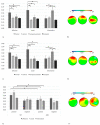
(A) A bar chart of alpha intra-brain connectivity for the three types of gestures (affective, social, and informative) in the frontal, central, temporo-parietal, and occipital areas. The bar chart shows the increase in frontal alpha intra-brain connectivity for affective and social gestures, and the increase in posterior alpha intra-brain connectivity for informative ones. Bars indicate ±1 SE. Statistically significant pairwise comparisons (p < 0.05) are marked by an asterisk. (B) Alpha intra-brain connectivity representations for affective (left side), social (middle), and informative (right side) gestures. The increase in alpha intra-brain connectivity is marked by the red area. (C) A bar chart of delta intra-brain connectivity for the three types of gestures (affective, social, and informative) in frontal, central, temporo-parietal, and occipital areas. The bar chart shows the increase in frontal delta intra-brain connectivity for the affective and social gestures, and the increase in posterior delta intra-brain connectivity for the informative gestures. Bars indicate ±1 SE. Statistically significant pairwise comparisons (p < 0.05) are marked by an asterisk. (D) Delta intra-brain connectivity representations for affective (left side), social (middle), and informative (right side) gestures. The increase in delta intra-brain connectivity is marked by the red area. (E) A bar chart of theta intra-brain connectivity for affective, social, and informative positive and negative gestures in the left and right cerebral side. The bar chart shows the increase in left frontal theta intra-brain connectivity for positive gestures. Bars indicate ±1 SE. Statistically significant pairwise comparisons (p < 0.05) are marked by an asterisk. (F) Theta intra-brain connectivity representations for affective (left side), social (middle), and informative (right side) gestures. The increase in theta intra-brain connectivity is marked by the red area.

References

    1. Esänger J., Emüller V., Elindenberger U. Intra- and interbrain synchronization and network properties when playing guitar in duets. Front. Hum. Neurosci. 2012;6:312. doi: 10.3389/fnhum.2012.00312. - DOI - PMC - PubMed
    1. Cabrera M.E., Novak K., Foti D., Voyles R., Wachs J.P. What Makes a Gesture a Gesture? Neural Signatures Involved in Gesture Recognition; Proceedings of the 12th IEEE International Conference on Automatic Face and Gesture Recognition and 1st International Workshop on Adaptive Shot Learning for Gesture Understanding and Production; Washington, DC, USA. 30 May–3 June 2017.
    1. Carpendale J.I., Carpendale A.B. The Development of Pointing: From Personal Directedness to Interpersonal Direction. Hum. Dev. 2010;53:110–126. doi: 10.1159/000315168. - DOI
    1. Kendon A. Reflections on the “gesture-first” hypothesis of language origins. Psychon. Bull. Rev. 2016;24:163–170. doi: 10.3758/s13423-016-1117-3. - DOI - PMC - PubMed
    1. Tomasello M., Carpenter M., Liszkowski U. A New Look at Infant Pointing. Child Dev. 2007;78:705–722. doi: 10.1111/j.1467-8624.2007.01025.x. - DOI - PubMed

LinkOut - more resources