Review
. 2021 May;70(5):1038-1050.
doi: 10.2337/dbi20-0034. Epub 2021 May 4.

Altered β-Cell Prohormone Processing and Secretion in Type 1 Diabetes

Affiliations
Review

Altered β-Cell Prohormone Processing and Secretion in Type 1 Diabetes

Teresa Rodriguez-Calvo et al. Diabetes. 2021 May.

Abstract

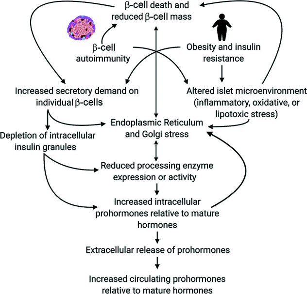
Analysis of data from clinical cohorts, and more recently from human pancreatic tissue, indicates that reduced prohormone processing is an early and persistent finding in type 1 diabetes. In this article, we review the current state of knowledge regarding alterations in islet prohormone expression and processing in type 1 diabetes and consider the clinical impact of these findings. Lingering questions, including pathologic etiologies and consequences of altered prohormone expression and secretion in type 1 diabetes, and the natural history of circulating prohormone production in health and disease, are considered. Finally, key next steps required to move forward in this area are outlined, including longitudinal testing of relevant clinical populations, studies that probe the genetics of altered prohormone processing, the need for combined functional and histologic testing of human pancreatic tissues, continued interrogation of the intersection between prohormone processing and autoimmunity, and optimal approaches for analysis. Successful resolution of these questions may offer the potential to use altered prohormone processing as a biomarker to inform therapeutic strategies aimed at personalized intervention during the natural history of type 1 diabetes and as a pathogenic anchor for identification of potential disease-specific endotypes.


Figures

Figure 1
Figure 1
Processing of proinsulin and processing of proIAPP follow similar steps. Insulin and IAPP follow similar pathways of biogenesis, and processing occurs through a mostly common repertoire of enzymes located within the secretory pathway of the β-cell (1,3). A: Insulin production begins with translation of preproinsulin mRNA to form preproinsulin, which is a polypeptide that contains an N-terminal signal peptide, A and B chains, and a connecting C-peptide and is 100 amino acids in length. Preproinsulin is subsequently translocated into the lumen of the ER, where the N-terminal signal peptide is cleaved and three disulfide bonds are formed between the B and A chains to yield proinsulin. Proinsulin is transported to the Golgi complex and is sorted into immature secretory granules. Within the secretory granules, proinsulin is proteolytically processed to form C-peptide and the mature insulin molecule. Proteolytic processing of proinsulin is the result of sequential cleavage steps by prohormone convertase 1/3 (PC1/3), prohormone convertase 2 (PC2), and carboxypeptidase E (CPE). The classically described pathway of proinsulin processing involves an initial cleavage by PC1/3 at the junction of the B chain and C-peptide (on the C-terminal side of two basic amino acids, Arg31 and Arg32), forming split 32,33 proinsulin. CPE trims dibasic residues at the C-terminal end of the split forms to yield des-31,32 proinsulin, followed by cleavage at the junction of the A chain and C-peptide by PC2 and trimming by CPE to yield insulin and C-peptide. Processing may also begin with PC2 cleavage (C-terminal to Lys64–Arg65) to form split 65,66 proinsulin. CPE trimming yields des-64,65 proinsulin, which is cleaved further by PC1/3 to form insulin and C-peptide. Ex vivo studies as well as analysis of circulating forms of proinsulin in humans, revealing higher levels of the des-31,32 proinsulin intermediate in comparison with des-64,65 proinsulin, support a model where initial cleavage by PC1/3 is strongly favored. B: Similar to preproinsulin, the signal peptide of pre-proIAPP is removed, creating the 67-residue IAPP precursor proIAPP. The C-terminal end of proIAPP is first cleaved by PC1/3 and CPE to produce the proIAPP1–48 intermediate, which is then cleaved at its N-terminal end by PC2. The C-terminus of IAPP is amidated by peptidyl α-amidating monooxygenase (PAM). Alternative processing of proIAPP may occur in states of β-cell dysfunction, and the plasma proIAPP1–48–to–total IAPP ratio is elevated in subjects with type 1 diabetes (6). Recent analyses have suggested that among human β-cells, PC1/3 may be more critical than PC2 for β-cell proinsulin processing, although impacts on human proIAPP processing remain to be tested (62).
Figure 2
Figure 2
Potential pathologic etiologies of increased circulating immature islet prohormones relative to mature hormones. Potential etiologies arising from sources extrinsic and intrinsic to the β-cell, as well as interactions, are displayed.
Figure 3
Figure 3
Immunofluorescent staining showing distribution of proinsulin, insulin, and IAPP in islets from a control donor without diabetes. Scale bars = 50 μm in the low-magnification image and 25 μm in the rest. Anti-proinsulin antibody (GS-9A8; Developmental Studies Hybridoma Bank) (detects the B-C junction and so cross-reactive with intact proinsulin, 65,66 split proinsulin, and des-64,65 proinsulin [validated in 63]) and anti-IAPP antibody (HPA053194; Atlas) were used.
Figure 4
Figure 4
Examples of islet prohormone phenotypes. A: Pancreas sections obtained from nPOD from three individuals (no diabetes, single islet Aab positive, and double islet Aab positive) showing representative examples of increases in proinsulin-positive area (staining further described and quantified in multiple donor samples in 5). Anti-proinsulin antibody was used (GS-9A8; Developmental Studies Hybridoma Bank) (detects the B-C junction and so cross-reactive with intact proinsulin, 65,66 split proinsulin, and des-64,65 proinsulin [validated in 63]). Sections were imaged with a Zeiss Axio Scan.Z1 slide scanner. Scale bars = 25 μm. B: Representative pancreas sections from three individuals obtained from the EADB and nPOD (no diabetes and donors with T1DE1 and T1DE2) showing increased colocalization of insulin and proinsulin in T1DE1 in comparison with no diabetes and T1DE2 (staining further described and quantified in multiple sections in 8). Anti-proinsulin primary antibody was used (ab8301; Abcam) (detects the B-C junction and so cross-reactive with intact proinsulin, 65,66 split proinsulin, and des-64,65 proinsulin). Sections were imaged with use of high-resolution confocal (Leica DMi8, SP8) microscopy. Scale bars = 50 μm.
Figure 5
Figure 5
Examples of islet prohormone phenotypes. A and B: Immunostaining of pancreas sections, obtained from individuals through nPOD, showing islets. A: From a donor without diabetes (scale bars = 20 μm) and a donor with type 1 diabetes with islets showing increased numbers of proinsulin-enriched, insulin-poor cells, indicated with yellow arrows (scale bars = 50 μm). B: From a donor without diabetes and a donor with type 1 diabetes with islets exhibiting staining for C-terminally extended proIAPP. Sections were imaged with a ZEISS LSM 700 confocal microscope (A) and a Leica SP5 confocal microscope (B). Scale bars = 50 μm. Staining in A is further described and quantified in . Anti-proinsulin antibody (GS-9A8; Developmental Studies Hybridoma Bank) (detects the B-C junction and so cross-reactive with intact proinsulin, 65,66 split proinsulin, and des-64,65 proinsulin [validated in 63]) and anti-proIAPP antibody (F063; gift from Medimmune) (raised to an epitope in the C-terminal flanking of peptide of human proIAPP and predicted to detect intact proIAPP) were used.
Figure 6
Figure 6
Key next steps in the field moving forward. Key next steps involve determining the natural history of abnormal relative prohormone expression in the islet and circulation during the progression of type 1 diabetes (T1D), better understanding the pathophysiology leading to these findings, defining the impact of disease treatment, and optimizing prohormone measurement.

References

    1. Vasiljević J, Torkko JM, Knoch KP, Solimena M.. The making of insulin in health and disease. Diabetologia 2020;63:1981–1989 - PMC - PubMed
    1. Westermark P, Andersson A, Westermark GT.. Islet amyloid polypeptide, islet amyloid, and diabetes mellitus. Physiol Rev 2011;91:795–826 - PubMed
    1. Weiss M, Steiner DF, Philipson LH.. Insulin biosynthesis, secretion, structure, and structure-activity relationships. In Endotext. Feingold KR, Anawalt B, Boyce A, et al., Eds. South Dartmouth, MA, 2000. - PubMed
    1. Wasserfall C, Nick HS, Campbell-Thompson M, et al. . Persistence of pancreatic insulin mRNA expression and proinsulin protein in type 1 diabetes pancreata. Cell Metab 2017;26:568–575.e3 - PMC - PubMed
    1. Rodriguez-Calvo T, Zapardiel-Gonzalo J, Amirian N, et al. . Increase in pancreatic proinsulin and preservation of β-cell mass in autoantibody-positive donors prior to type 1 diabetes onset. Diabetes 2017;66:1334–1345 - PMC - PubMed

Publication types

MeSH terms