

Examining age, sex, and race characteristics of velopharyngeal structures in 4- to 9-year old children using magnetic resonance imaging
- PMID: 33948051
- PMCID: PMC8092075
- DOI: 10.1177/1055665617718549
Examining age, sex, and race characteristics of velopharyngeal structures in 4- to 9-year old children using magnetic resonance imaging
Abstract
Objective: The purpose of this study was to quantify the growth of the various craniofacial and velopharyngeal structures and examine sex and race effects.
Methods: Eight-five healthy children (53 White and 32 Black) with normal velopharyngeal anatomy between 4 and 9 years of age who met the inclusion criteria and successfully completed the MRI scans were included in the study.
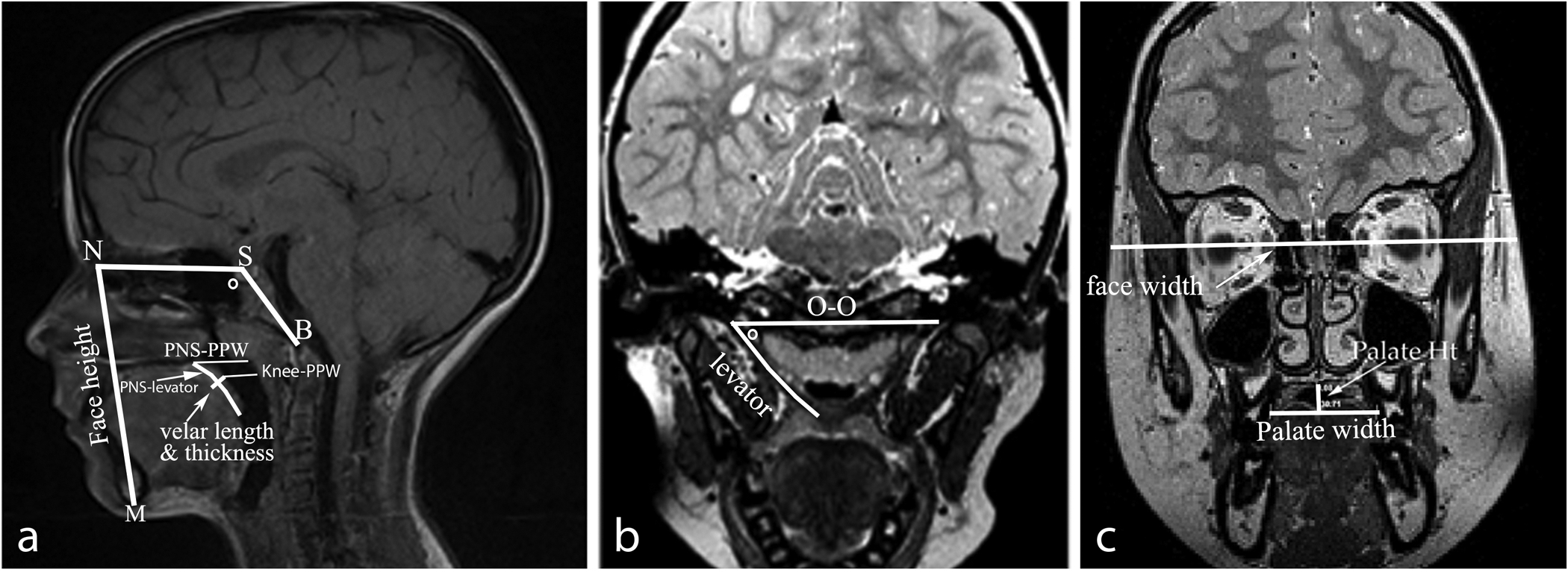
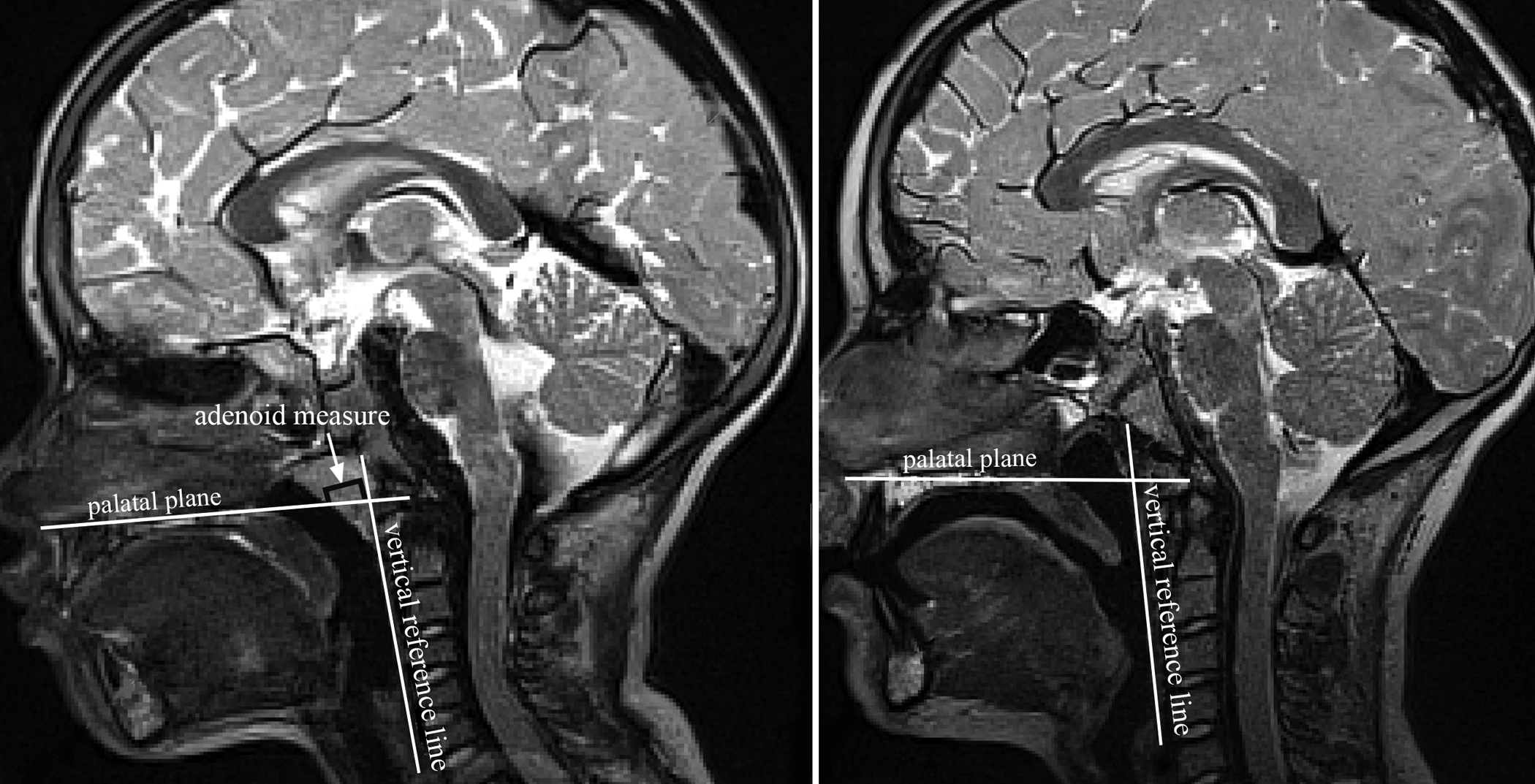
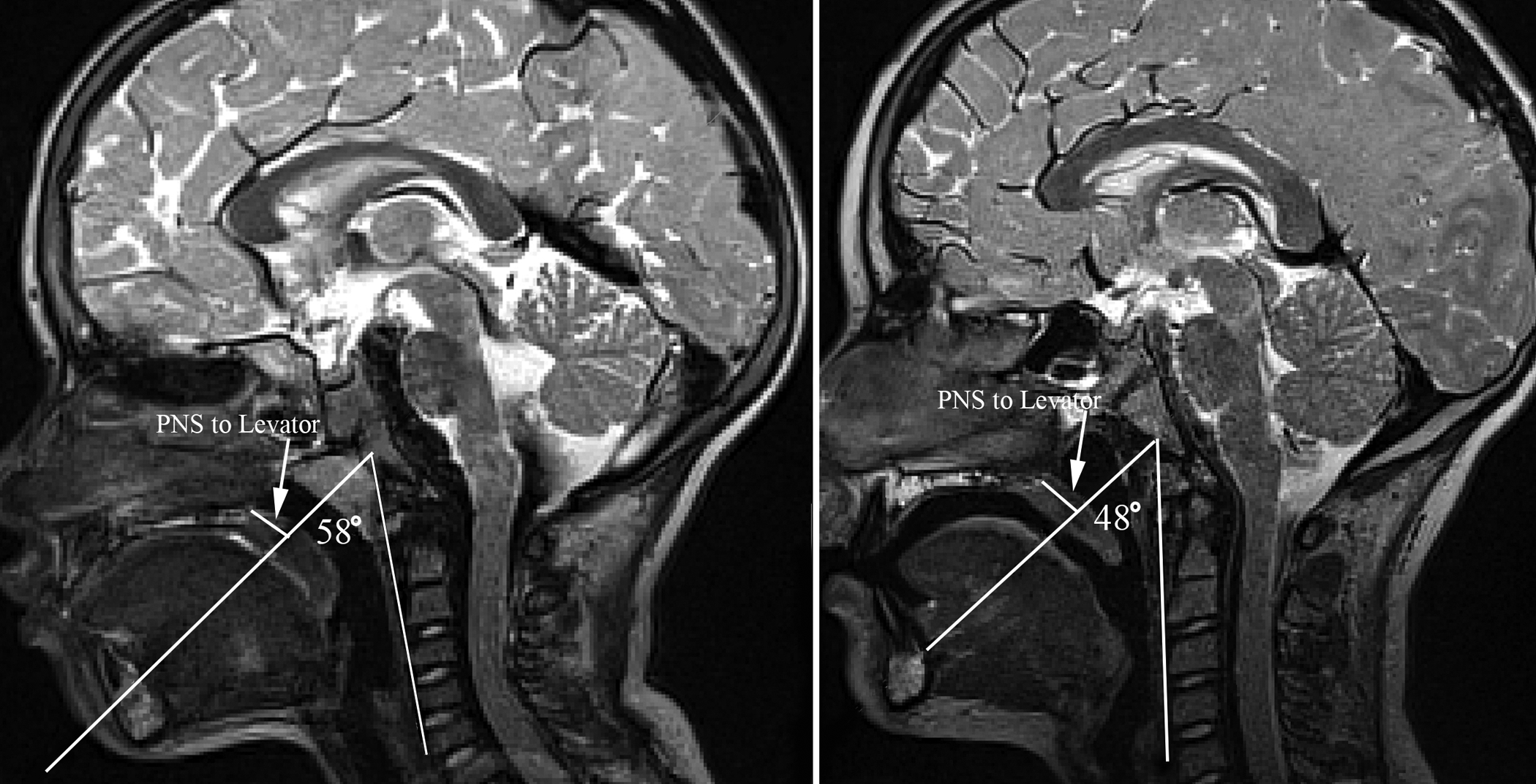
Results: Developmental normative mean values for selected craniometric and velopharyngeal variables by race and sex are reported. Cranial variables (face height, nasion to sella, sella to basion, palate height, palate width) and velopharyngeal variables (levator muscle length, angle of origin, sagittal angle, velar length, velar thickness, velar knee to posterior pharyngeal wall, and posterior nasal spine to levator muscle) demonstrated a trend toward a decrease in angle measures and increase in linear measures as age increased (with the exception of PNS to levator muscle). Only hard palate width and levator muscle length showed a significant sex effect. However, two cranial and six velopharyngeal variables showed a significant race effect. The interactions between sex, race, and age were not statistically significant across all variables, with the exception of posterior nasal spine to posterior pharyngeal wall.
Conclusion: Findings established a large age and race-specific normative reference for craniometiric and velopharyngeal variables. Data reveal minimal sexual dimorphism variables used in the present study; however, significant racial effects were observed.
Keywords: MRI; craniometrics; levator veli palatine; sex differences; velopharyngeal.
Figures
References
-
- Behrents RG. The biological basis for understanding craniofacial growth during adulthood. Prog Clin Biol Res. 1985;187:307–319. - PubMed
-
- Bishara SE, Jakobsen JR, Hession TJ, Treder JE. Soft tissue profile changes from 5 to 45 years of age. Am J Orthod Dentofac. 1998;114:698–706. - PubMed
-
- Bishara SE, Treder JE, Jakobsen JR. Facial and dental changes in adulthood. Am J Orthod Dentofac. 1994;106:175–186. - PubMed
-
- Cheung LK, Chua HD, Bendeus M. Distraction or osteotomy for the correction of maxillary cleft deformities: which is better? Ann R Australas Coll Dent Surg. 2004;17:57–63. - PubMed
Grants and funding
LinkOut - more resources
Full Text Sources
Miscellaneous
