Review
. 2021 Apr 16:13:1759720X211004326.
doi: 10.1177/1759720X211004326. eCollection 2021.

The sonographic identification of cortical bone interruptions in rheumatoid arthritis: a morphological approach

Affiliations
Review

The sonographic identification of cortical bone interruptions in rheumatoid arthritis: a morphological approach

Edoardo Cipolletta et al. Ther Adv Musculoskelet Dis. .

Abstract

Bone erosions are the hallmark of structural damage in rheumatoid arthritis (RA). Among imaging techniques, ultrasonography (US) has emerged as an accurate, reliable, repeatable, low-cost and non-invasive imaging modality to detect erosive changes in RA. However, small interruptions of the cortical bone detectable by last generation US equipment do not necessarily represent bone erosions. According to the available data, in addition to cortical bone interruption itself, only a few morphological US findings have been proposed to define RA bone erosions. However, other additional features may be considered to facilitate the interpretation of US cortical bone interruptions in RA. These could be summarised using the following four domains: size, site, shape and scenery. This hypothesis article provides a critical literature review of US features characteristic of RA bone erosions and pictorial evidence supporting the potential role of a morphological analysis in the US identification of bone erosions in RA patients.

Plain language summary: The ultrasonographic morphology of cortical interruptions is helpful for the identification of bone erosions in rheumatoid arthritis: the "four Ss" approach Bone erosions are characteristic features of rheumatoid arthritis. They are associated with a more aggressive disease and with irreversible physical disability. In recent years, ultrasonography has emerged as an accurate and reliable technique for the detection of bone erosions, that appear as interruptions of the cortical bone with variable size. However, cortical bone interruptions do not necessarily represent bone erosions. Since bone erosions represent the earliest evidence of the destructive behaviour of RA, their identification is crucial.Besides the cortical interruption itself, only a few morphological ultrasonographic features were proposed to characterise bone erosions in rheumatoid arthritis.We believe that a morphological approach, including size, site, shape and scenery, may be considered to facilitate the interpretation of ultrasonographic cortical bone interruptions in rheumatoid arthritis.In this hypothesis article we carried out a critical review of the scientific literature and provided extensive pictorial evidence of the ultrasonographic spectrum of cortical interruptions supporting the potential role of considering the "four Ss" for the ultrasonographic identification of bone erosions in rheumatoid arthritis.

Keywords: OMERACT; bone erosions; rheumatoid arthritis; structural damage; ultrasonography.


Conflict of interest statement

Conflict of interest statement: E.F. has received speaking fees from AbbVie, Bristol-Myers Squibb, Celgene, Janssen-Cilag, Novartis, Pfizer, Roche and Union Chimique Belge Pharma. M.D.C. has received speaking fees from AbbVie, Novartis, Pfizer and Sanofi Aventis. W.G. has received speaking fees from AbbVie, Celgene, Grünenthal, Pfizer and Union Chimique Belge Pharma. All other authors have declared no conflict of interest.

Figures

Figure 1.
Figure 1.
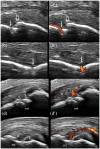
(a–f) Normal cortical bone in healthy subjects. Metacarpophalangeal joint. Longitudinal (a, c) and transverse (b, d) scans obtained at the dorsal (a, b) and the radial (c, d) aspects using a 18 MHz probe. Proximal interphalangeal joint. Longitudinal (e) and transverse (f) scans obtained at the dorsal aspect using a 22 MHz probe. When perpendicularly insonated, normal cortical bone appears as a sharp, regular, continuous and highly hyperechoic line with a posterior acoustic shadowing. mc, metacarpal bone; mp, middle phalanx; pp, proximal phalanx.
Figure 2.
Figure 2.
(a–d) Pseudo-erosions. Metacarpophalangeal joint. Longitudinal (a, a’) and transverse (b, b’) scans obtained at the dorsal aspect using a 22 MHz probe with (a’, b’) and without (a, b) power Doppler. Physiological cortical vascular channels (arrows) are characterised by an interruption of the bony cortex with sharp, parallel and straight margins, submillimetric size and the absence of any sign of synovial inflammation, with or without the presence of a linear Doppler signal that appears as a curved or straight line crossing the bony cortex. Note the step-up appearance of the osteophyte (open arrows) generating a posterior acoustic shadowing and an erosion-like feature (arrowhead) at the metacarpal bone level. Metatarsophalangeal joint. Longitudinal (c, c’) and transverse (d, d’) scans obtained at the medial aspect of the first metatarsal head using a 18 MHz probe with (c’, d’) and without (c, d) power Doppler. Note the presence of a cortical interruption (arrowheads) in a patient with hallux valgus. mc, metacarpal bone; mt, metatarsal bone.
Figure 3.
Figure 3.
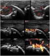
(a–c) Size and shape. Metacarpophalangeal joint. Longitudinal (a) and transverse (a’) scans obtained at the lateral aspect of the metacarpal head using a 18 MHz probe. Note the presence of a ‘hot’ bone erosion (arrowheads) characterised by a large diameter (3.4 mm) and power Doppler within the crater. Metacarpophalangeal joint. Longitudinal (b, b’) and transverse (c, c’) scans obtained at the dorsal side of the metacarpal head using a 18 MHz probe, showing smaller (ranging from 0.5 to 1.2 mm), multiple ‘hot’ bone erosions with inflamed synovial tissue within the erosive crater. Note the irregular margins of the bone erosions. mc, metacarpal bone; pp, proximal phalanx.
Figure 4.
Figure 4.
(a–d) Site. Metacarpophalangeal joint. Longitudinal (a, a’, c, c’) and transverse (b, d) scans obtained at the dorsal side of the metacarpal head using a 22 MHz probe with (a’, c’) and without (a, b, c, d) power Doppler, revealing active submillimetric [0.4 mm (a, b); 1.1 mm (c, d)] subchondral cortical breaks (arrowheads), indicative of bone erosions. mc, metacarpal bone; pp, proximal phalanx.
Figure 5.
Figure 5.
Scenery. (a, b) Metacarpophalangeal joint. Longitudinal (a, a’) and transverse (b, b’) scans obtained at the dorsal aspect using a 18 MHz probe with (a’, b’) and without (a, b) power Doppler. Note a cortical break (arrowheads) in the area of the metacarpal depression, which is a common site for pseudo-erosions. The surrounding scenery, characterised by florid synovial hypertrophy (+) showing power Doppler signal in contact with, and invading into, the cortical break (open arrows), provides additional information supporting the diagnosis of bone erosion. The deep echoes (curved arrows) in b and b’ suggest the presence of bone erosions despite submillimetric small cortical interruptions (0.2 and 0.6 mm) (arrowheads). mc, metacarpal bone; pp, proximal phalanx.
Figure 6.
Figure 6.
(a–f) Microerosions and other atypical cortical interruptions. Metatarsophalangeal joint. Transverse (a, b) and longitudinal (c, d) scans obtained at the lateral aspect of the metatarsal head of the 5th digit using a 18 MHz probe showing a Ω-shaped bone erosion characterised by Doppler signal and deep echoes (curved arrows) with a small cortical break (0.24 mm) (arrowheads). Proximal interphalangeal join. Transverse (e, e’) scans obtained at the dorsal aspect with (e’) and without (e) power Doppler using a 22 MHz probe revealing a loss of sharpness of the bony cortex suggestive for a pre-erosive change (white arrow) and a definite cortical break indicative of a microerosion (arrowhead). Note the presence of highly vascularised synovial hypertrophy contacting the bone surface (open arrows). Metatarsophalangeal joint. Longitudinal (f and f’) scans obtained at the lateral aspect of the metatarsal head of the 5th digit with (f’) and without (f) power Doppler using a 18 MHz probe showing both a cortical defect without a definite cortical interruption and a loss of sharpness of the bone surface. The presence of Doppler signal contacting the bony cortex (open arrows) and the absence of this notch in the contralateral 5th metatarsal head are highly indicative of an atypical bone erosion. mt, metatarsal bone; pp, proximal phalanx.

Similar articles

Cited by

References

    1. McInnes IB, Schett G. The pathogenesis of rheumatoid arthritis. N Engl J Med 2011; 365: 2205–2219. - PubMed
    1. Cipolletta E, Hurnakova J, Di Matteo A, et al.. Prevalence and distribution of cartilage and bone damage at metacarpal head in healthy subjects. Clin Exp Rheumatol, Epub ahead of print 2 March 2021. - PubMed
    1. Hurnakova J, Filippucci E, Cipolletta E, et al.. Prevalence and distribution of cartilage damage at the metacarpal head level in rheumatoid arthritis and osteoarthritis: an ultrasound study. Rheumatology (Oxford) 2019; 58: 1206–1213. - PubMed
    1. Cipolletta E, Filippucci E, Di Matteo A, et al.. The reliability of ultrasound in the assessment of hyaline cartilage in rheumatoid arthritis and healthy metacarpal heads. Ultraschall Med. Epub ahead of print 30 October 2020. DOI: 10.1055/a-1285-4602. - DOI - PubMed
    1. Aletaha D, Funovits J, Smolen JS. Physical disability in rheumatoid arthritis is associated with cartilage damage rather than bone destruction [published correction appears in Ann Rheum Dis 2011; 70: 1880]. Ann Rheum Dis 2011; 70: 733–739. - PubMed

LinkOut - more resources