. 2021 Apr 15:12:2040622320987348.
doi: 10.1177/2040622320987348. eCollection 2021.

EZH2 is involved in psoriasis progression by impairing miR-125a-5p inhibition of SFMBT1 and leading to inhibition of the TGFβ/SMAD pathway

Affiliations

EZH2 is involved in psoriasis progression by impairing miR-125a-5p inhibition of SFMBT1 and leading to inhibition of the TGFβ/SMAD pathway

Shengming Qu et al. Ther Adv Chronic Dis. .

Abstract

Aims: In this study, we aimed to decipher the impact of enhancer of zeste homolog 2 (EZH2) in psoriasis as well as the underlying mechanism.

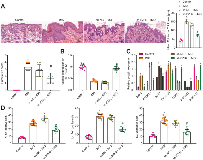
Methods: A mouse model of psoriasis was developed by means of imiquimod induction, with the expression of EZH2, microRNA-125a-5p (miR-125a-5p), and SFMBT1 determined. The role of EZH2, miR-125a-5p, and SFMBT1 in malignant phenotypes of HaCaT cells and the development of psoriasis in vivo was subsequently investigated through gain- and loss-of-function experiments. Chromatin immunoprecipitation assay and dual-luciferase reporter assay were conducted to explore the relationship between EZH2 or SFMBT1 and miR-125a-5p. Finally, the effects of EZH2 and miR-125a-5p on the transforming growth factor β (TGFβ)/SMAD pathway were analyzed.

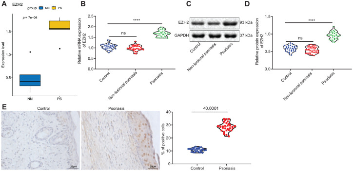
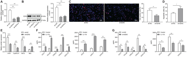
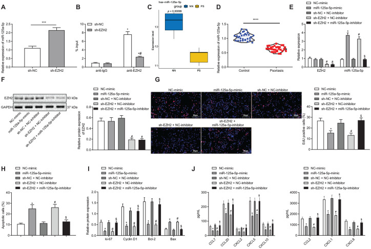
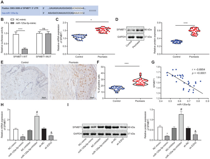
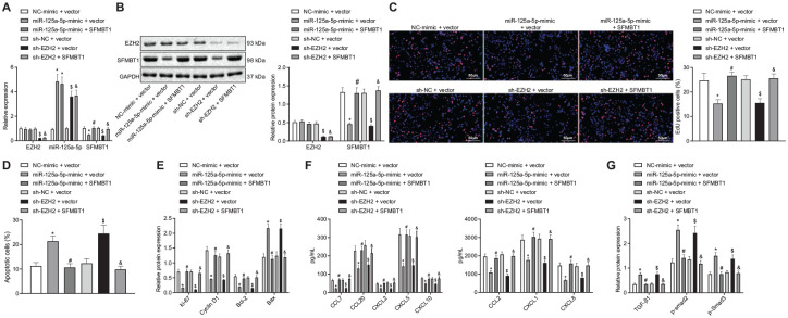
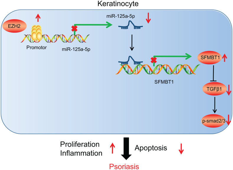
Results: Overexpressed SFMBT1 and EZH2 was detected while miR-125a-5p were downregulated in psoriasis tissues and human keratinocyte (HaCaT) cells. EZH2 increased the levels of IL-17A-induced cytokines and promoted the malignant phenotypes of HaCaT cells. Functionally, EZH2 reduced miR-125a-5p expression while miR-125a-5p targeted SFMBT1 to activate the TGFβ/SMAD pathway in vitro. Knockdown of EZH2 or up-regulation of miR-125a-5p inhibited cell proliferation and the levels of IL-17A-induced cytokines, but increased the expression of TGFβ1 and the extent of smad2 and smad3 phosphorylation in HaCaT cells. Notably, EZH2 contributed to the development of psoriasis in vivo by inhibiting the TGFβ/SMAD pathway via impairment of miR-125a-5p-mediated SFMBT1 inhibition.

Conclusion: Taken together, the results of the current study highlight the ability of EZH2 to potentially inactivate the TGFβ/SMAD pathway via upregulation of miR-125a-5p-dependent SFMBT1during the progression of psoriatic lesions.

Keywords: EZH2; SFMBT1; TGFβ/SMAD pathway; microRNA-125a-5p; psoriasis.


Conflict of interest statement

Conflict of interest statement: The authors declare that there is no conflict of interest.

Figures

Figure 1.
Figure 1.
EZH2 was highly expressed in psoriasis. (A) Expression of EZH2 in the microarray database (NN on the horizontal axis represents normal skin tissues and PS represents psoriatic skin tissues). (B) Reverse transcription-quantitative polymerase chain reaction determination of the EZH2 expression in psoriatic skin tissues and normal skin tissues in non-lesional skin and lesional skin tissues. (C) Western blot analysis of the EZH2 expression in psoriatic skin tissues and normal skin tissues in non-lesional skin and lesional skin tissues normalized to GAPDH. (D) Quantitation of panel C. (E) Immunohistochemistry detection of the EZH2 expression in psoriatic skin tissues and normal skin tissues (×400). The results of the data were measurement data, which were presented as the mean ± standard deviation and analyzed by independent-sample t-test, N = 30. EZH2, enhancer of zeste homolog 2; GAPDH, glyceraldehyde-3-phosphate dehydrogenase; mRNA, messenger RNA.
Figure 2.
Figure 2.
Down-regulation of EZH2 suppressed keratinocyte proliferation and IL-17A-induced cytokine levels. (A) Reverse transcription-quantitative polymerase chain reaction determination of EZH2 expression in human keratinocyte (HaCaT) cells after sh-EZH2-1 and sh-EZH2-2 treatment. (B) Western blot analysis of the EZH2 expression in HaCaT cells after sh-EZH2-1 and sh-EZH2-2 treatment normalized to GAPDH. (C) EdU detection of HaCaT cell proliferation after sh-EZH2 treatment (×200). (D) HaCaT cell apoptosis after sh-EZH2 treatment assessed by flow cytometry. (E) Western blot analysis of the expression of Ki-67, Cyclin D1, Bcl-2, and Bax in HaCaT cells after sh-EZH2 treatment normalized to GAPDH. (F) CCL2, CCL7, CCL20, CXCL1, CXCL2, CXCL5, CXCL8, and CXCL10 levels in HaCaT cells induced by IL-17A. G, CCL2, CCL7, CCL20, CXCL1, CXCL2, CXCL5, CXCL8, and CXCL10 levels in HaCaT cells after EZH2 knockdown. In panels A–G, *p < 0.05 compared with treatment of sh-NC, and in panel F, *p < 0.05 compared with control. The results of the data were measurement data, which were presented as the mean ± standard deviation and analyzed by independent-sample t-test. Also, data between multiple groups were compared with one-way analysis of variance. *, *** and **** indicate comparison between the two groups. EdU, 5-ethynyl-2′-deoxyuridine; EZH2, enhancer of zeste homolog 2; sh, short hairpin RNA; GAPDH, glyceraldehyde-3-phosphate dehydrogenase; NC, negative control; mRNA, messenger RNA.
Figure 3.
Figure 3.
Down-regulation of EZH2 increased the expression of miR-125a-5p to impede the proliferation of keratinocytes and decreased IL-17A-induced cytokine levels. (A) Reverse transcription-quantitative polymerase chain reaction (RT-qPCR) determination of the expression of miR-125a-5p in human keratinocyte (HaCaT) cells after sh-EZH2 treatment, *p < 0.05 compared with the treatment of sh-NC, ***p < 0.001. (B) ChIP results of EZH2 enrichment in the miR-125a-5p promoter region in HaCaT cells after sh-EZH2 treatment, *p < 0.05 compared with treatment of anti-IgG, and #p < 0.05 compared with treatment of anti-EZH2. (C) Expression of hsa-miR-125a-5p in the microarray database (NN in the abscissa indicates normal skin tissue and PS indicates psoriatic skin tissue). (D) RT-qPCR determination of the miR-125a-5p expression in psoriatic skin tissues and normal skin tissues, *p < 0.05 compared with normal skin tissues, ****p < 0.0001. (E) RT-qPCR determination of the EZH2 and miR-125a-5p expression in HaCaT cells after miR-125a-5p mimic, sh-EZH2, or miR-125a-5p inhibitor treatment. (F) Western blot analysis of the EZH2 expression in HaCaT cells after miR-125a-5p mimic, sh-EZH2, or miR-125a-5p inhibitor treatment normalized to GAPDH. (G) EdU assay of HaCaT cell proliferation after miR-125a-5p mimic, sh-EZH2, or miR-125a-5p inhibitor treatment (×200). (H) HaCaT cell apoptosis after miR-125a-5p mimic, sh-EZH2, or miR-125a-5p inhibitor treatment assessed by flow cytometry. (I) Western blot analysis of the expression of Ki-67, Cyclin D1, Bcl-2, and Bax in HaCaT cells after miR-125a-5p mimic, sh-EZH2, or miR-125a-5p inhibitor treatment normalized to GAPDH. (J) Cytokine levels in HaCaT cells after miR-125a-5p mimic, sh-EZH2, or miR-125a-5p inhibitor treatment. In panels D–I, *p < 0.05 compared with treatment of NC-mimic, #p < 0.05 compared with treatment of sh-NC + NC-inhibitor, and $p < 0.05 compared with treatment of sh-EZH2 + NC-inhibitor. The results of the data were measurement data, which were presented as the mean ± standard deviation and analyzed by independent-sample t-test. Also, data between multiple groups were compared with one-way analysis of variance. EdU, 5-ethynyl-2′-deoxyuridine; EZH2, enhancer of zeste homolog 2; sh, short hairpin RNA,; GAPDH, glyceraldehyde-3-phosphate dehydrogenase; miR-125a-5p, microRNA-125a-5p; NC, negative control; sh, short hairpin RNA.
Figure 4.
Figure 4.
EZH2 inhibited the expression of miR-125a-5p to up-regulate SFMBT1 expression. (A) Binding sites between miR-125a-5p and SFMBT1 predicted through the Targetscan database. (B) Dual-luciferase reporter assay results of interaction between miR-125a-5p and SFMBT1, ****p < 0.0001 compared with the treatment of wild-type NC. (C) Reverse transcription-quantitative polymerase chain reaction (RT-qPCR) determination of SFMBT1 expression in psoriatic skin tissues and controls, *p < 0.05 compared with control, N = 30. (D) Western blot analysis of SFMBT1 expression in psoriatic skin tissues and controls normalized to GAPDH, ****p < 0.0001 compared with control, N = 30. (E) Immunohistochemistry of SFMBT1 expression in psoriatic skin tissues and controls (×400). (F) Quantitation of panel E. ****p < 0.0001 compared with control, N = 30. (G) Correlation analysis between miR-125a-5p expression and SFMBT1 expression in clinical skin tissue samples of psoriasis. (H) RT-qPCR determination of SFMBT1 expression in human keratinocyte (HaCaT) cells after miR-125a-5p-mimic, miR-125a-5p-inhibitor, or sh-EZH2 treatment, *p < 0.05 compared with treatment of NC-mimic, #p < 0.05 compared with treatment of NC-inhibitor, and $p < 0.05 compared with treatment of sh-NC. (I) Western blot analysis of SFMBT1 expression in HaCaT cells after miR-125a-5p-mimic, miR-125a-5p-inhibitor, or sh-EZH2 treatment normalized to GAPDH, *p < 0.05 compared with treatment of NC-mimic, #p < 0.05 compared with treatment of NC-inhibitor, and p < 0.05 compared with treatment of sh-NC, &p < 0.05 compared with treatment of sh-NC. The data were measurement data, which were presented as the mean ± standard deviation. Data between the two groups were compared using an independent-sample t-test while data between multiple groups were compared using one-way analysis of variance. Pearson’s analysis was used to determine the correlation between miR-125a-5p and SFMBT1. EZH2, enhancer of zeste homolog 2; GAPDH, glyceraldehyde-3-phosphate dehydrogenase; miR-125a-5p, microRNA-125a-5p; MUT, mutant type; NC, negative control; SFMBT1, Scm (Sex comb on midleg) with four MBT (malignant brain tumor) domains 1; sh, short hairpin RNA; WT, wild type; mRNA, messenger RNA.
Figure 5.
Figure 5.
EZH2 inactivated the TGFβ/SMAD pathway by regulating the expression of miR-125a-5p-mediated SFMBT1 to promote keratinocyte proliferation and increase IL-17A-induced cytokine levels. (A) Reverse transcription-quantitative polymerase chain reaction determination of the expression of EZH2, miR-125a-5p, and SFMBT1 in human keratinocyte (HaCaT) cells after miR-125a-5p-mimic, SFMBT1, or sh-EZH2 treatment. (B) Western blot analysis of the EZH2 and SFMBT1 expression in HaCaT cells after miR-125a-5p-mimic, oe-SFMBT1, or sh-EZH2 treatment, normalized to GAPDH. (C) HaCaT cell proliferation assessed by EdU assay after miR-125a-5p-mimic, SFMBT1, or sh-EZH2 treatment (×200). (D) HaCaT cell apoptosis assessed by flow cytometry after miR-125a-5p-mimic, SFMBT1, or sh-EZH2 treatment. (E) Western blot analysis of the Ki-67, Cyclin D1, Bcl-2, and Bax expression in HaCaT cells after miR-125a-5p-mimic, SFMBT1, or sh-EZH2 treatment, normalized to GAPDH. (F) Levels of cytokines CCL2, CCL7, CCL20, CXCL1, CXCL2, CXCL5, CXCL8, and CXCL10 in HaCaT cells after miR-125a-5p-mimic, SFMBT1, or sh-EZH2 treatment. (G) Western blot analysis of the expression of TGFβ1, and the extent of smad2 and smad3 phosphorylation in HaCaT cells after miR-125a-5p-mimic, SFMBT1, or sh-EZH2 treatment, normalized to GAPDH. *p < 0.05 compared with treatment of NC-mimic + vector, #p < 0.05 compared with treatment of miR-125a-5p-mimic + vector, $p < 0.05 compared with treatment of sh-NC + vector, and &p < 0.05 compared with treatment of sh-EZH2 + vector. The results of the data were measurement data and presented as the mean ± standard deviation. Data between multiple groups were compared using one-way analysis of variance. EZH2, enhancer of zeste homolog 2; GAPDH, glyceraldehyde-3-phosphate dehydrogenase; miR-125a-5p, microRNA-125a-5p; NC, negative control; SFMBT1, Scm (Sex comb on midleg) with four MBT (malignant brain tumor) domains 1; sh, short hairpin RNA; TGFβ, transforming growth factor-β.
Figure 6.
Figure 6.
Knockdown of EZH2 in vivo activated the TGFβ/SMAD pathway through regulation of miR-125a-5p-mediated SFMBT1 to inhibit psoriasis. (A) Representative macroscopic images of dorsal skin samples and hematoxylin-eosin (HE) staining of skin sections obtained from mice on the fifth day from the start of lentiviral injection, and the cumulative score of epidermal thickness, and erythema of HE-stained skin sections of mice after IMQ induction or IMQ-induced mice with sh-EZH2 treatment was quantified (×200), N = 8. (B) Reverse transcription-quantitative polymerase chain reaction determination of the miR-125a-5p expression in skin tissues of mice after IMQ induction or IMQ-induced mice with sh-EZH2 treatment. (C) Western blot analysis of the expression of EZH2, SFMBT1, Ki-67, Cyclin D1, Bcl-2, and TGFβ1, along with the extent of smad2 and smad3 phosphorylation in skin tissues of mice after IMQ induction or IMQ-induced mice with sh-EZH2 treatment normalized to GAPDH. (D) Immunohistochemistry measurement of Ki-67, IL-17A and EZH2 expression in skin tissues of mice after IMQ induction or IMQ-induced mice with sh-EZH2 treatment. *p < 0.05 compared with control, #p < 0.05 compared with IMQ-induced mice with sh-NC treatment. The results of the data were measurement data and presented by the mean ± standard deviation. Data between multiple groups were compared using one-way analysis of variance. EZH2, enhancer of zeste homolog 2; GAPDH, glyceraldehyde-3-phosphate dehydrogenase; IMQ, imiquimod; miR-125a-5p, microRNA-125a-5p; NC, negative control; p, phosphorylated; SFMBT1, Scm (Sex comb on midleg) with four MBT (malignant brain tumor) domains 1; sh, short hairpin RNA; TGFβ, transforming growth factor-β.
Figure 7.
Figure 7.
Schematic diagram of the proposed mechanism. EZH2 is enriched in the miR-125a-5p promoter and inhibits miR-125a-5p expression, thereby increasing the expression of the miR-125a-5p target SFMBT1. SFMBT1 further inhibits the expression of TGFβ1, and the extent of smad2 and smad3 phosphorylation (p-smad2/3), and consequently impedes keratinocyte apoptosis and promotes keratinocyte proliferation as well as the inflammatory reactions, eventually exacerbating psoriasis. EZH2, enhancer of zeste homolog 2; miR-125a-5p, microRNA-125a-5p; SFMBT1, Scm (Sex comb on midleg) with four MBT (malignant brain tumor) domains 1; TGFβ, transforming growth factor-β.

Similar articles

Cited by

References

    1. Nestle FO, Kaplan DH, Barker J. Psoriasis. N Engl J Med 2009; 361: 496–509. - PubMed
    1. Boehncke WH, Schon MP. Psoriasis. Lancet 2015; 386: 983–994. - PubMed
    1. Albanesi C, Madonna S, Gisondi P, et al.. The interplay between keratinocytes and immune cells in the pathogenesis of psoriasis. Front Immunol 2018; 9: 1549. - PMC - PubMed
    1. Furue M, Furue K, Tsuji G, et al.. Interleukin-17A and keratinocytes in psoriasis. Int J Mol Sci 2020; 21: 1275. - PMC - PubMed
    1. Lebwohl M. Psoriasis. Ann Intern Med 2018; 168: ITC49–ITC64. - PubMed

LinkOut - more resources