Review
. 2021;4(3):675-687.
doi: 10.1007/s42242-021-00125-8. Epub 2021 Apr 30.

Three-dimensional printing for heart diseases: clinical application review

Affiliations
Review

Three-dimensional printing for heart diseases: clinical application review

Yanyan Ma et al. Biodes Manuf. 2021.

Abstract

Heart diseases remain the top threat to human health, and the treatment of heart diseases changes with each passing day. Convincing evidence shows that three-dimensional (3D) printing allows for a more precise understanding of the complex anatomy associated with various heart diseases. In addition, 3D-printed models of cardiac diseases may serve as effective educational tools and for hands-on simulation of surgical interventions. We introduce examples of the clinical applications of different types of 3D printing based on specific cases and clinical application scenarios of 3D printing in treating heart diseases. We also discuss the limitations and clinically unmet needs of 3D printing in this context.

Keywords: Cardiac imaging techniques; Congenital heart disease; Heart diseases; Three-dimensional printing; Transcatheter aortic valve replacement.


Conflict of interest statement

Conflict of interestThe authors declare that they have no conflict of interest.

Figures

Fig. 1
Fig. 1
Three-dimensional printing flowchart of the cardiac system. MRI: magnetic resonance imaging, CTA: computed tomography angiography, TEE: transesophageal echocardiography, 3D: three-dimensional
Fig. 2
Fig. 2
Function of three-dimensional printing in the cardiovascular field. 3D: three-dimensional, PVL: paravalvular leak, LVOT: left ventricular outflow tract
Fig. 3
Fig. 3
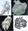
Three-dimensional (3D)-printed model of congenital heart diseases. a Computer modeling shows the internal structural profile of complex congenital heart diseases and coarctation of the aorta. The ascending aorta and descending aorta remain normal. b 3D-printed model of cor triatriatum. The left atrium of the patient is separated into false and true chambers by a septum; the structures of the left ventricle, the right atrium, and the right ventricle remain normal. c 3D-printed model of a double outlet right ventricle. The right ventricle of the patient is connected to both the aorta and the pulmonary artery. d 3D-printed model of a coronary fistula. A giant right coronary artery fistula drained to the left ventricle is shown. AAo: ascending aorta, CAF: coronary artery fistula, CoA: coarctation of the aorta, DAo: descending aorta, Ao: aorta, LAA: left atrial appendage, LA: left atrium, LV: left ventricle, RA: right atrium, RV: right ventricle. Image data, 3D computer reconstructions, and 3D-printed models are from the Department of Cardiovascular Surgery, Xijing Hospital
Fig. 4
Fig. 4
Three-dimensional (3D)-printed models of congenital heart diseases used for training. a 3D-printed model of a ventricular septal defect. b Training for the basic operative skills of suturing using a 3D-printed model. c Training young doctors and medical students using a 3D-printed model. d Evaluation of the suture technique using a 3D-printed model. VSD: ventricular septal defect, IVC: inferior vena cava, LV: left ventricle, RV: right ventricle, TV: tricuspid valve. The 3D-printed models are from the Department of Cardiovascular Surgery, West China Hospital
Fig. 5
Fig. 5
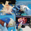
Three-dimensional (3D)-printed heart model to mimic the radiofrequency ablation of atrial fibrillation and a mitral valve repair operation in a patient with mirror-image dextrocardia. a 3D-printed heart model with planned ablation route; b surgical planning of the ablation route; c using the atrial fibrillation clamp to mimic the ablation procedure; d 3D printing technique-guided radiofrequency ablation of atrial fibrillation and mitral valve repair operation. IVC: inferior vena cava, LSPV: left superior pulmonary vein, LIPV: left inferior pulmonary vein, LV: left ventricle, RIPV: right inferior pulmonary vein, RSPV: right superior pulmonary vein, RV: right ventricle, SVC: superior vena cava. Image data, 3D computer reconstruction, and 3D-printed model are from the Department of Cardiovascular Surgery, Xiangya Second Hospital of Central South University
Fig. 6
Fig. 6
Three-dimensional (3D)-printed heart model to mimic the transcatheter aortic valve replacement (TAVR) procedure. a Transducing system across the valve; b releasing the step 1 button; c fully releasing the stented valve; d left ventricle view of the model to evaluate the results of the TAVR procedure. Ao: aorta, LCA: left coronary artery, LCC: left coronary cusp, LVOT: left ventricular outflow tract, NCC: non-coronary cup, RCA: right coronary artery, RCC: right coronary cups. Image data, 3D computer reconstruction, and 3D-printed model are from the Department of Cardiovascular Surgery, Xijing Hospital
Fig. 7
Fig. 7
Three-dimensional (3D) digital model to evaluate the risk of coronary artery occlusion during TAVR. ac Computer 3D model shows the calcification of the valve leaflets and the height of the coronary artery in patients at risk of coronary artery occlusion. d 3D-printed model shows the calcification of the valve leaflets and the height of the coronary artery. e In vitro balloon dilation using the 3D-printed model to evaluate the possibility of coronary obstruction. f In vitro valve implantation using the 3D-printed model to evaluate the possibility of coronary obstruction. LCA: left coronary artery, RCA: right coronary artery. Image data, 3D computer reconstruction, and 3D-printed model are from the Department of Cardiovascular Surgery, Xijing Hospital
Fig. 8
Fig. 8
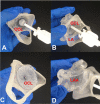
Three-dimensional (3D)-printed simulated left atrial appendage occlusion. a Partially released device using the 3D-printed left atrial appendage model; b push–pull experiment to test the stability of the device, c fully released device using the 3D-printed left atrial appendage model, d left atrial appendage view of the model to evaluate the results of the procedure. LAA: left atrial appendage, LA: left atrium, OCL: occluder. Image data, 3D computer reconstruction, and 3D-printed model are from the Department of Cardiovascular Surgery, the First Affiliated Hospital of Xi’an Jiaotong University
Fig. 9
Fig. 9
Ultrasound-guided percutaneous transluminal myocardial septal radiofrequency ablation (Liwen procedure) for the treatment of obstructive hypertrophic cardiomyopathy. a Computed tomography angiography (CTA) two-dimensional imaging display of the hypertrophic septum and the surgical path of radiofrequency ablation. The red line is a simulated electroacupuncture approach. The approach is shown to avoid the surface coronary artery and to ablate the septal branches during the operation. b Ultrasound images display the hypertrophic septum; the computer section shows the myocardium. c Digital three-dimensional (3D) model based on CTA images of the hypertrophic septum and the inner structure of the heart. d Digital 3D model based on CTA image display of the hypertrophic septum and the outer structure of the heart. e 3D cardiac printed model based on CTA images of the hypertrophic septum and the coronary artery branch. f Determination of the optimal puncture site and the proper electrode needle path using the 3D model. IVS: interventricular septum, LA: left atrium, LV: left ventricle, RA: right atrium, RV: right ventricle. Image data, computer 3D reconstruction, and 3D-printed models from the National Innovation Center for Additive Materials Manufacturing and the Department of Cardiovascular Surgery, Xijing Hospital

Similar articles

Cited by

References

    1. Baumgartner H, Falk V, Bax JJ, De Bonis M, Hamm C, Holm PJ, Iung B, Lancellotti P, Lansac E, Munoz DR, Rosenhek R, Sjogren J, Tornos Mas P, Vahanian A, Walther T, Wendler O, Windecker S, Zamorano JL, Group ESCSD 2017 ESC/EACTS Guidelines for the management of valvular heart disease. Eur Heart J. 2017;38:2739–2791. doi: 10.1093/eurheartj/ehx391. - DOI - PubMed
    1. White JM, Kodali SK. Atrial fibrillation and transcatheter aortic valve replacement: the burden of advanced cardiovascular disease in aortic stenosis. JACC Cardiovasc Interv. 2015;8:1356–1358. doi: 10.1016/j.jcin.2015.07.004. - DOI - PubMed
    1. McGurk M, Amis AA, Potamianos P, Goodger NM. Rapid prototyping techniques for anatomical modelling in medicine. Ann R Coll Surg Engl. 1997;79:169–174. - PMC - PubMed
    1. Gittard SD, Narayan RJ, Lusk J, Morel P, Stockmans F, Ramsey M, Laverde C, Phillips J, Monteiro-Riviere NA, Ovsianikov A, Chichkov BN. Rapid prototyping of scaphoid and lunate bones. Biotechnol J. 2009;4:129–134. doi: 10.1002/biot.200800233. - DOI - PubMed
    1. Potamianos P, Amis AA, Forester AJ, McGurk M, Bircher M. Rapid prototyping for orthopaedic surgery. Proc Inst Mech Eng [H] 1998;212:383–393. doi: 10.1243/0954411981534150. - DOI - PubMed

LinkOut - more resources