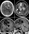
Giant chondrosarcoma of the falx in an adolescent: A case report
- PMID: 33948308
- PMCID: PMC8088535
- DOI: 10.25259/SNI_898_2020
Giant chondrosarcoma of the falx in an adolescent: A case report
Abstract
Background: Intracranial chondrosarcomas are slowly growing malignant cartilaginous tumors that are especially rare in adolescents.
Case description: A 19-year-old woman with no medical history presented with symptoms of intermittent facial twitching and progressive generalized weakness for 6 months. The patient's physical examination was unremarkable. Imaging revealed a large bifrontal mass arising from the falx cerebri, with significant compression of both cerebral hemispheres and downward displacement of the corpus callosum. The patient underwent a bifrontal craniotomy for gross total resection of tumor. Neuropathologic examination revealed a bland cartilaginous lesion most consistent with low-grade chondrosarcoma. Her postoperative course was uneventful, and she was discharged to home on postoperative day 3.
Conclusion: This is an unusual case of an extra-axial, non-skull base, low-grade chondrosarcoma presenting as facial spasm in an adolescent patient.
Keywords: Adolescent; Chondrosarcoma; Falx cerebri; Intracranial; Pediatric.
Copyright: © 2021 Surgical Neurology International.
Conflict of interest statement
There are no conflicts of interest.
Figures


Similar articles
-
Falcine Myxoid Chondrosarcoma: A Rare Aggressive Case.Asian J Neurosurg. 2018 Jan-Mar;13(1):68-71. doi: 10.4103/1793-5482.181116. Asian J Neurosurg. 2018. PMID: 29492125 Free PMC article.
-
Parafalcine chondrosarcoma: an unusual localization for a classical variant. Case report and review of the literature.Surg Neurol. 2001 Mar;55(3):174-9. doi: 10.1016/s0090-3019(01)00329-9. Surg Neurol. 2001. PMID: 11311919 Review.
-
Intracranial mesenchymal chondrosarcoma: case report and literature review.Br J Neurosurg. 2012 Dec;26(6):912-4. doi: 10.3109/02688697.2012.697219. Epub 2012 Jun 25. Br J Neurosurg. 2012. PMID: 22731866 Review.
-
Primary Extraskeletal Falcine Myxoid Chondrosarcoma-A Case Report and Review of Literature.Asian J Neurosurg. 2024 May 13;19(2):280-285. doi: 10.1055/s-0043-1772764. eCollection 2024 Jun. Asian J Neurosurg. 2024. PMID: 38974434 Free PMC article.
-
Chondroid tumors arising from the meninges--report of 2 cases and review of the literature.Clin Neuropathol. 2004 Jul-Aug;23(4):149-53. Clin Neuropathol. 2004. PMID: 15328878
References
-
- Gerszten PC, Pollack IF, Hamilton RL. Primary parafalcine chondrosarcoma in a child. Acta Neuropathol. 1998;95:111–4. - PubMed
-
- Güneş M, Günaldi O, Tuğcu B, Tanriverdi O, Güler AK, Cöllüoğlu B. Intracranial chondrosarcoma: A case report and review of the literature. Minim Invasive Neurosurg. 2009;52:238–41. - PubMed
-
- Lin HS, Tsai CC, Chang CK, Chen SJ. Giant intracranial mesenchymal chondrosarcoma with uncal herniation. Formos J Surg. 2012;45:93–6.
-
- Nagata S, Sawada K, Kitamura K. Chondrosarcoma arising from the falx cerebri. Surg Neurol. 1986;25:505–9. - PubMed
-
- Omezine S, Bouali S, Taallah M, Zehani A, Kallel J, Jemel H. Distinguishing falcine chondrosarcomas from their mimics and management. World Neurosurg. 2018;118:279–83. - PubMed
Publication types
LinkOut - more resources
Full Text Sources
Other Literature Sources
