

Lycorine hydrochloride inhibits melanoma cell proliferation, migration and invasion via down-regulating p21Cip1/WAF1
- PMID: 33948364
- PMCID: PMC8085853
Lycorine hydrochloride inhibits melanoma cell proliferation, migration and invasion via down-regulating p21Cip1/WAF1
Erratum in
-
Erratum: Lycorine hydrochloride inhibits melanoma cell proliferation, migration and invasion via down-regulating p21Cip1/WAF1.Am J Cancer Res. 2022 Jul 15;12(7):3491-3494. eCollection 2022. Am J Cancer Res. 2022. PMID: 35968327 Free PMC article.
-
Erratum: Lycorine hydrochloride inhibits melanoma cell proliferation, migration and invasion via down-regulating p21Cip1/WAF1.Am J Cancer Res. 2024 Sep 25;14(9):4683-4685. doi: 10.62347/UGBF6070. eCollection 2024. Am J Cancer Res. 2024. PMID: 39417165 Free PMC article.
Abstract
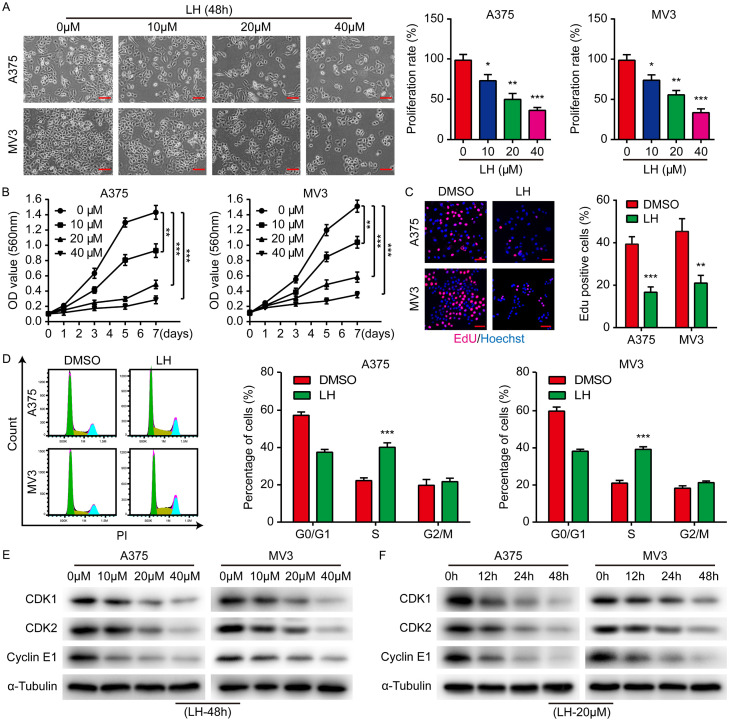
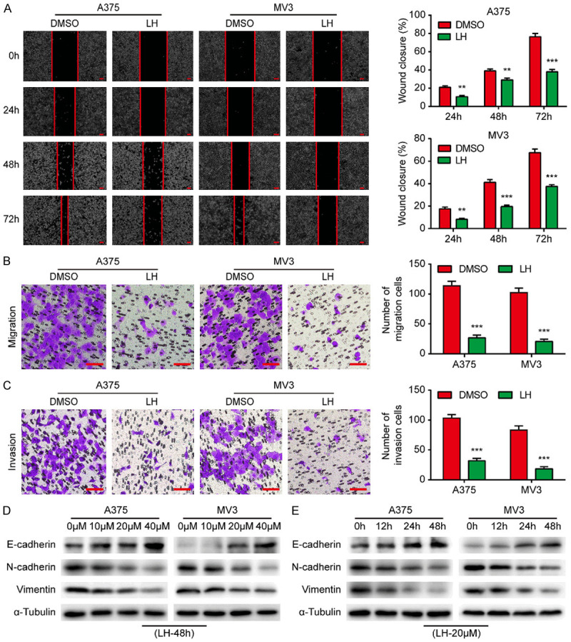
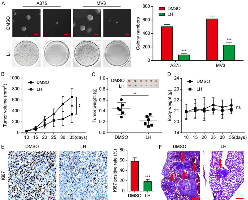
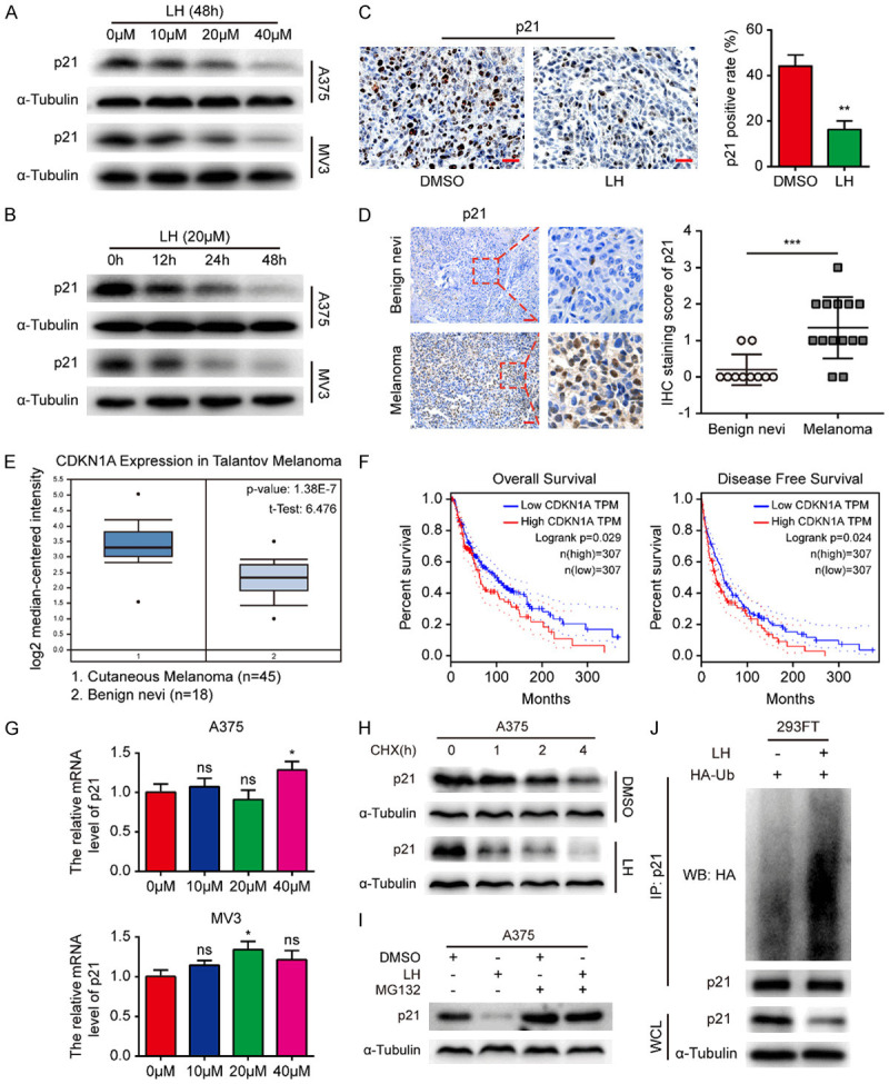
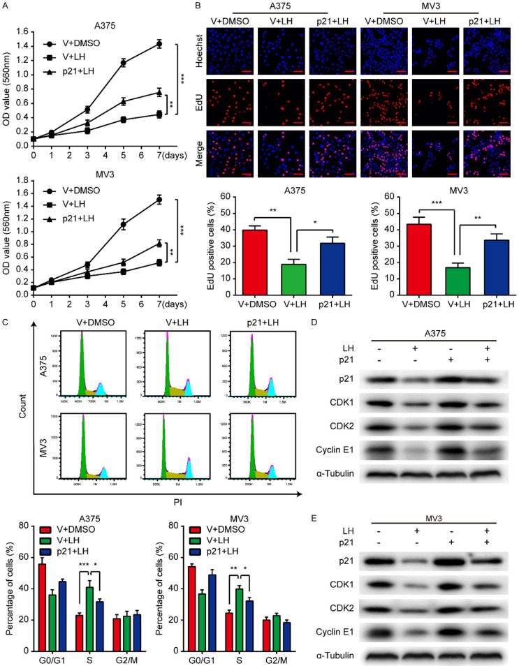
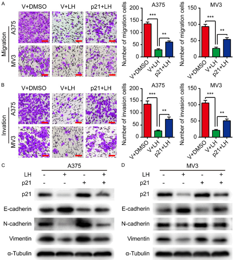
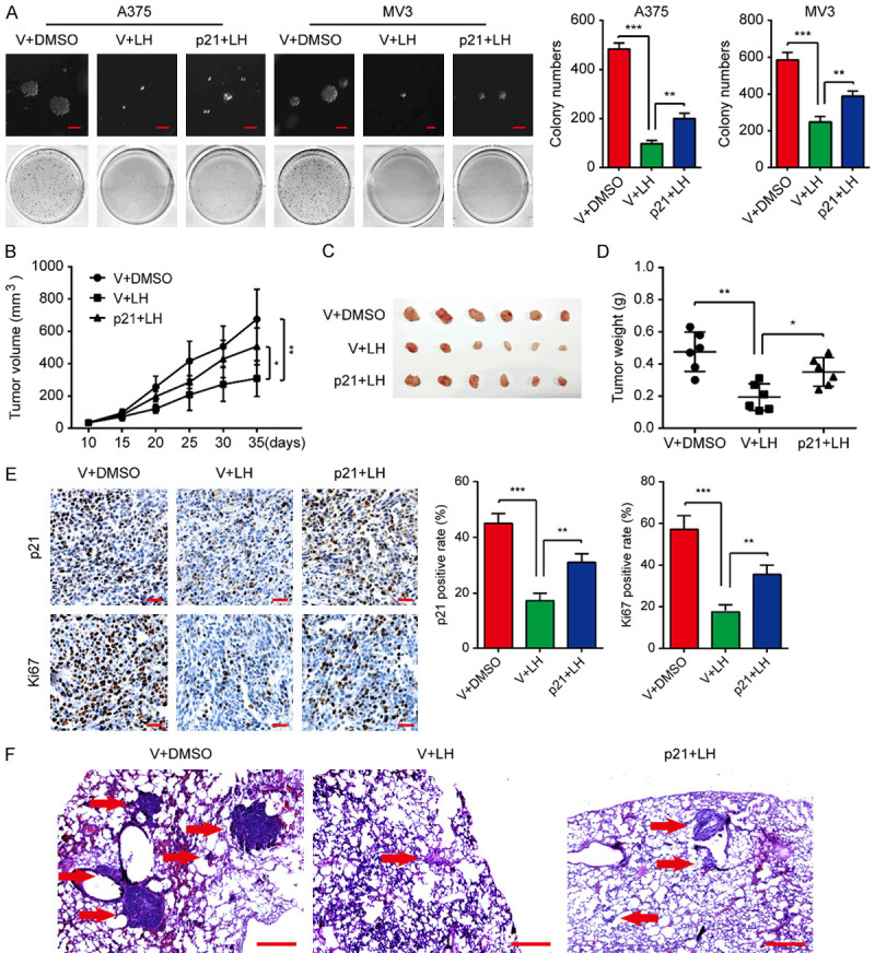
Lycorine hydrochloride (LH) is an active ingredient sourced from the medicinal herb Lycoris radiata. Previous studies have suggested that LH exerts tumor suppression activity in several human cancers. However, the anti-cancer effect of LH in melanoma and the potential molecular mechanisms still need to be further studied. p21Cip1/WAF1, unlike its traditional cyclin-dependent kinase (CDK) inhibitor role, is believed to act as an oncogene under certain cellular conditions. In this research, an increased expression of p21Cip1/WAF1 was found in human melanoma tissues and positively related to the tumor invasion depth. High level of p21Cip1/WAF1 was found to correlate with bad outcomes of melanoma patients by Kaplan-Meier survival analysis. Functional experiments demonstrated that the proliferation, migration and invasion ability of A375 and MV3 melanoma cells was powerfully inhibited by LH through inducing S phase cell cycle arrest and regulating epithelial-mesenchymal transition (EMT). In NOD/SCID mice model, LH effectively inhibited the xenograft tumor growth and lung metastasis of A375 cells. Further research revealed that LH reduced p21Cip1/WAF1 protein by accelerating its ubiquitination. Importantly, the LH-induced suppression of cell proliferation and metastasis was rescued by p21Cip1/WAF1 overexpression, both in vitro an in vivo. Taken together, LH, which suppresses the proliferation and metastasis of melanoma cells via down-regulating p21Cip1/WAF1, is expected to be developed as an effective medicine for melanoma therapy.
Keywords: Melanoma; cell proliferation; invasion; lycorine hydrochloride; migration; p21Cip1/WAF1.
AJCR Copyright © 2021.
Conflict of interest statement
None.
Figures
Similar articles
-
Antibiotic drug tigecycline inhibits melanoma progression and metastasis in a p21CIP1/Waf1-dependent manner.Oncotarget. 2016 Jan 19;7(3):3171-85. doi: 10.18632/oncotarget.6419. Oncotarget. 2016. PMID: 26621850 Free PMC article.
-
Induction of G2/M Cell Cycle Arrest via p38/p21Waf1/Cip1-Dependent Signaling Pathway Activation by Bavachinin in Non-Small-Cell Lung Cancer Cells.Molecules. 2021 Aug 25;26(17):5161. doi: 10.3390/molecules26175161. Molecules. 2021. PMID: 34500594 Free PMC article.
-
Down-regulation of miRNA-106b inhibits growth of melanoma cells by promoting G1-phase cell cycle arrest and reactivation of p21/WAF1/Cip1 protein.Oncotarget. 2014 Nov 15;5(21):10636-49. doi: 10.18632/oncotarget.2527. Oncotarget. 2014. PMID: 25361006 Free PMC article.
-
p21Cip1/Waf1 protein and its function based on a subcellular localization [corrected].J Cell Biochem. 2011 Dec;112(12):3502-6. doi: 10.1002/jcb.23296. J Cell Biochem. 2011. PMID: 21815189 Review.
-
Differential effects of cell cycle regulatory protein p21(WAF1/Cip1) on apoptosis and sensitivity to cancer chemotherapy.Drug Resist Updat. 2003 Aug;6(4):183-95. doi: 10.1016/s1368-7646(03)00044-x. Drug Resist Updat. 2003. PMID: 12962684 Review.
Cited by
-
HER-2 Receptor and αvβ3 Integrin Dual-Ligand Surface-Functionalized Liposome for Metastatic Breast Cancer Therapy.Pharmaceutics. 2024 Aug 27;16(9):1128. doi: 10.3390/pharmaceutics16091128. Pharmaceutics. 2024. PMID: 39339166 Free PMC article.
-
Modulation of hypoxia-inducible factor-1 signaling pathways in cancer angiogenesis, invasion, and metastasis by natural compounds: a comprehensive and critical review.Cancer Metastasis Rev. 2024 Mar;43(1):501-574. doi: 10.1007/s10555-023-10136-9. Epub 2023 Oct 4. Cancer Metastasis Rev. 2024. PMID: 37792223 Review.
-
Magnetic propelled hydrogel microrobots for actively enhancing the efficiency of lycorine hydrochloride to suppress colorectal cancer.Front Bioeng Biotechnol. 2024 Feb 21;12:1361617. doi: 10.3389/fbioe.2024.1361617. eCollection 2024. Front Bioeng Biotechnol. 2024. PMID: 38449675 Free PMC article.
-
Nanodelivery Approaches of Phytoactives for Skin Cancers: Current and Future Perspectives.Curr Pharm Biotechnol. 2025;26(5):631-653. doi: 10.2174/0113892010300081240329033208. Curr Pharm Biotechnol. 2025. PMID: 38616742 Review.
-
Uncover the anticancer potential of lycorine.Chin Med. 2024 Sep 8;19(1):121. doi: 10.1186/s13020-024-00989-9. Chin Med. 2024. PMID: 39245716 Free PMC article. Review.
References
-
- Wu Y, Wang Y, Wang L, Yin P, Lin Y, Zhou M. Burden of melanoma in China, 1990-2017: findings from the 2017 global burden of disease study. Int J Cancer. 2020;147:692–701. - PubMed
-
- Ross MI, Gershenwald JE. Evidence-based treatment of early-stage melanoma. J Surg Oncol. 2011;104:341–353. - PubMed
-
- O’Neill CH, Scoggins CR. Melanoma. J Surg Oncol. 2019;120:873–881. - PubMed
LinkOut - more resources
Full Text Sources
Research Materials