. 2021 Jun;8(6):1183-1199.
doi: 10.1002/acn3.51290. Epub 2021 May 5.

Selective vulnerability to atrophy in sporadic Creutzfeldt-Jakob disease

Affiliations

Selective vulnerability to atrophy in sporadic Creutzfeldt-Jakob disease

Kyan Younes et al. Ann Clin Transl Neurol. 2021 Jun.

Abstract

Objective: Identification of brain regions susceptible to quantifiable atrophy in sporadic Creutzfeldt-Jakob disease (sCJD) should allow for improved understanding of disease pathophysiology and development of structural biomarkers that might be useful in future treatment trials. Although brain atrophy is not usually present by visual assessment of MRIs in sCJD, we assessed whether using voxel-based morphometry (VBM) can detect group-wise brain atrophy in sCJD.

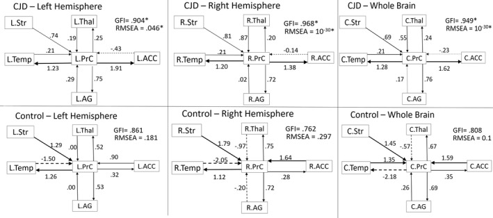
Methods: 3T brain MRI data were analyzed with VBM in 22 sCJD participants and 26 age-matched controls. Analyses included relationships of regional brain volumes with major clinical variables and dichotomization of the cohort according to expected disease duration based on prion molecular classification (i.e., short-duration/Fast-progressors (MM1, MV1, and VV2) vs. long-duration/Slow-progressors (MV2, VV1, and MM2)). Structural equation modeling (SEM) was used to assess network-level interactions of atrophy between specific brain regions.

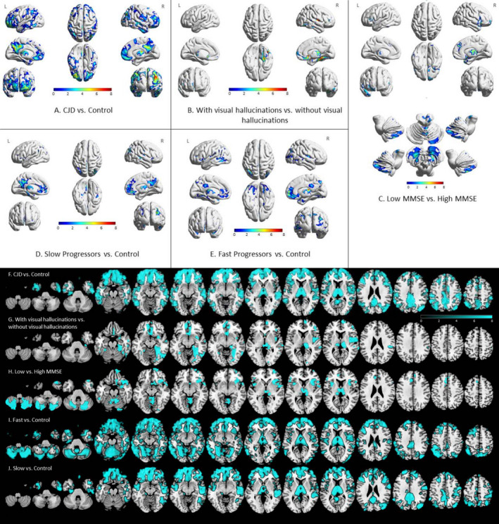
Results: sCJD showed selective atrophy in cortical and subcortical regions overlapping with all but one region of the default mode network (DMN) and the insulae, thalami, and right occipital lobe. SEM showed that the effective connectivity model fit in sCJD but not controls. The presence of visual hallucinations correlated with right fusiform, bilateral thalami, and medial orbitofrontal atrophy. Interestingly, brain atrophy was present in both Fast- and Slow-progressors. Worse cognition was associated with bilateral mesial frontal, insular, temporal pole, thalamus, and cerebellum atrophy.

Interpretation: Brain atrophy in sCJD preferentially affects specific cortical and subcortical regions, with an effective connectivity model showing strength and directionality between regions. Brain atrophy is present in Fast- and Slow-progressors, correlates with clinical findings, and is a potential biomarker in sCJD.


Conflict of interest statement

MDG receives/received research support on prion disease from the NIH/NIA (R01‐AG031189; R56‐AG055619; R01AG062562), the Michael J. Homer Family Fund and Alliance Biosecure. He has consulted for Adept Field Consulting (Backbay consulting), Advanced Medical Inc., Anderson Boutwell Traylor, Acsel Health LLC, Best Doctors Inc., Blade Therapeutics, Biohaven Pharmaceuticals, Bioscience Pharma Partners, LLC (BPP), ClearView HealthCare Partners, Grand Rounds Inc./Second Opinion Inc., Gerson Lehrman Group (GLG) Inc., Guidepoint Global LLC, Market Plus, InThought Consulting, LifeSci Capital LLC, Maupin Cox Legoy, MEDACorp, Quest Diagnostics, 3M Communications (Microvention Terumo), Smith & Hennessey LLC, TeleDoc Health Inc., and Trinity Partners LLC. He has received speaking honoraria for various medical center lectures and from Oakstone Publishing. He has received past research support from CurePSP, the Tau Consortium, Quest Diagnostics, and NIH. Dr. Geschwind serves on the board of directors for San Francisco Bay Area Physicians for Social Responsibility and on the editorial board of Dementia & Neuropsychologia. JCR is a site PI for clinical trials supported by Eli Lilly and receives support from NIH. IIG is supported by the Rio Hortega grant (CM17/00074) from “Acción Estratégica en Salud 2013‐2016” and the Global Brain Health Institute.

Figures

Figure 1
Figure 1
Regional gray matter atrophy in sporadic Creutzfeldt‐Jakob disease. A‐E show a 3D rendering, whereas F‐J show the same data rendered in axial view. All results shown in color passed permutations‐based correction for multiple comparisons p < 0.05. Orientation is neurological (e.g., left side is left brain). Redder colors (A‐E) signify higher level of significance (higher t‐stat). For F‐J (axial views), color bar represents various t‐scores. Only regions of t‐scores > 2 (i.e., > 2 SD away from the mean) are shown; blue regions color have significantly greater atrophy than the comparison group. Clusters with volume reductions in sCJD compared to Controls were found in the bilateral frontopolar, mesial and inferior frontal, mesial and lateral parietal, bilateral lateral temporal and left mesial temporal, and inferior posterior right occipital regions (A, F). sCJD participants with visual hallucinations had significant volume loss in the bilateral thalami, medial orbitofrontal, rectus gyri, and right fusiform compared to participants without visual hallucinations (B, G). The sCJD group with more severe cognitive impairment (based on dichotomization by the median MMSE score) showed volume reduction in the bilateral mesial and inferior frontal, cerebellum, left orbitofrontal, and right mesial temporal regions compared to the group with less cognitive impairment (C, H). Volume differences between Slow‐progressors (based on molecular classification subtype) and Controls were present in the bilateral mesial and lateral frontal, bilateral precuneal, middle temporal, postcentral, and occipitoparietal regions (Slow‐progressors = 4 MM2, 5 MV2) (D, J). Volume differences in Fast‐progressors, based on molecular classification, and Controls were found in bilateral mesial and lateral frontal, bilateral precuneal, middle temporal, postcentral, and occipitoparietal regions as well as occipital and temporal (Fast‐progressors = 3 MV1, 2 VV2) (E, I). No volume differences were found between comparison of Fast‐progressors versus Slow‐progressors (not shown; see text).
Figure 2
Figure 2
Sporadic Creutzfeldt‐Jakob disease selectively changes the effective connectivity between specific cortical and subcortical brain regions that overlap with the default mode network nodes. The figure shows the models of brain effective connectivity when brain volume data are tested in a network of cortical and subcortical regions usually noted by the authors to be commonly affected clinically on diffusion imaging in sCJD, specifically the default mode network plus the striatum and thalamus. Two key take‐away points from this figure are (1) the model fit in sCJD but not in Controls, and (2) that the precuneus (PrC) seems to play a central role in influencing volumetric changes in other regions. In the following text, we explain the SEM model and the meaning of the arrows from a mathematical standpoint. The graphs represent anatomical nodes in boxes connected by paths of trophic influence (arrows) that determine the regional volumetric influence on the target nodes. The effective connectivity (i.e., direction of the trophic effect) is represented by the arrow direction. Connectivity strength (i.e., strength of an effect) is represented by path coefficients (i.e., beta coefficient) displayed by the number over each arrow, with higher numbers meaning stronger tropic influence. The thickness of the arrow is a visual representation of the strength of the correlation and the dashed lines representing a negative correlation. Positive values indicate induction of atrophy in the direction of the arrow, whereas negative values indicate induction of increased volume. Goodness‐of‐fit statistics (GFIs) > .900 are considered significant with the p value equivalent shown by root mean square error of approximation (RMSEA)—only the models in sCJD, and none of the models in Controls, were significant (significant results are indicated with an *). In the whole brain and the right hemisphere models, and partially in the left hemisphere model, the precuneus exerts a large and disproportionate effect on the anterior cingulate (ACC), angular gyrus (AG), and temporal lobe (Temp). For example, in the whole brain model, one‐unit change in the precuneus volume results in 1.62, .76, and 1.28 points change in the ACC, AG, and Temp, respectively. Conversely, changes in the ACC, AG, and Temp volumes results in −.23, .17, and .21 unit change, respectively, in the precuneus. Interestingly, compared to the tropic influence of the precuneus, the effects were more balanced between the thalamus (Thal) and the precuneus and were unidirectional from the striatum (Str) to the precuneus. Models that included bidirectional effect between the precuneus and the Str did not meet the goodness‐of‐fit and the statistical significance parameters. This suggests that the striatum influenced atrophy of the precuneus, but not the reverse. L = left hemisphere, R = right hemisphere, C = combined or bilateral structure.

References

    1. Geschwind MD. Prion diseases. Continuum (Minneap Minn) 2016;21(16 Neuroinfectious Disease); 1612–38. 10.1212/CON.0000000000000251. - DOI - PMC - PubMed
    1. Kovanen J, Erkinjuntti T, Iivanainen M, et al. Cerebral MR and CT imaging in Creutzfeldt‐Jakob disease. J Comput Assist Tomogr 1985;9(1):125–128. 10.1097/00004728-198501000-00023. - DOI - PubMed
    1. Hayashi R, Hanyu N, Kuwabara T, Moriyama S. Serial computed tomographic and electroencephalographic studies in Creutzfeldt‐Jakob disease. Acta Neurol Scand 1992;85(3):161–165. 10.1111/j.1600-0404.1992.tb04020.x. - DOI - PubMed
    1. Finkenstaedt M, Szudra A, Zerr I, et al. MR imaging of Creutzfeldt‐Jakob disease. Radiology 1996;199(3):793–798. 10.1148/radiology.199.3.8638007. - DOI - PubMed
    1. Parchi P, Giese A, Capellari S, et al. Classification of sporadic Creutzfeldt‐Jakob disease based on molecular and phenotypic analysis of 300 subjects. Ann Neurol 1999;46(2):224–233. https://www.ncbi.nlm.nih.gov/pubmed/10443888. - PubMed

Publication types

MeSH terms

Supplementary concepts