. 2021 Jul 5;220(7):e202012101.
doi: 10.1083/jcb.202012101. Epub 2021 May 5.

Elimination of nurse cell nuclei that shuttle into oocytes during oogenesis

Affiliations

Elimination of nurse cell nuclei that shuttle into oocytes during oogenesis

Zehra Ali-Murthy et al. J Cell Biol. .

Abstract

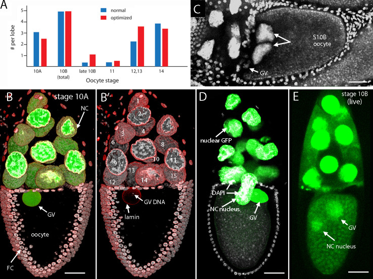
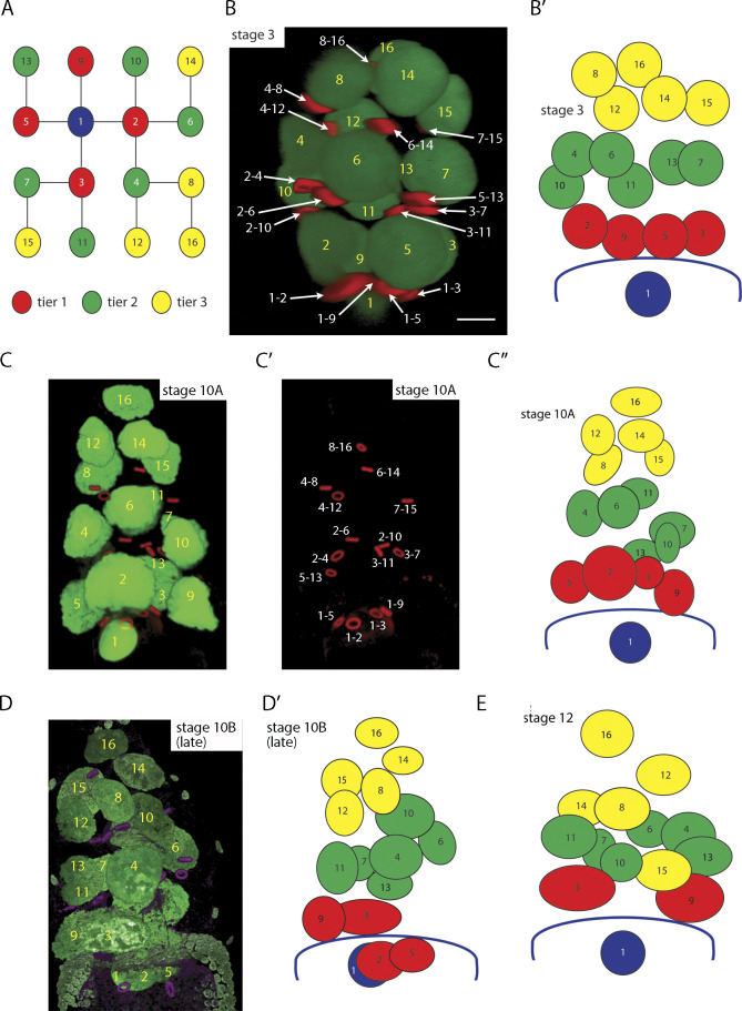
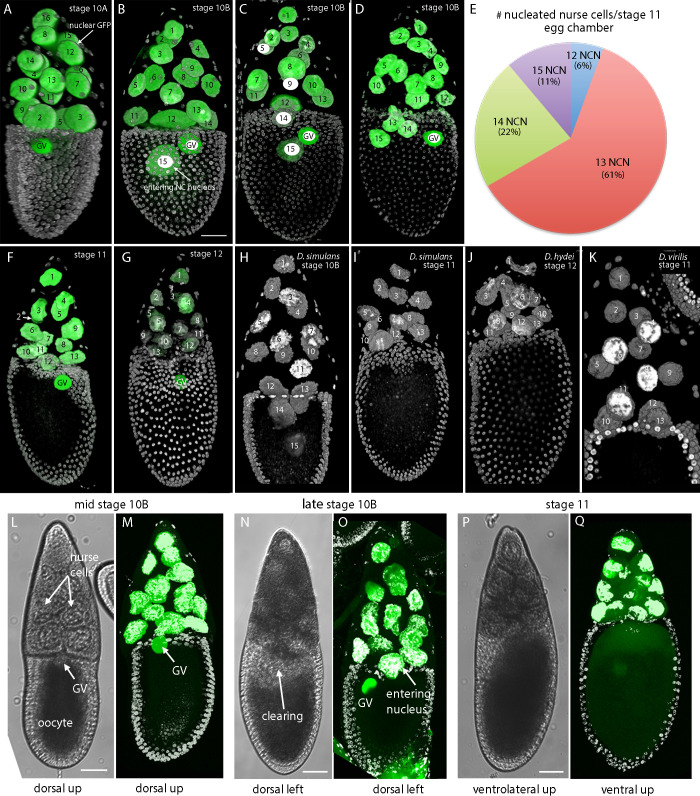
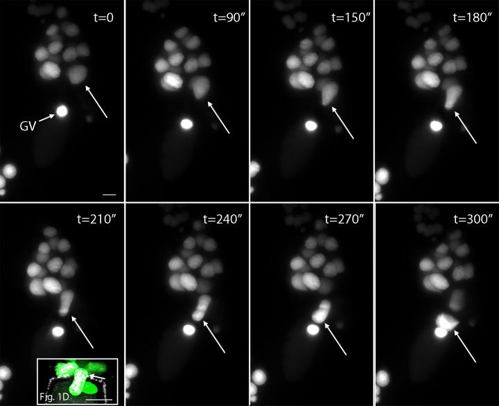
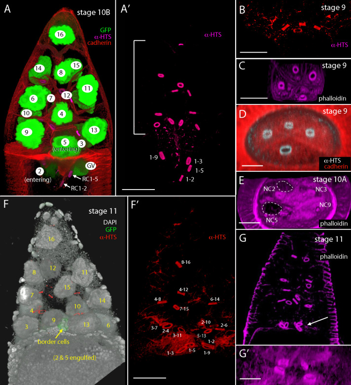
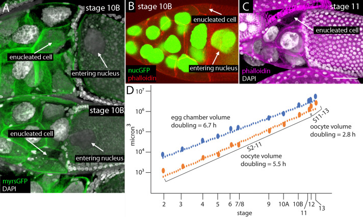
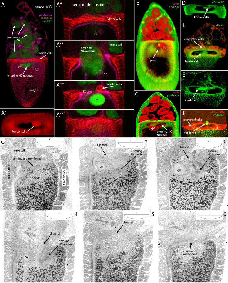
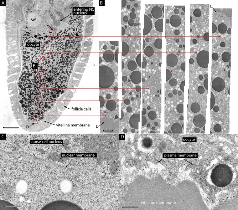
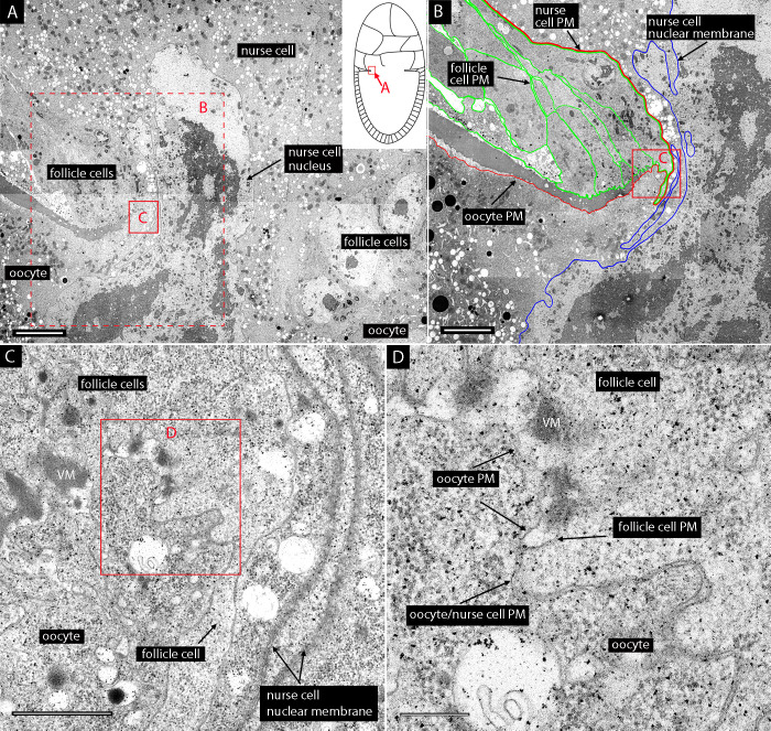
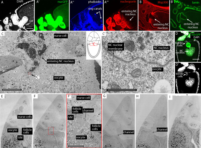
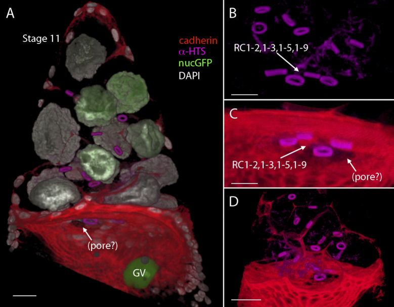
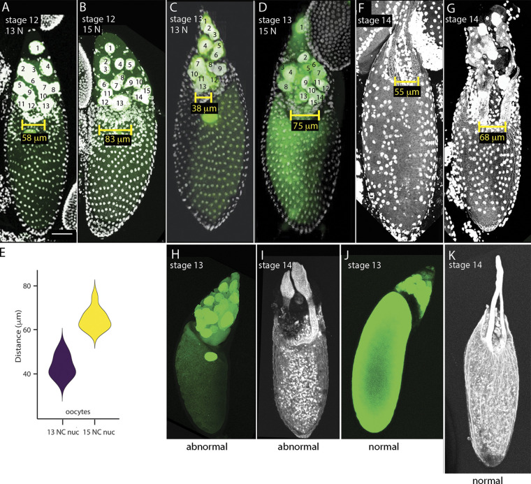
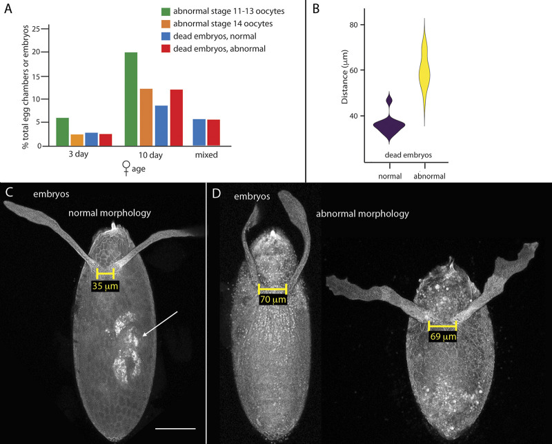
Drosophila oocytes develop together with 15 sister germline nurse cells (NCs), which pass products to the oocyte through intercellular bridges. The NCs are completely eliminated during stages 12-14, but we discovered that at stage 10B, two specific NCs fuse with the oocyte and extrude their nuclei through a channel that opens in the anterior face of the oocyte. These nuclei extinguish in the ooplasm, leaving 2 enucleated and 13 nucleated NCs. At stage 11, the cell boundaries of the oocyte are mostly restored. Oocytes in egg chambers that fail to eliminate NC nuclei at stage 10B develop with abnormal morphology. These findings show that stage 10B NCs are distinguished by position and identity, and that NC elimination proceeds in two stages: first at stage 10B and later at stages 12-14.


Figures

Figure 1.
Figure 1.
NCN in stage 10 oocytes.(A) Graph tabulating egg chamber stages in WT ovaries of females raised under normal conditions (59 ovary lobes) or conditions optimized for stages late 10B (with NCN in ooplasm) and 11 (54 ovary lobes). (B and B′) Stage 10A egg chambers with germline expression of nuclear GFP (green) in 15 NCN and GV, stained with α-lamin antibody (red) and DAPI (white); orientation anterior up. NC numbering: 1–15 from most anterior. (C) Stage 10B WT egg chamber, fixed and stained with DAPI; arrows indicate GV and NCN in ooplasm; orientation anterior left. (D) Fixed stage 10B egg chamber stained with DAPI (white) with germline expression of nuclear GFP, and one entering NC nucleus (arrow); orientation anterior up. (E) Unfixed (live) stage 10B egg chamber with germline expression of nuclear GFP (green) in 15 NCN, one in ooplasm (arrow); orientation anterior up. Scale bars: 50 µm.
Figure 2.
Figure 2.
Defined positions of NCN.(A) Lineage tree of NCs; oocyte (1; blue), oocyte daughters (2, 3, 5, 9; tier 1, red), oocyte granddaughters (4, 6, 7, 10, 11, 13; tier 2, green), oocyte great-granddaughters (8, 12, 14, 15, 16; tier 3, yellow); tiers 1, 2, and 3 are touching the oocyte, in middle NC region, farthest from oocyte, respectively. (B–E) Orientation anterior up; number cell numbering in all panels relate to lineage indicated in A. (B) Stage 3 egg chamber with nuclei and RCs identities marked. (B′) Cartoon of egg chamber in B. (C–C″) Stage 10A egg chamber with identities of nuclei (C) and RCs marked (C′) and depicted in cartoon (C″). (D and D′) Stage 10B depicted in 3D projection (D) and cartoon (D′). (E) Stage 12 egg chamber depicted in cartoon. Genotype: MTD-Gal4 UASp-nucGFP.
Figure 3.
Figure 3.
NC content in Drosophila egg chambers.(A–G) Analysis of D. melanogaster stage 10A–12 egg chambers expressing nuclear GFP (green) and stained with DAPI (white); orientation anterior up. (A) Stage 10A oocyte has no ectopic nuclei; numbering indicates lineage as defined in Fig. 2 A. (B–D) Stage 10B with one (B), two (C), or three (D) ectopic oocyte nuclei; NC numbering: 1–15 from most anterior, not according to lineage. (E) Pie chart indicating fraction of 18 stage 11 egg chambers that have 12, 13, 14, or 15 NCN. (F and G) Stage 11 (F) and stage 12 (G) egg chambers with 13 NCN and no ectopic nuclei in the oocyte. NC numbering: 1–15 from most anterior. (H–K) D. simulans, D. hydei, and D. virilis egg chambers stained with DAPI (white) with two ectopic NCN in the stage 10B D. simulans oocyte (H) and 13 NCN in the D. simulans (I), D. hydei (J), and D. virilis (K) stage 11 egg chambers. Scale bar: 50 µm. (L–Q) Midstage 10 (L and M), late stage 10 (N and O), and stage 11 (P and Q) egg chambers (MTD-Gal4 UASp-NLSGFP) viewed with transmitted light (L, N, and P) and fluorescence microscopy (M, O, and Q). Orientation anterior to top or figure and as indicated. Egg chambers were viewed under a dissecting microscope during ex vivo incubation and distinguished both by relative size of the oocyte and by opacity of anterior ooplasm for identification of GV at midstage 10 (L), entering NCN at late stage 10B (N), and stage 11 (P). Egg chambers were photographed at the three indicated stages or were fixed, stained with DAPI, and mounted for imaging with a confocal microscope. Timing was ≤10 min after isolation (L and M), 10–20 min after appearance of GV at anterior oocyte membrane (N and O), and 7–17 min after appearance of broad anterior clearing (P and Q). Scale bars: 50 µm.
Figure S1.
Figure S1.
Frames from time-lapse fluorescence images of the early stages of NC nuclear entry captured by lattice light sheet microscopy. Arrows indicate the entering NC nucleus. Inset from Fig. 1 D shows similar shapes of the entering NCN in the live fluorescence image (time 240 min) and after fixation. Scale bars: 50 µm.
Figure 4.
Figure 4.
Ring canals precede NCN into oocyte.(A and A′) Stage 10B egg chamber expressing nuclear GFP (green), stained with α-HTS antibody to mark RCs and α-cadherin antibody to mark cell boundaries, with nuclei identified. Ring canals linking oocyte with NC 2 (RC1–2) and NC 5 (RC1–5) are positioned in the oocyte posterior to the entering nuclei of NCs 2 and 5; other RCs are bracketed; orientation anterior up. (B–E) Transverse sections created from 3D projections of confocal optical sections to show arrangements of RCs (marked with α-HTS antibody [B and D] or phalloidin fluorescence [C and E]) at the anterior face of stage 9 and stage 10 oocytes. (E) Gaps in fluorescence-marked anterior oocyte face are at sites of NCs 2 and 5 (dashed boundaries). (F and F′) Stage 11 egg chamber lacks NC 2 and 5 nuclei; stained with DAPI (white) and with α-HTS antibody to mark RCs, all of which are anterior to the oocyte; nuclear GFP marks border cells (green; 3745-Gal4); orientation anterior up. (G) RCs marked with phalloidin staining in stage 11 egg chamber, with four RCs at the anterior oocyte face (arrow); orientation anterior up. (G′) Higher-magnification image of region with four oocyte-linked RCs. Scale bars: 50 mm.
Figure 5.
Figure 5.
Enucleated NCs in stage 10B and stage 11 egg chambers.(A and B) Enucleated NCs indicated (arrows) in stage 10B chambers expressing myristoylated-GFP and stained with DAPI (A) or expressing nuclear-GFP and stained with phalloidin (B). (C) Enucleated NC of stage 11 egg chamber stained with phalloidin and DAPI. (D) Graph showing volumes of stage 2–13 egg chambers (80 egg chambers analyzed; data in Table S2). Scale bar: 50 µm for A–C.
Figure 6.
Figure 6.
Anatomy of the anterior face of the stage 10B oocyte.(A–A″″′) Stage 10B egg chamber marked with nuclear GFP (green), phalloidin fluorescence (purple), and α-cadherin staining (red) identifies oocyte, NCs, NCN, follicle cells, border cells, and RCs, but phalloidin or cadherin staining is absent at the anterior face of the oocyte at the site of entering NC nucleus. Frontal optical sections, orientation anterior up (A and A″–A″″′) and transverse section created from a 3D projection of confocal optical sections (A′). (B–F) 3D projection images of stage 10B egg chambers with channel at oocyte anterior face, rotated for partially axial orientations, marked with membrane-tethered GFP (green) and α-lamin (red; B), α-lamin (red) and phalloidin fluorescence (C), phalloidin fluorescence (D), and Dpp:Cherry (red) and α-cadherin (green; E, E′, and F). Arrow indicates channel (B). (G) EM images of successive sections of a stage 10B oocyte showing continuous plasma membrane across the anterior oocyte face (1 and 6) and channel through which a NC nucleus enters (2–5). Orientation anterior up (B, C, and G). Cartoons depict sectioned egg chamber. Scale bars: 50 µm (bar in A for A–D and F; bar in A′ for A′–A″″′, E, and E′); 20 µm (G).
Figure S2.
Figure S2.
Fine-structure images of ooplasm.(A and B) EM of a stage 10B egg chamber with an entering NC nucleus showing the oocyte at low magnification (A) and a montage of high-magnification images (B) showing the ooplasm devoid of membrane between the entering NC nucleus at the anterior and plasma oocyte membrane juxtaposed to vitelline membrane at the posterior pole. (C and D) Fine structure of the nuclear membrane of the entering NC (C) and oocyte plasma membrane (D). Scale bars: 20 µm (A and B); 5 µm (C and D).
Figure 7.
Figure 7.
Contiguous plasma membrane of the oocyte and NC.(A) EM of an entering NC nucleus. Cartoon indicates position in egg chamber. (B) Dashed red-lined box in A imaged at higher magnification with traced plasma membranes (PM) of follicle cells (green), contiguous plasma membranes of oocyte and NC (red), and nuclear membrane of entering NC (blue). (C) Red-lined box in B imaged at higher magnification. (D) Red-lined box in C imaged at higher magnification. Scale bars: 5 µm (A and B); 1 µm (C); 0.25 µm (D).
Figure 8.
Figure 8.
Anatomy of the nuclear membrane of the entering NC nucleus and fine structure of anterior face of a stage 11 oocyte.(A–A″′) The entering NC nucleus of the stage 10B oocyte is imaged with DAPI (white, A), nuclear GFP (green, A′), phalloidin (blue, A″), and α-nucleoporin, which stains only the portion of the nucleus that has not entered (A″′). (B and B′) Entering NC nucleus stained with α-Msp300 (red) and α-lamin (green) antibodies. (C) EM of entering NC nucleus (position in egg chamber indicated by cartoon) with red-lined region shown at high magnification in C′. (D and D′) Nuclear GFP (green) and DAPI (white) marks NC nucleus in the ooplasm, border cells, and GV. (E–I) Successive sections imaged by electron microscopy of a stage 11 egg chamber showing contiguous plasma membrane at the anterior oocyte face in outer sections (E and I) and channel in intermediate sections (F–H). (F′) Higher-magnification image of red-lined boxed region in F showing NC, follicle cells, vitelline membrane (VM), oocyte, and channel. Scale bars: 50 µm (bar in A for A–B′, bar in D for D and D′); 20 µm (E–I); 10 µm (C); 1 µm (C′); 10 µm (F′).
Figure S3.
Figure S3.
Images from a 3D reconstruction of a stage 11 egg chamber that expressed nucGFP and was stained with DAPI and with antibodies against cadherin and HTS.(A–D) The RCs that connect the oocyte with NCs are visible at the surface of the anterior face of the oocyte (A–C), but these images did not resolve whether the apparent discontinuity of cadherin staining in this region (A, C, and D) was due to the presence of a channel or to the complex morphology. Scale bars: 50 µm.
Figure 9.
Figure 9.
Abnormalities of egg chambers that do not eliminate NCN.(A–D) Stage 12 (A and B) and stage 13 (C and D )egg chambers marked with nuclear GFP fluorescence (green) and stained with DAPI (white) with 13 (A and C) and 15 (B and D) NCN, respectively, with distance between developing dorsal appendages marked. (E) Violin plot showing distributions of distances between dorsal appendages of stage 12/13 oocytes with either 13 or 15 NCN (NC nuc). (F and G) Stage 14 egg chambers with normal (F) and abnormal morphology (G). (H–K) Egg chambers (MTD-Gal4 UASp-NLSGFP, DAPI) identified at stage 13 as morphologically abnormal (H) or normal (J) and then fixed for imaging, or cultured for 5+ h at RT after morphological characterization as abnormal (I) or normal (K). Scale bar: 50 µm (bar in A for A–D and F–K).
Figure S4.
Figure S4.
Nondeveloping embryos with abnormalities characteristic of egg chambers that do not eliminate NCN.(A) Graph indicating percentage of stage 11–13 and stage 14 egg chambers with abnormal morphology laid by 3- and 10-d-old females, and percentage of unhatched embryos laid by females of mixed age, 3 and 10 d after eclosion with normal or abnormal morphology. (B) Violin plot showing distributions of distances between dorsal appendages of inviable embryos that either developed to organogenesis stages (normal) or appeared to be unfertilized (abnormal). (C and D) Embryos with normal (C) and abnormal (D) morphology, with distance between developing dorsal appendages marked and autofluorescent internal organs indicated by arrow. Scale bar: 100 µm (bar in C for C and D).
Figure 10.
Figure 10.
NC nuclear elimination in stage 10B egg chambers. Summary diagram illustrating the principal features of nuclear elimination. Subsequent to the anterodorsal localization of the GV (purple) in close proximity to the border cells (yellow; a), RCs (green) migrate into the oocyte and a pore forms that directly connects the oocyte with adjacent NCs (b), NCN translocate into the oocyte (c), and the GV migrates posteriorly (d). The NCN are absent from egg chambers in which the RCs have returned to the interface of the oocyte with enucleated NCs.

References

    1. Alexandrova, O., Schade M., Böttger A., and David C.N.. 2005. Oogenesis in Hydra: nurse cells transfer cytoplasm directly to the growing oocyte. Dev. Biol. 281:91–101. 10.1016/j.ydbio.2005.02.015 - DOI - PubMed
    1. Baum, J.S., Arama E., Steller H., and McCall K.. 2007. The Drosophila caspases Strica and Dronc function redundantly in programmed cell death during oogenesis. Cell Death Differ. 14:1508–1517. 10.1038/sj.cdd.4402155 - DOI - PubMed
    1. Brown, E.H., and King R.C.. 1964. Studies on the events resulting in the formation of an egg chamber in Drosophila melanogaster. Growth. 28:41–81. - PubMed
    1. Cardona, A., Saalfeld S., Schindelin J., Arganda-Carreras I., Preibisch S., Longair M., Tomancak P., Hartenstein V., and Douglas R.J.. 2012. TrakEM2 software for neural circuit reconstruction. PLoS One. 7:e38011. 10.1371/journal.pone.0038011 - DOI - PMC - PubMed
    1. Chapman, R.F.1999. The Insects. Cambridge University Press, Cambridge, UK.

Publication types

LinkOut - more resources