. 2021 Nov 29;144(10):3005-3019.
doi: 10.1093/brain/awab182.

Engineered microRNA-based regulatory element permits safe high-dose miniMECP2 gene therapy in Rett mice

Affiliations

Engineered microRNA-based regulatory element permits safe high-dose miniMECP2 gene therapy in Rett mice

Sarah E Sinnett et al. Brain. .

Abstract

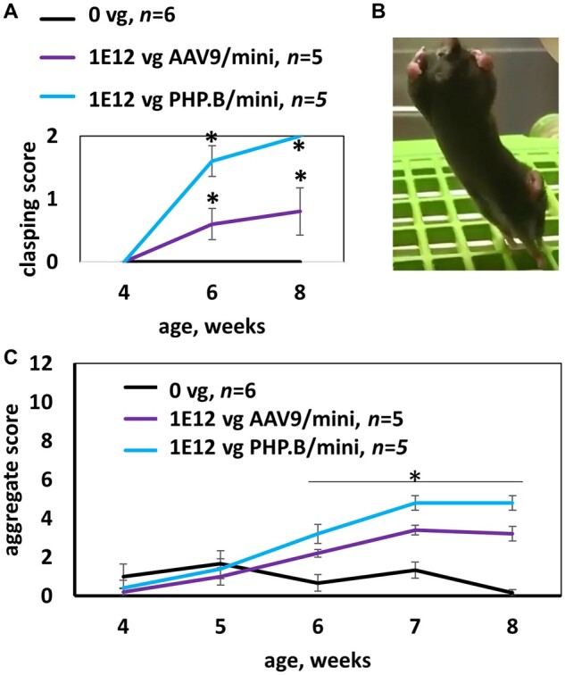
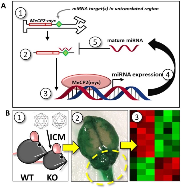
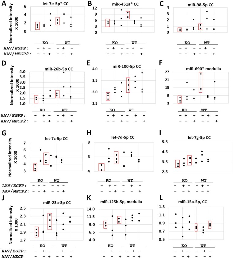
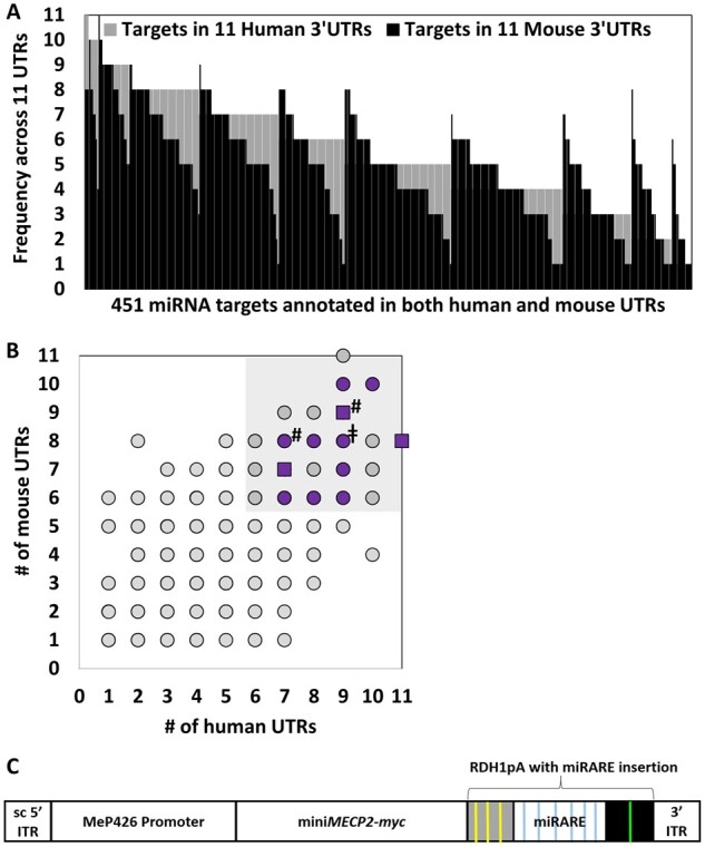
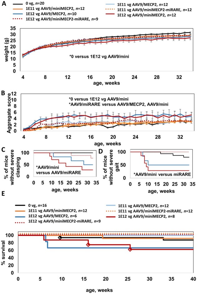
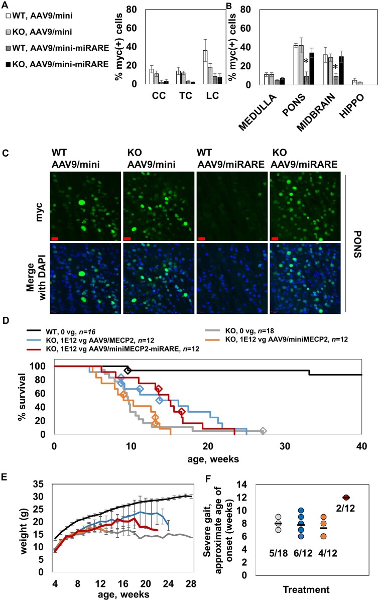
MECP2 gene transfer has been shown to extend the survival of Mecp2-/y knockout mice modelling Rett syndrome, an X-linked neurodevelopmental disorder. However, controlling deleterious overexpression of MECP2 remains the critical unmet obstacle towards a safe and effective gene therapy approach for Rett syndrome. A recently developed truncated miniMECP2 gene has also been shown to be therapeutic after AAV9-mediated gene transfer in knockout neonates. We show that AAV9/miniMECP2 has a similar dose-dependent toxicity profile to that of a published second-generation AAV9/MECP2 vector after treatment in adolescent mice. To overcome that toxicity, we developed a risk-driven viral genome design strategy rooted in high-throughput profiling and genome mining to rationally develop a compact, synthetic microRNA target panel (miR-responsive auto-regulatory element, 'miRARE') to minimize the possibility of miniMECP2 transgene overexpression in the context of Rett syndrome gene therapy. The goal of miRARE is to have a built-in inhibitory element responsive to MECP2 overexpression. The data provided herein show that insertion of miRARE into the miniMECP2 gene expression cassette greatly improved the safety of miniMECP2 gene transfer without compromising efficacy. Importantly, this built-in regulation system does not require any additional exogenous drug application, and no miRNAs are expressed from the transgene cassette. Although broad applications of miRARE have yet to be determined, the design of miRARE suggests a potential use in gene therapy approaches for other dose-sensitive genes.

Keywords: AAV; MECP2; Rett; intrathecal; microRNA.


Figures

Figure 1
Figure 1
MiniMECP2 vectors cause side effects in wild-type mice. (A) In an acute toxicity study, overexpression of miniMECP2 caused significant increases in abnormal clasping scores. n = 5–6 mice per group. Saline versus virus treatments; and pre- versus post-injection: #P < 0.05. (B) Severe hindlimb clasping observed 3 days after treatment with PHP.B/miniMECP2 [1 × 10¹² vg/mouse, intracisterna magna (ICM) administration]. In healthy mice, hindlimbs extend outwards. (C) Both AAV9/ and PHP.B/miniMECP2 significantly increase aggregate phenotype severity scores within 2 weeks post-injection (P < 0.05). Figure 1 shows data for mice housed and treated at UNC-Chapel Hill. All other mice in this paper were tested at UTSW. (A and C) Data are means ± SEM.
Figure 2
Figure 2
Schematic of strategy used to rationally synthesize a miRNA target panel tailored for Rett syndrome. (A) The first five steps of a conceptual negative feedback loop are indicated: (1) viral genome in nucleus; (2) exogenous MECP2-myc mRNA in cytoplasm; (3) translation and nuclear localization of MECP2-myc; (4) exogenous MECP2-myc upregulates miRNA expression; (5) mature miRNAs bind to non-coding targets in the 3ʹ UTR of an exogenous MECP2-myc transcript. After mature miRNAs bind to targets in the exogenous MECP2 mRNA, endogenous RNAi machinery (not shown) may conditionally silence exogenous MECP2 expression. The targets inserted into the viral genome may match targets found in the endogenous Mecp2 mRNA. Conceptually, this model could be adapted for other potentially toxic transcription factors that drive the expression of miRNAs in vivo. Alternatively, insertion of miRNA targets into the viral genome could improve the safety of gene therapies encoding potentially toxic cytoplasmic proteins, provided that those proteins indirectly upregulate miRNAs. (B) To identify translationally relevant miRNAs upregulated by toxic MECP2 gene transfer, (1) adolescent wild-type (WT) and knockout (KO) mice were treated intracisternally with saline, 1 × 10¹² vg AAV9/MECP2, or 1 × 10¹² vg AAV9/EGFP. (2) Two to three weeks later, CNS tissue nearest to the injection site (cerebellum, medulla and cervical cord; see dashed circle) was harvested from treated mice. The image shown in step 2 illustrates the spread of dye after ICM administration. The relatively concentrated dye localization near the cerebellum indicates how vulnerable tissue near the intracisternal site may be after treatment with high titre, toxic virus. (3) RNA was purified from dissected tissue, frozen and shipped to LC Sciences for miRNA profiling. Data from this screen were subsequently used to rank candidate miRNA targets from a secondary bioinformatics approach.
Figure 3
Figure 3
Putative positive hits upregulated in correlation with endogenous or exogenous MECP2 according to miRNA microarray. (AF) Significant differences between saline-treated knockout (KO) and saline-treated wild-type (WT) mice. (GL) Significant differences between saline-treated and AAV9/MECP2-treated mice. (AL) None of the 12 miRNAs listed here were significantly upregulated by AAV9/EGFP (versus saline-treated knockout or wild-type mice; P > 0.05). Each data-point represents the average of two screening replicates; error bars for each mean are not shown. n = 3 mice per group. P < 0.05 between the groups boxed in red. *A ‘reg1’ miRNA target panel featuring binding sites for miR-451a, miR-690, and let-7e-5p failed to improve the safety of miniMECP2 gene transfer (data not shown). As discussed in the main text, negative results (P > 0.05) should be interpreted with caution, as miRNAs upregulated at the cell-type level may be masked by noise from other cell types in the same tissue. Furthermore, low normalized fluorescence intensities may reflect low miRNA expression levels in a tissue sample but may fail to reveal high expression levels within a single cell type within said tissue. In general, many of the significant differences in normalized fluorescence intensity are for relatively small increases in mean signal between the indicated treatment groups. These subtle changes, along with the general noisiness of the data, warranted a secondary technique to justify the use of specific miRNA targets within a new target panel design. Finally, miRNAs significantly upregulated by AAV9/EGFP (versus saline-treated knockout mice) are miR-99a-5p, miR-107-3p and let-7f-5p in cervical cord (CC; data not shown). MiRNAs upregulated by AAV9/EGFP (versus saline-treated wild-type mice) are miR-669a-3p in cervical cord; let-7j in cerebellum; and miR-669f-3p, miR-669p-3p, miR-30c-5p and miR-669a-3p in medulla (data not shown).
Figure 4
Figure 4
Microarray expression data were used to rank targets that appear frequently across a curated list of 3ʹ UTRs for genes mediating intellectual disability. (A) Many miRNA targets appear frequently across the curated list of 3ʹ UTRs. Among 2491 human targets and 1831 mouse targets, 451 targets had identical annotation across both mouse and human 3ʹ UTR datasets. (B) The scatter plot shows the same data as those shown in A. Targets that are annotated in over half of the examined 3ʹ UTR sequences for both species were prioritized for target panel design (shaded area). Within the shaded area, purple data-points indicate targets corresponding to miRNAs expressed at moderate to high levels in dissected cervical cord, cerebellar, and/or medullar tissue (signal intensity >500). Square data-points indicate targets for putative MECP2-responsive miRNAs indicated in Fig. 3A, C, D and G–J as well as two additional miRNAs (miR-9-5p and miR-27a-3p; indicated by a hash symbol), which showed trends in increased expression in cervical cord when data for MECP2− and MECP2+ treatment groups were aggregated together (data not shown). MECP2− groups, for example, would include both saline-treated and AAV9/EGFP-treated knockout mice. For clarity, we have limited notation (purple and square data-points) to the shaded area only. The double dagger symbol (‡) indicates the target for miR-218-5p included in the new panel design (‘miRARE’) for the purpose of making miRARE more broadly applicable for multiple gene therapy applications. Expression of miR-218-5p did not appear to be MECP2-responsive in our high throughput screening data. (C) Cartoon of the miniMECP2-miRARE viral genome cassette. Yellow and green lines indicate miRNA targets that are part of the previously published RDH1pA. Blue lines indicate miRARE targets that were inserted into RDH1pA. ITR = inverted terminal repeat; sc = mutated self-complementary ITR sequence.
Figure 5
Figure 5
AAV9/miniMECP2-miRARE is well-tolerated after intrathecal administration in wild-type adolescents. Throughout this study, all treatments were administered between 4 and 5 weeks of age. (A) AAV9/miniMECP2-treated wild-type mice (1 × 10¹² vg/mouse) had significantly lower weight than that of saline-treated wild-type mice beginning at 15 weeks of age (P < 0.05). No significant difference was observed between saline- and AAV9/miniMECP2-miRARE-treated wild-type mice (1 × 10¹² vg/mouse). The same legend also applies to B. (B) AAV9/MECP2- and AAV9/miniMECP2-treated wild-type mice had a significantly higher mean aggregate behavioural severity score versus that observed for saline-treated mice (P < 0.05; at 6–30 and 7–27 weeks of age, respectively). AAV9/miniMECP2-miRARE-treated wild-type mice had a significantly lower mean aggregate severity score versus those of AAV9/MECP2- and AAV9/miniMECP2-treated mice at most time points from 11–19 and 9–20 weeks of age, respectively. No significant difference was observed between saline- and AAV9/miniMECP2-miRARE-treated wild-type mice (1 × 10¹² vg/mouse). (C) Most wild-type mice treated with AAV9/miniMECP2-miRARE did not develop severe clasping abnormalities (severe clasping for n = 2/9 mice). (D) Wild-type mice treated with AAV9/miniMECP2-miRARE did not develop severe gait abnormalities (n = 0/9 mice). (C and D) Severe abnormal gait and severe clasping are each scored as a 2 on a scale of 0–2. All vectors were administered at 1 × 10¹² vg/mouse. Mice that were euthanized early before developing severe gait or clasping scores (E) were excluded from data in C and D. (C and D) *P < 0.05 using Gehan–Breslow–Wilcoxon test, which can be used to evaluate significance for Kaplan–Meier plots. A one-way ANOVA of mean age of onset for D is not encouraged, as there are no time points listed here at which AAV9/miniMECP2-miRARE-treated wild-type mice develop severe gait abnormalities. (E) No early deaths have been observed for AAV9/miniMECP2-miRARE-treated wild-type mice. MiRARE groups are offset for clarity. Diamonds indicate veterinarian-requested euthanasias for bullying-related injuries (saline-treated) and for prolapses (AAV9/miniMECP2-treated). Prolapses were observed in 8–17% of AAV9/miniMECP2-treated wild-type mice (1 × 10¹¹ to 1 × 10¹² vg, respectively). Four AAV9/MECP2-treated mice were dissected at 8 weeks of age and were therefore excluded from the survival plot. The survival data for saline-treated wild-type mice also appears in Fig. 6D and Supplementary Fig. 9, as these mice were evaluated in parallel. (A and B) For simplicity only statistical differences between saline- and high-dose-treated wild-type mice were analysed. Data are means ± SEM. *P < 0.05.
Figure 6
Figure 6
AAV9/miniMECP2-miRARE extends the survival of knockout mice despite regulation. (A) Percentage of myc+ cells is shown for cervical cord (CC), thoracic cord (TC) and lumbar cord (LC). MiRARE provided robust regulation across host genotypes. n = 3–5 mice per group. (B) Percentage of myc+ cells is shown. MiRARE significantly decreased expression in the pons and midbrain of AAV9/miniMECP2-miRARE-treated wild-type mice. P < 0.05 versus all other groups. The same legend applies for A and B. (C) Representative pons images. Scale bars = 20 µm. (D) Median survivals for saline- and AAV9/miniMECP2-miRARE-treated knockout mice are 9.6 and 15.0 weeks (P < 0.02). Compare to lower doses shown in Supplementary Fig. 9. Diamonds indicate veterinarian-requested euthanasias, which were primarily for severe tail lesions or tail self-amputations. (E) None of the treatments affected knockout weight. Any trends in increased weight for virus-treated knockout mice (versus saline-treated knockout mice) are insignificant because few mice are alive at later time points. n per group in E: wild-type, 0 vg (20); knockout, 0 vg (18); knockout, 1 × 10¹² vg AAV9/MECP2 (12); knockout, 1 × 10¹² vg AAV9/miniMECP2 (12); and knockout, 1 × 10¹² vg AAV9/miniMECP2-miRARE (12). (D and E) The same wild-type control data appear in Fig. 5 as these mice were treated in parallel. (F) The frequency of achieving severe gait (score = 2) among saline-, AAV9/MECP2-, AAV9/miniMECP2- and AAV9/miniMECP2-miRARE-treated mice were 28%, 50%, 33% and 17%. Each mouse was administered 1 × 10¹² vg. Black bars indicate the mean age of onset for severe clasping. Each data-point represents one mouse. Treatment groups are colour-coded as shown in D and E. The fraction of mice (subset versus entire group) is listed for each treatment group. AAV9/miniMECP2-miRARE-treated mice developing severe gait (n = 2 of 12) did so 4–5 weeks after other groups. Although this delay is significantly different, the number of data-points are too few to draw firm conclusions. Figure 5D and F show the same type of data. In general, Kaplan–Meier plots are the clearest way to visually communicate age-of-onset data (Fig. 5D), but these plots lend themselves to the assumption that all mice retaining normal gait live for the entire time period specified on the x-axis. This is true for wild-type mice (Fig. 5D) but not for knockout mice (F). Importantly, few AAV9/miniMECP2-miRARE-treated knockout mice developed severe gait, despite having an extended survival. For a graphical comparison, see Supplementary Fig. 8 for wild-type scatter plot data (also presented in Fig. 5D). (A, B and E) Data are mean ± SEM.

References

    1. Chahil G, Bollu PC. Rett syndrome. In: StatPearls [Internet]. StatPearls Publishing; 2019. - PubMed
    1. Van den Veyver IB, Zoghbi HY. Genetic basis of Rett syndrome. Ment Retard Dev Disabil Res Rev . 2002;8(2):82–86. - PubMed
    1. Mnatzakanian GN, Lohi H, Munteanu I, et al. A previously unidentified MECP2 open reading frame defines a new protein isoform relevant to Rett syndrome. Nat Genet . 2004;36(4):339–341. - PubMed
    1. Gadalla KKE, Vudhironarit T, Hector RD, et al. Development of a novel AAV gene therapy cassette with improved safety features and efficacy in a mouse model of Rett syndrome. Mol Ther Methods Clin Dev . 2017;5:180–190. - PMC - PubMed
    1. Sinnett SE, Gray SJ. Recent endeavors in MECP2 gene transfer for gene therapy of Rett syndrome. Discov Med . 2017;24(132):153–159. - PubMed

Publication types