Review
. 2021 Jun;78(12):5107-5122.
doi: 10.1007/s00018-021-03831-9. Epub 2021 May 5.

Cardiac regenerative capacity: an evolutionary afterthought?

Affiliations
Review

Cardiac regenerative capacity: an evolutionary afterthought?

Phong D Nguyen et al. Cell Mol Life Sci. 2021 Jun.

Abstract

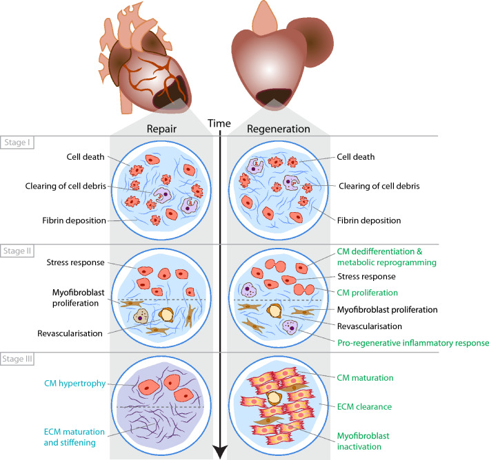
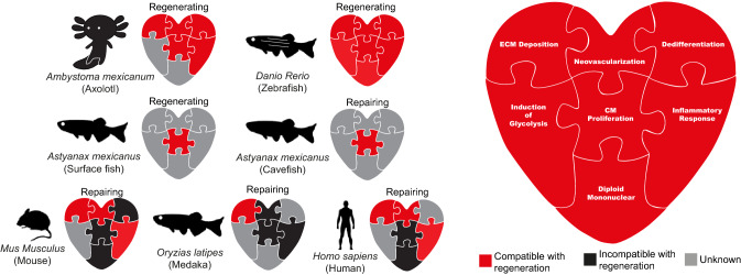
Cardiac regeneration is the outcome of the highly regulated interplay of multiple processes, including the inflammatory response, cardiomyocyte dedifferentiation and proliferation, neovascularization and extracellular matrix turnover. Species-specific traits affect these injury-induced processes, resulting in a wide variety of cardiac regenerative potential between species. Indeed, while mammals are generally considered poor regenerators, certain amphibian and fish species like the zebrafish display robust regenerative capacity post heart injury. The species-specific traits underlying these differential injury responses are poorly understood. In this review, we will compare the injury induced processes of the mammalian and zebrafish heart, describing where these processes overlap and diverge. Additionally, by examining multiple species across the animal kingdom, we will highlight particular traits that either positively or negatively affect heart regeneration. Last, we will discuss the possibility of overcoming regeneration-limiting traits to induce heart regeneration in mammals.

Keywords: Cardiomyocyte; Evolution; Extracellular matrix; Inflammatory response; Proliferation; Regeneration; Repair; Scar.


Conflict of interest statement

The authors declare that they have no competing interests.

Figures

Fig. 1
Fig. 1
Comparison of repair vs regenerative response following cardiac injury. Schematic summary of the main processes and their response in animals that can either repair or regenerate following cardiac injury. This is also summarized in Table 1
Fig. 2
Fig. 2
Regenerative traits viewed as pieces of a jigsaw that facilitates cardiac regenerative capacity. Summary of the current literature in regards to a specific regenerative trait and whether it can facilitate normal adult cardiac regeneration in Axolotl, Zebrafish, Human, Medaka, Mouse and Cavefish. Red represents confirmed trait involved in facilitating regeneration. Black represents confirmed trait that is incompatible with regeneration. Grey represents traits in which have not been directly tested. References for these traits are summarized in Table 2

References

    1. Bely AE, Nyberg KG. Evolution of animal regeneration: re-emergence of a field. Trends Ecol Evol. 2010;25(3):161–170. doi: 10.1016/j.tree.2009.08.005. - DOI - PubMed
    1. Forbes SJ, Rosenthal N. Preparing the ground for tissue regeneration: from mechanism to therapy. Nat Med. 2014;20(8):857–869. doi: 10.1038/nm.3653. - DOI - PubMed
    1. Lai SL, Marín-Juez R, Moura PL, Kuenne C, Lai JKH, Tsedeke AT, Guenther S, Looso M, Stainier DY. Reciprocal analyses in zebrafish and medaka reveal that harnessing the immune response promotes cardiac regeneration. Elife. 2017 doi: 10.7554/eLife.25605. - DOI - PMC - PubMed
    1. Ito K, Morioka M, Kimura S, Tasaki M, Inohaya K, Kudo A. Differential reparative phenotypes between zebrafish and medaka after cardiac injury. Dev Dyn. 2014;243(9):1106–1115. doi: 10.1002/dvdy.24154. - DOI - PubMed
    1. Stockdale WT, Lemieux ME, Killen AC, Zhao J, Hu Z, Riepsaame J, Hamilton N, Kudoh T, Riley PR, van Aerle R, Yamamoto Y, Mommersteeg MTM. Heart regeneration in the mexican cavefish. Cell Rep. 2018;25(8):1997–2007.e7. doi: 10.1016/j.celrep.2018.10.072. - DOI - PMC - PubMed