

Regional Brain and Spinal Cord Volume Loss in Spinocerebellar Ataxia Type 3
- PMID: 33951232
- PMCID: PMC9521507
- DOI: 10.1002/mds.28610
Regional Brain and Spinal Cord Volume Loss in Spinocerebellar Ataxia Type 3
Abstract
Background: Given that new therapeutic options for spinocerebellar ataxias are on the horizon, there is a need for markers that reflect disease-related alterations, in particular, in the preataxic stage, in which clinical scales are lacking sensitivity.
Objective: The objective of this study was to quantify regional brain volumes and upper cervical spinal cord areas in spinocerebellar ataxia type 3 in vivo across the entire time course of the disease.
Methods: We applied a brain segmentation approach that included a lobular subsegmentation of the cerebellum to magnetic resonance images of 210 ataxic and 48 preataxic spinocerebellar ataxia type 3 mutation carriers and 63 healthy controls. In addition, cervical cord cross-sectional areas were determined at 2 levels.
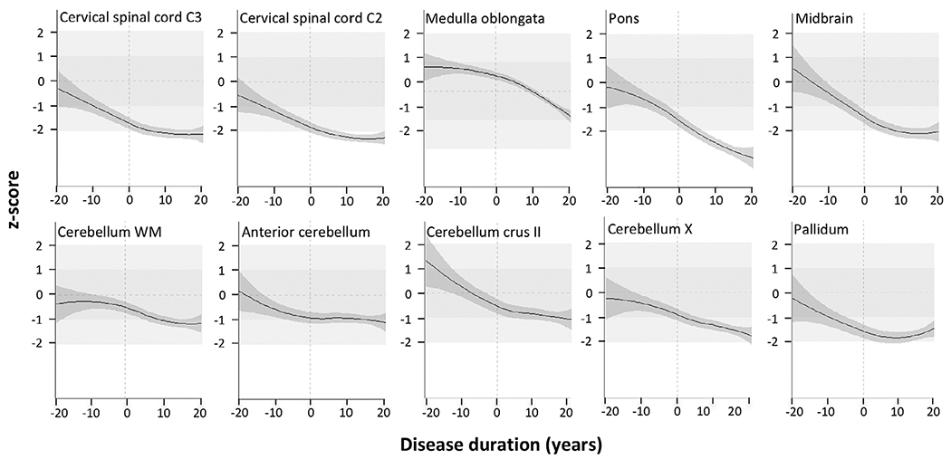
Results: The metrics of cervical spinal cord segments C3 and C2, medulla oblongata, pons, and pallidum, and the cerebellar anterior lobe were reduced in preataxic mutation carriers compared with controls. Those of cervical spinal cord segments C2 and C3, medulla oblongata, pons, midbrain, cerebellar lobules crus II and X, cerebellar white matter, and pallidum were reduced in ataxic compared with nonataxic carriers. Of all metrics studied, pontine volume showed the steepest decline across the disease course. It covaried with ataxia severity, CAG repeat length, and age. The multivariate model derived from this analysis explained 46.33% of the variance of pontine volume.
Conclusion: Regional brain and spinal cord tissue loss in spinocerebellar ataxia type 3 starts before ataxia onset. Pontine volume appears to be the most promising imaging biomarker candidate for interventional trials that aim at slowing the progression of spinocerebellar ataxia type 3. © 2021 The Authors. Movement Disorders published by Wiley Periodicals LLC on behalf of International Parkinson and Movement Disorder Society.
Trial registration: ClinicalTrials.gov NCT01470729.
Keywords: MRI; biomarker; spinocerebellar ataxia; volumetry.
© 2021 The Authors. Movement Disorders published by Wiley Periodicals LLC on behalf of International Parkinson and Movement Disorder Society.
Figures
References
-
- Ruano L, Melo C, Silva MC, Coutinho P. The global epidemiology of hereditary ataxia and spastic paraplegia: a systematic review of prevalence studies. Neuroepidemiology 2014;42(3):174–183. - PubMed
-
- Rub U, Schols L, Paulson H, et al. Clinical features, neurogenetics and neuropathology of the polyglutamine spinocerebellar ataxias type 1, 2, 3, 6 and 7. Prog Neurobiol 2013;104:38–66. - PubMed
-
- Schmitz-Hubsch T, Coudert M, Bauer P, et al. Spinocerebellar ataxia types 1, 2, 3, and 6: disease severity and nonataxia symptoms. Neurology 2008;71(13):982–989. - PubMed
-
- Jacobi H, du Montcel ST, Bauer P, et al. Long-term disease progression in spinocerebellar ataxia types 1, 2, 3, and 6: a longitudinal cohort study. Lancet Neurol 2015;14(11):1101–1108. - PubMed
Publication types
MeSH terms
Associated data
Grants and funding
LinkOut - more resources
Full Text Sources
Other Literature Sources
Medical
Miscellaneous
