. 2021 Aug 15:237:118135.
doi: 10.1016/j.neuroimage.2021.118135. Epub 2021 May 2.

A high-resolution interactive atlas of the human brainstem using magnetic resonance imaging

Affiliations

A high-resolution interactive atlas of the human brainstem using magnetic resonance imaging

Syed M Adil et al. Neuroimage. .

Abstract

Conventional atlases of the human brainstem are limited by the inflexible, sparsely-sampled, two-dimensional nature of histology, or the low spatial resolution of conventional magnetic resonance imaging (MRI). Postmortem high-resolution MRI circumvents the challenges associated with both modalities. A single human brainstem specimen extending from the rostral diencephalon through the caudal medulla was prepared for imaging after the brain was removed from a 65-year-old male within 24 h of death. The specimen was formalin-fixed for two weeks, then rehydrated and placed in a custom-made MRI compatible tube and immersed in liquid fluorocarbon. MRI was performed in a 7-Tesla scanner with 120 unique diffusion directions. Acquisition time for anatomic and diffusion images were 14 h and 208 h, respectively. Segmentation was performed manually. Deterministic fiber tractography was done using strategically chosen regions of interest and avoidance, with manual editing using expert knowledge of human neuroanatomy. Anatomic and diffusion images were rendered with isotropic resolutions of 50 μm and 200 μm, respectively. Ninety different structures were segmented and labeled, and 11 different fiber bundles were rendered with tractography. The complete atlas is available online for interactive use at https://www.civmvoxport.vm.duke.edu/voxbase/login.php?return_url=%2Fvoxbase%2F. This atlas presents multiple contrasting datasets and selected tract reconstruction with unprecedented resolution for MR imaging of the human brainstem. There are immediate applications in neuroanatomical education, with the potential to serve future applications for neuroanatomical research and enhanced neurosurgical planning through "safe" zones of entry into the human brainstem.

Keywords: Atlas; Brainstem; Diffusion tractography; Human brain; Magnetic resonance imaging.


Conflict of interest statement

Declaration of Competing Interest None

Figures

Fig. 1.
Fig. 1.
Postmortem specimen including the full brainstem and partial volumes of the medial, deep forebrain used for magnetic resonance histology. (A) ventral view. (B) dorsal view. Photographs were taken after dissection, vascular flushing and tissue fixation.
Fig. 2.
Fig. 2.
Axial GRE image slice of the brainstem at the level of the rostral pons obtained in a transverse plane, with respect to the longitudinal axis of the brainstem. The image is oriented to be consistent with most conventional histological atlases of the brainstem where ventral is toward bottom of image (inverted with respect to in vivo MR imaging in the axial plane). Note the visibility of descending fiber bundles of the corticospinal tracts amidst the nuclei and transverse fibers of the basilar pons.
Fig. 3.
Fig. 3.
3D-rendered segmentation model used to guide tractography. Each color represents a different gray or white matter structure that could be used as a seed region or ROI for diffusion tractography. (A) Anterior surface view. (B) Posterior surface view with opacity reduced and thalamus and fourth ventricle removed for greater visibility of underlying structures.
Fig. 4.
Fig. 4.
DWI image at the level of the mesencephalon demonstrating segmentation of various gray and white matter structures; image plane and orientation as in Fig. 2. Cpd, cerebral peduncle; MB, mamillary body; ML, medial lemniscus; MTT, mammillothalamic tract; Opt, optic tract; PB, pineal body; PG, periaqueductal gray; SC, superior colliculus; SN, substantia nigra.
Fig. 5.
Fig. 5.
FA image from the mid-pons at the level of the root of the trigeminal nerve; image plane and orientation as in Fig. 2. FA images are useful for visualizing white matter tracts such as the cerebellar peduncles, cranial nerve roots, and major longitudinal white matter tracts of the brainstem. 4 V, fourth ventricle; CNV, cranial nerve 5; CSP, corticospinal tract; MCP, middle cerebellar peduncle; ML, medial lemniscus.
Fig. 6.
Fig. 6.
Multiple image contrasts obtained by magnetic resonance histology of a postmortem human brainstem at the level of the rostral midbrain; image plane as in Fig. 2. (A) GRE, (B) DWI, (C) AD, (D) RD, (E) FA, (F) color FA.
Fig. 7.
Fig. 7.
Human brainstem viewed in custom-specified longitudinal planes through the FA dataset. (A) An oblique longitudinal plane acquired by rotating approximately 60° clockwise from the axial plane. (B) Parasagittal plane. Custom planes such as these were used to more fully visualize and segment the brainstem. Delineations made in one plane transposed to any other plane.
Fig. 8.
Fig. 8.
Simultaneous visualization of selected nuclei and white matter pathways. Deep brain stimulation for Parkinson’s disease patients targets the subthalamic nuclei (blue) but should avoid the corticospinal tracts (fibers). Note the decussation of the corticospinal tract at the inferior end of the brainstem specimen.
Fig. 9.
Fig. 9.
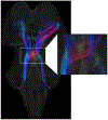
Crossing fibers in the decussation of the superior cerebellar peduncle are resolved, given the high angular resolution of imaging for magnetic resonance histology. Inset shows this decussation at higher magnification.
Fig. 10.
Fig. 10.
Interactive atlas viewing sections at the level of the red nucleus. Upper left shows overview of the brainstem in the midsagittal plane with interactive sliders defining location and angle of the cross-sections shown in the two lower panes. Bottom two panes show these two chosen contrasts: DWI (left) and color FA (right). The outline of the red nucleus is displayed in each of these windows. Upper right provides a pane for user to upload their own datasets or image files; the axial ICBM 152 dataset from the MNI dataset is loaded here with the level set to closely approximate the level selected for display in the lower panes.
Fig. 11.
Fig. 11.
Interactive atlas viewing sections and representative DT. Upper left shows overview of the brainstem in the midsagittal plane with interactive sliders defining location and angle of the cross-sections shown in the two lower panes. Bottom two panels show two chosen contrasts at different longitudinal levels through the specimen: DWI at the level of the caudal diencephalon (left) and color FA at the level of the caudal pons (right). Upper right provides a pane for viewing in 3D a reconstructed tract derived from DT, with the sections from the other panes displayed for contextual reference. In this example, the corticospinal tracts are displayed bilaterally with the transverse sections illustrating passage of corticospinal tract fibers through the posterior limb of internal capsule into the cerebral peduncle (DWI image) and the more caudal intercalation of fibers among pontine nuclei and pontocerebellar fibers (color FA image). Users may interact with these images and tracts and vary rotational perspective, translation and magnification.

References

    1. Afshar F, Watkins ES, Yap JC, 1978. Stereotaxic Atlas of the Human Brainstem and Cerebellar Nuclei. A Variability Study. Raven Press, New York.
    1. Aggarwal M, Zhang J, Pletnikova O, Crain B, Troncoso J, Mori S, 2013. Feasibility of creating a high-resolution 3D diffusion tensor imaging based atlas of the human brainstem: a case study at 11.7 T. Neuroimage 74, 117–127. - PMC - PubMed
    1. Alexander DC, Hubbard PL, Hall MG, Moore EA, Ptito M, Parker GJ, Dyrby TB, 2010. Orientationally invariant indices of axon diameter and density from diffusion MRI. Neuroimage 52, 1374–1389. - PubMed
    1. Axer H, Beck S, Axer M, Schuchardt F, Heepe J, Flucken A, Axer M, Prescher A, Witte OW, 2011. Microstructural analysis of human white matter architecture using polarized light imaging: views from neuroanatomy. Front. Neuroinform 5, 28. - PMC - PubMed
    1. Baete SH, Cloos MA, Lin YC, Placantonakis DG, Shepherd T, Boada FE, 2019. Fingerprinting Orientation Distribution Functions in diffusion MRI detects smaller crossing angles. Neuroimage 198, 231–241. - PMC - PubMed

Publication types

MeSH terms