. 2022 Apr 1;107(4):816-824.
doi: 10.3324/haematol.2020.270421.

Interleukin 4 promotes phagocytosis of murine leukemia cells counteracted by CD47 upregulation

Affiliations

Interleukin 4 promotes phagocytosis of murine leukemia cells counteracted by CD47 upregulation

Pablo Peña-Martínez et al. Haematologica. .

Abstract

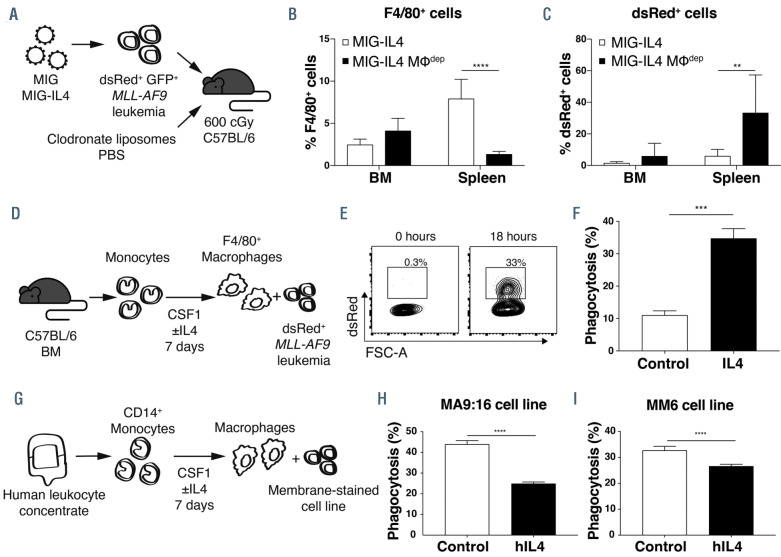
Cytokines are key regulators of tumor immune surveillance by controlling immune cell activity. Here, we investigated whether interleukin 4 (IL4) has antileukemic activity via immune-mediated mechanisms in an in vivo murine model of acute myeloid leukemia driven by the MLL-AF9 fusion gene. Although IL4 strongly inhibited leukemia development in immunocompetent mice, the effect was diminished in immune-deficient recipient mice, demonstrating that the antileukemic effect of IL4 in vivo is dependent on the host immune system. Using flow cytometric analysis and immunohistochemistry, we revealed that the antileukemic effect of IL4 coincided with an expansion of F4/80+ macrophages in the bone marrow and spleen. To elucidate whether this macrophage expansion was responsible for the antileukemic effect, we depleted macrophages in vivo with clodronate liposomes. Macrophage depletion eliminated the antileukemic effect of IL4, showing that macrophages mediated the IL4-induced killing of leukemia cells. In addition, IL4 enhanced murine macrophage-mediated phagocytosis of leukemia cells in vitro. Global transcriptomic analysis of macrophages revealed an enrichment of signatures associated with alternatively activated macrophages and increased phagocytosis upon IL4 stimulation. Notably, IL4 concurrently induced Stat6-dependent upregulation of CD47 on leukemia cells, which suppressed macrophage activity. Consistent with this finding, combining CD47 blockade with IL4 stimulation enhanced macrophage-mediated phagocytosis of leukemia cells. Thus, IL4 has two counteracting roles in regulating phagocytosis in mice; enhancing macrophage-mediated killing of leukemia cells, but also inducing CD47 expression that protects target cells from excessive phagocytosis. Taken together, our data suggest that combined strategies that activate macrophages and block CD47 have therapeutic potential in acute myeloid leukemia.


Figures

Figure 1.
Figure 1.
Interleukin-4 has antileukemic activity in a microenvironment-dependent manner. (A) dsRed+ c-Kit+ MLL–AF9 acute myeloid leukemia (AML) cells were transduced with retroviral vectors coexpressing green fluorescent protein (GFP) and a murine interleukin 4 cDNA (MIG–IL4) or an empty control vector (MIG). Two days later, sorted GFP+ AML cells were transplanted into sublethally irradiated mice. (B) Transplantation of 10,000 leukemia cells into C57BL/6 mice. Kaplan-Meier survival curves (9 mice per group, pooled from 2 independent experiments), and percentage of leukemia (dsRed+) cells in the bone marrow (BM) of mice at the time of sacrifice. (C) Transplantation of 30,000 leukemia cells into NOD/SCID mice. Kaplan-Meier survival curves (6 mice per group) and percentage of leukemia cells in the BM of mice at the time of sacrifice. (D) Transplantation of 30,000 leukemia cells into NSG mice. Kaplan-Meier survival curves (14 mice per group, pooled from 2 independent experiments), and percentage of leukemia cells in the BM of mice at the time of sacrifice. ***P<0.001; ****P<0.0001.
Figure 2.
Figure 2.
Interleukin 4 stimulation increases the frequency of macrophages in vivo. C57BL/6 mice were transplanted with 30,000 sorted green fluorescent protein (GFP)+ MLL-AF9 acute myeloid leukemia (AML) cells 2 days after transduction with retroviral vectors co-expressing GFP and a murine interleukin 4 cDNA (MIG–IL4) or a control vector (MIG). (A) Percentages of blood cell populations within dsRed cells 19 days after transplantation (n=3). (B) Percentage of leukemia (dsRed+ ) cells in the peripheral blood on day 19 after transplantation (n=3). (C) White blood cell counts at days 12 and 27 for MIG–IL4 and non-transplanted irradiated control mice (IL4 group, n=4; controls, n=3). (D) Percentage of F4/80+ cells within dsRed cells in bone marrow and spleens of mice at the time of sacrifice (controls, n=4; IL4 group, n=5). (E) Representative immunohistochemistry staining of F4/80+ cells in bone marrow (40×; scale bar, 20 mm) and spleens (10x; scale bar, 100 mm). BM: bone marrow; N.D.: not detected; PB: peripheral blood; WBC: white blood cell; IHC: immunohistochemistry. **P<0.01; ***P<0.001; ****P<0.0001.
Figure 3.
Figure 3.
Interleukin 4 stimulation causes macrophage-mediated depletion of leukemia cells in vivo. (A) C57BL/6 mice were transplanted with 30,000 sorted green fluorescent protein (GFP)+ MLL-AF9 acute myeloid leukemia (AML) cells transduced with retroviral vectors expressing a murine interleukin 4 cDNA (MIG–IL4) or GFP only (MIG; data presented in Online Supplementary Figure S2). One day prior to transplantation, mice received intraperitoneal (i.p.) injections of clodronate liposomes (MΦdep group; n=4) or phosphate-buffered saline as control (n=5). Every tenth day, new i.p. injections were performed. (B) Percentage of F4/80+ cells and (C) leukemia cells in bone marrow (BM) and spleens at the time of sacrifice in the IL4 group. (D) Monocytes were isolated from mouse BM and differentiated into macrophages in culture with mCSF1 (25 ng/mL) and mIL4 (20 ng/mL) for 7 days, and then MLL-AF9 dsRed+ AML cells were co-cultured with the macrophages. (E) Representative flow cytometry contour plots showing dsRed+ cells within F4/80+ cells in freshly mixed cultures (0 h) and after 18 h of co-culture with macrophages and dsRed+ leukemia cells. (F) Phagocytosis assay with dsRed+ AML cells and murine macrophages (n=3). The percentage of dsRed+ cells within F4/80+ cells is presented. (G) CD14+ cells were isolated from human blood and differentiated into macrophages in culture with human (h)CSF1 (25 ng/mL) and hIL4 (20 ng/mL) for 7 days and then co-cultured with membrane-stained AML cell lines. (H) Phagocytosis assay with PKH67+ MA9:16 cells and PKH26+ human macrophages (n=4). The percentage of PKH67+ cells within PKH26+ cells is presented. (I) Phagocytosis assay with PKH67+ Mono Mac 6 cells and PKH26+ human macrophages (n=5). BM, bone marrow; MM6, Mono Mac 6; MΦ, macrophage. **P<0.01; ***P<0.001; ****P<0.0001.
Figure 4.
Figure 4.
Interleukin 4 expands macrophages enriched for gene expression signatures associated with alternative activation of macrophages and phagocytosis. RNA sequencing was performed on murine macrophages generated from monocytes in vitro, and on sorted dsRed-F4/80+ macrophages from mice in the interleukin 4 (IL4) and control groups. (A) Volcano plots displaying differential gene expression between IL4-stimulated macrophages and control macrophages in vitro (left plot), and macrophages from mice in the IL4 or control group (right plot). The y-axis corresponds to the –log(q-value) and the x-axis to the log of the gene expression fold change. Green dots represent significantly differentially expressed genes with a q-value <0.05 and fold change >2.0. (B) Heatmap showing expression of genes associated with upregulation in tumor-associated macrophages. IL4-stimulated macrophages and control macrophages were harvested from mice. (C) Gene set enrichment analysis revealed enrichment of phagocytosis and MHC protein complex signatures in macrophages harvested from mice. FDR, false discovery rate; GO: gene ontology; MΦ, macrophage; NES, normalized enrichment score; TAM: tumor-associated macrophage.
Figure 5.
Figure 5.
Combined interleukin 4 stimulation and CD47 blockade result in enhanced macrophage-mediated phagocytosis of acute myeloid leukemia cells. (A) Representative histograms showing CD47 expression on acute myeloid leukemia (AML) cells in bone marrow (BM) and spleens of mice transplanted with dsRed+ leukemia cells transduced with the MIG–interleukin 4 (MIG-IL4) or control (MIG) vectors. (B) CD47 expression on AML cells following IL4 stimulation for 24 h. (C) Cd47 expression shown as FPKM values of normalized reads from RNA sequencing data of c-Kit+ dsRed+ leukemia cells and c-Kit+ normal BM cells stimulated with IL4 for 18 h. Data are presented as box and whiskers diagrams; the line indicates median, box limits are first and third quartiles, and bars indicate maximum and minimum values. (D) CD47 expression measured by flow cytometry after 24 h of stimulation with murine (m)IL4 (100 ng/mL) in cells transduced with lentiviral vectors expressing Stat6 or control sgRNA. (E) Phagocytosis assay with macrophages derived from murine BM monocytes stimulated with mCSF1 (25 ng/mL) and mIL4 (20 ng/mL) for 7 days. The AML cells were treated with mIL4 (100 ng/mL) or no IL4 (control) for 24 h prior to co-culture (n=3). Phagocytosis is presented as the percentage of dsRed+ cells within F4/80+ cells. (F) Phagocytosis assay with mouse BM monocyte-derived macrophages stimulated for 7 days with mCSF1 (25 ng/mL) and mIL4 (20 ng/mL) or mCSF1 only (n=3). AML cells were cultured for 1 h with a blocking anti-CD47 antibody or corresponding isotype control and then mixed with the macrophages. FPKM, fragments per kilobase million; gMFI, geometric mean fluorescence intensity; NBM, normal bone marrow. *P<0.05; **P<0.01; ***P<0.001; ****P<0.0001.

References

    1. Costello RT, Sivori S, Marcenaro E, et al. . Defective expression and function of natural killer cell-triggering receptors in patients with acute myeloid leukemia. Blood. 2002;99(10):3661-3667. - PubMed
    1. Jaiswal S, Jamieson CH, Pang WW, et al. . CD47 is upregulated on circulating hematopoietic stem cells and leukemia cells to avoid phagocytosis. Cell. 2009; 138(2):271-285. - PMC - PubMed
    1. Al-Matary YS, Botezatu L, Opalka B, et al. . Acute myeloid leukemia cells polarize macrophages towards a leukemia supporting state in a growth factor independence 1 dependent manner. Haematologica. 2016; 101(10):1216-1227. - PMC - PubMed
    1. Carlsten M, Järås M. Natural killer cells in myeloid malignancies: immune surveillance, NK cell dysfunction, and pharmacological opportunities to bolster the endogenous NK cells. Front Immunol. 2019; 10:2357. - PMC - PubMed
    1. Paczulla AM, Rothfelder K, Raffel S, et al. . Absence of NKG2D ligands defines leukaemia stem cells and mediates their immune evasion. Nature. 2019; 572(7768): 254-259. - PMC - PubMed