. 2021 Apr 6;21(1):193.
doi: 10.1186/s12935-021-01854-7.

Pentraxin 3 is a diagnostic and prognostic marker for ovarian epithelial cancer patients based on comprehensive bioinformatics and experiments

Affiliations

Pentraxin 3 is a diagnostic and prognostic marker for ovarian epithelial cancer patients based on comprehensive bioinformatics and experiments

Xiaoying Chang et al. Cancer Cell Int. .

Abstract

Background: Ovarian epithelial cancer is one of the leading malignant tumors in gynecology and lacks effective diagnostic and prognostic markers. Our study aims to screen and verify ovarian epithelial cancer biomarkers.

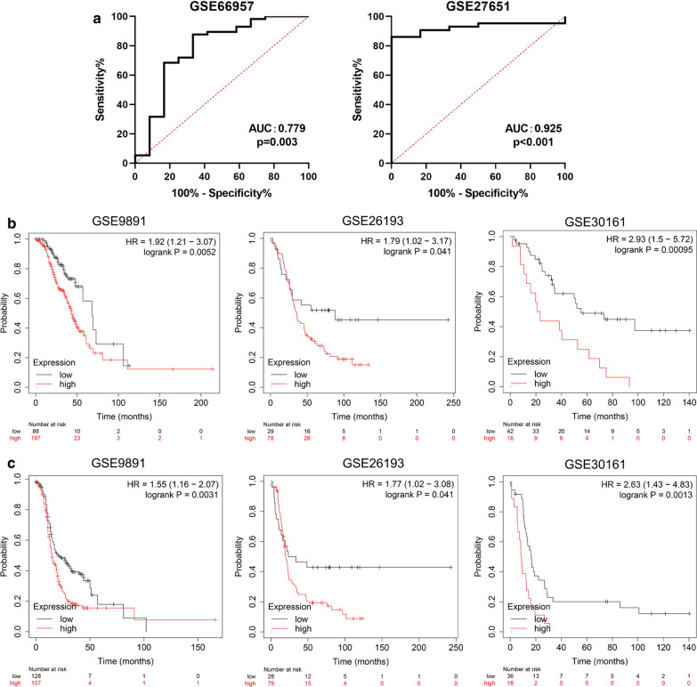
Methods: GSE18520 and GSE26712 were downloaded from the GEO database. The "limma" and "WGCNA" packages were used to explore hub genes. The Kaplan-Meier Plotter database was used for survival analysis of the hub genes. Immunohistochemical analysis was used to identify the expression level of Pentraxin 3 in ovarian epithelial cancer samples.

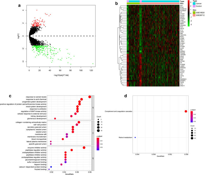
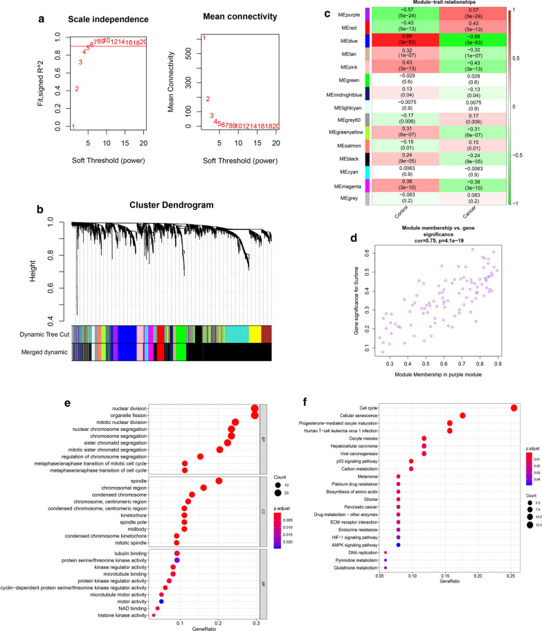
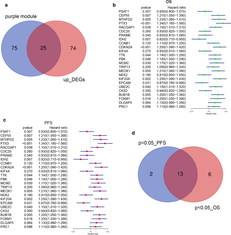
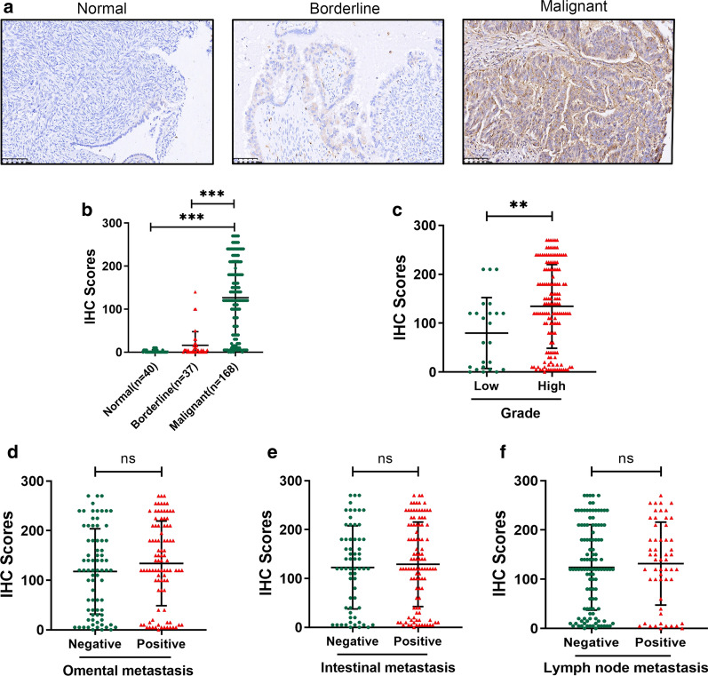
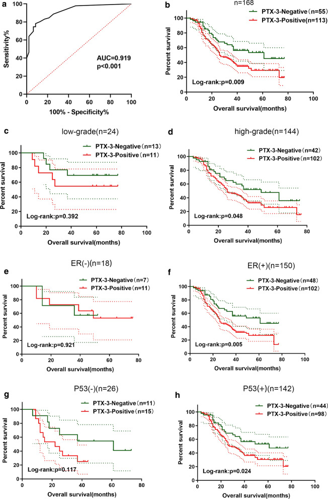
Results: In this study, we integrated and analyzed two datasets, GSE18520 and GSE26712, and a total of 238 differentially expressed genes (DEGs) were screened out. Enrichment analysis showed that these DEGs were related to collagen-containing extracellular matrix and other pathways. Further application of WGCNA (weighted gene coexpression network analysis) identified 15 gene modules, with the purple module showing the highest correlation with ovarian epithelial cancer. Twenty-five genes were shared between the purple module and DEGs, 13 genes were related to the prognosis of ovarian epithelial cancer patients, and the PTX3 gene had the highest hazardous risk (HR) value. We performed immunohistochemical analyses on the 255 Pentraxin-3 (PTX3)-based clinical samples. PTX3 was found to be overexpressed in ovarian epithelial cancer and related to the degree of differentiation. The Cox proportional hazard model indicates that high PTX3 expression is an independent risk factor for the prognosis of ovarian epithelial cancer patients.

Conclusions: In conclusion, through WGCNA and a series of comprehensive bioinformatics analyses, PTX3 was first identified as a novel diagnostic and prognostic biomarker for ovarian epithelial cancer.

Keywords: Biomarker; Ovarian epithelial cancer; PTX3; Prognosis; WGCNA.


Conflict of interest statement

All authors declare that no competing interests exist.

Figures

Fig. 1
Fig. 1
Expression values between DEGs and the enrichment analysis results. a Ovarian cancer and normal tissues identified from GSE18520 and GSE26712. Red represents upregulation, and blue shows downregulation. b Top 50 differential genes heat map. c GO enrichment analyses of DEGs. d KEGG enrichment analysis of DEGs
Fig. 2
Fig. 2
WGCNA. a Analyze the scale-free fit index and average connectivity of the 1–20 soft threshold power (β). b Genes are grouped into various modules by hierarchical clustering, and different colors represent different modules. c Heatmap shows correlations of module eigengenes with features. d A scatter plot of the genes in the purple module. e GO enrichment analyses of genes in purple module. f KEGG enrichment analysis of genes in the purple module
Fig. 3
Fig. 3
Survival analysis of hub genes. a up_DEGs and module gene Venn diagram. b Prognostic values of 25 genes in ovarian tumors (OS in Kaplan–Meier plotter). c Prognostic values of 25 genes in ovarian tumors (PFS in Kaplan–Meier plotter). d Genes were significantly different in both OS and PFS
Fig. 4
Fig. 4
PTX3 protein expression was overexpressed in ovarian tumor tissues. a Representative images of PTX3 staining in human normal ovary tissue, borderline tissue and malignant ovarian tumor; b The IHC scores of PTX3 were significantly increased in malignant ovarian tissues compared to ovary tissues and borderline tissues; c The IHC scores of PTX3 were increased in high-grade compared with low-grade patients; df The IHC scores of PTX3 show no significant difference in differential status of omental metastasis, intestinal metastasis and lymph node metastasis
Fig. 5
Fig. 5
Diagnostic and survival value of PTX3 in ovarian epithelial cancer. a ROC analysis for PTX3 diagnostic value in ovarian epithelial cancer; b Kaplan–Meier analysis for overall survival in ovarian epithelial cancer patients; c Kaplan–Meier analysis for overall survival in low-grade ovarian epithelial cancer patients; d Kaplan–Meier analysis for overall survival in high-grade ovarian epithelial cancer patients; e Kaplan–Meier analysis for overall survival in ER(−) ovarian epithelial cancer patients; f Kaplan–Meier analysis for overall survival in ER(+) ovarian epithelial cancer patients; g Kaplan–Meier analysis for overall survival in P53(−) ovarian epithelial cancer patients; h Kaplan–Meier analysis for overall survival in P53(+) ovarian epithelial cancer patients
Fig. 6
Fig. 6
Relationship of PTX3 expression with ovarian epithelial cancer prognosis. a Forest map based on univariate Cox regression analysis of the OS of ovarian epithelial cancer patients. b Forest map based on multivariate Cox regression analysis of the OS of ovarian epithelial cancer patients
Fig. 7
Fig. 7
Validation of the diagnostic and prognostic performance of the PTX-3 gene in an external GEO dataset.  a ROC analysis for PTX3 diagnostic value in ovarian epithelial cancer based on GSE66957 and GSE27651; b prognostic values (OS) of the PTX-3 gene in ovarian tumors based on GSE989, GSE26193 and GSE30161; c prognostic values (PFS) of the PTX-3 gene in ovarian tumors based on GSE989, GSE26193 and GSE30161

References

    1. Lheureux S, Braunstein M, Oza AM. Epithelial ovarian cancer: Evolution of management in the era of precision medicine. Cancer J Clin. 2019;69:280–304. - PubMed
    1. Gourley C, Bookman MA. Evolving concepts in the management of newly diagnosed epithelial ovarian cancer. J Clin Oncol. 2019;37:2386–97. doi: 10.1200/JCO.19.00337. - DOI - PubMed
    1. Pujade-Lauraine E, Banerjee S, Pignata S. Management of platinum-resistant, relapsed epithelial ovarian cancer and new drug perspectives. J Clin Oncol. 2019;37:2437–48. doi: 10.1200/JCO.19.00194. - DOI - PubMed
    1. Lheureux S, Gourley C, Vergote I, Oza AM. Epithelial ovarian cancer. Lancet. 2019;393:1240–53. doi: 10.1016/S0140-6736(18)32552-2. - DOI - PubMed
    1. van Dam S, Võsa U, van der Graaf A, Franke L, de Magalhães JP. Gene co-expression analysis for functional classification and gene-disease predictions. Brief Bioinform. 2018;19:575–92. - PMC - PubMed

LinkOut - more resources