. 2021 May 5;13(1):104.
doi: 10.1186/s13148-021-01091-9.

Methylated markers accurately distinguish primary central nervous system lymphomas (PCNSL) from other CNS tumors

Affiliations

Methylated markers accurately distinguish primary central nervous system lymphomas (PCNSL) from other CNS tumors

Bradley M Downs et al. Clin Epigenetics. .

Abstract

Background: Definitive diagnosis of primary central nervous system lymphoma (PCNSL) requires invasive surgical brain biopsy, causing treatment delays. In this paper, we identified and validated tumor-specific markers that can distinguish PCNSL from other CNS tumors in tissues. In a pilot study, we tested these newly identified markers in plasma.

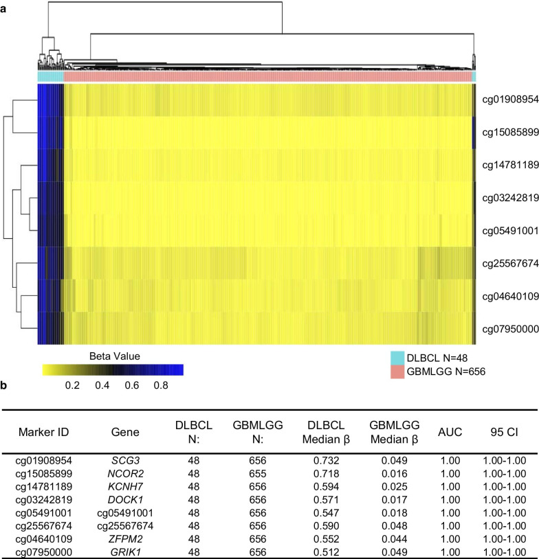
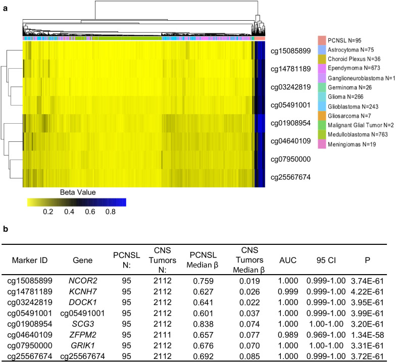
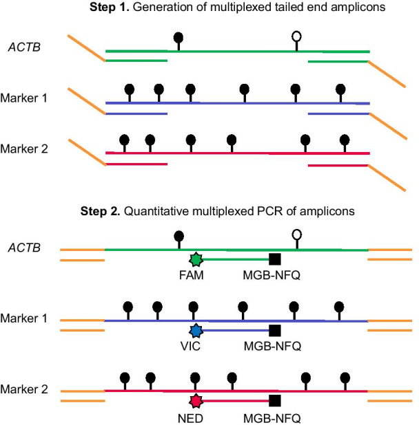
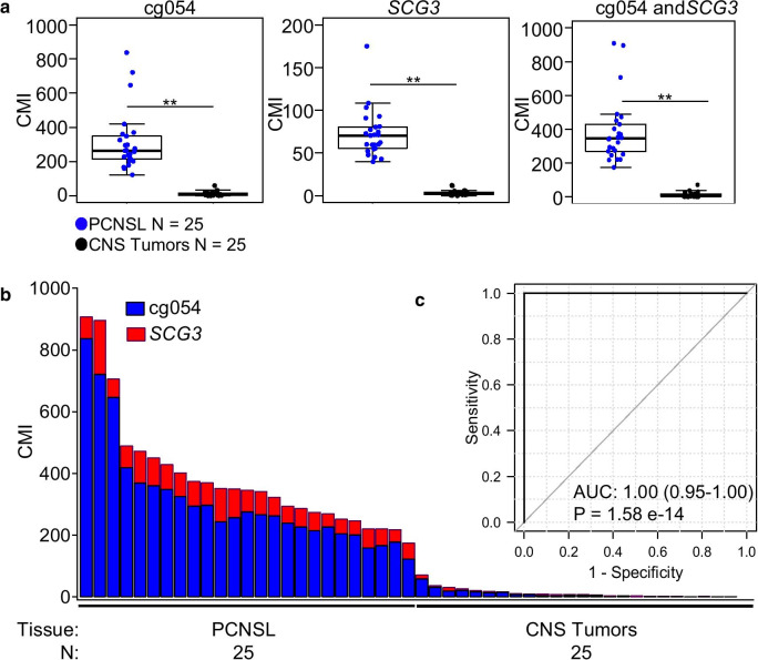
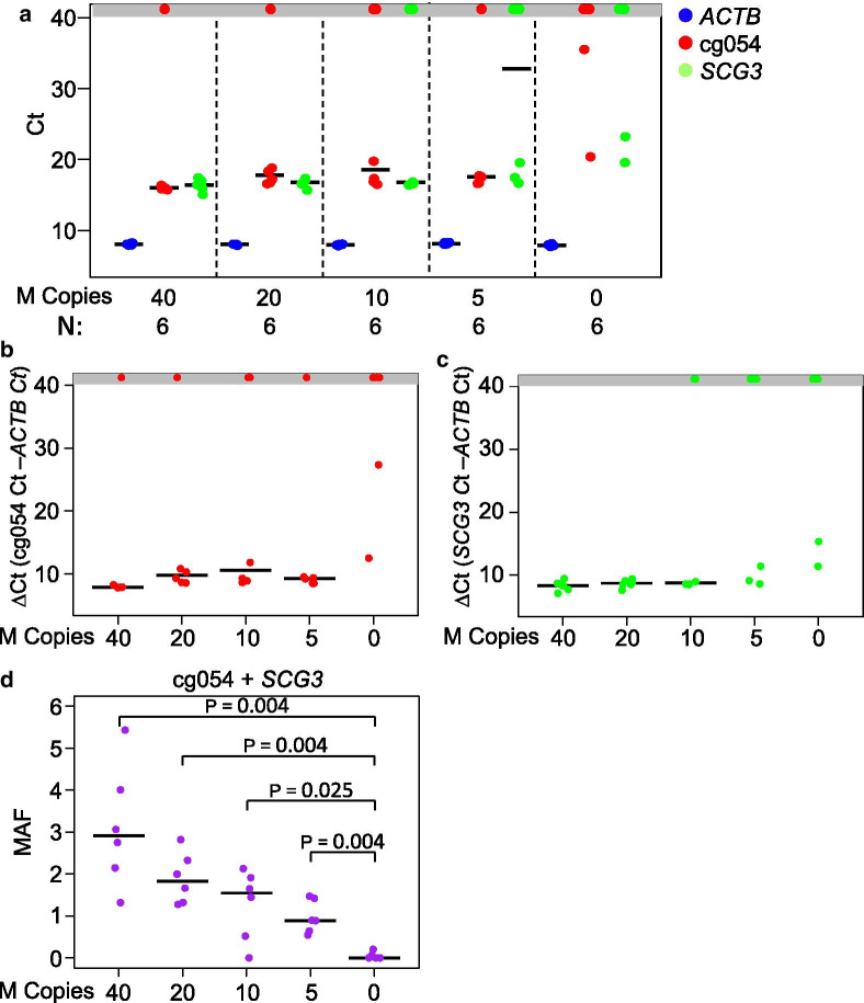
Results: The Methylation Outlier Detector program was used to identify markers in TCGA dataset of 48 diffuse large B-cell lymphoma (DLBCL) and 656 glioblastomas and lower-grade gliomas. Eight methylated markers clearly distinguished DLBCL from gliomas. Marker performance was verified (ROC-AUC of ≥ 0.989) in samples from several GEO datasets (95 PCNSL; 2112 other primary CNS tumors of 11 types). Next, we developed a novel, efficient assay called Tailed Amplicon Multiplexed-Methylation-Specific PCR (TAM-MSP), which uses two of the methylation markers, cg0504 and SCG3 triplexed with ACTB. FFPE tissue sections (25 cases each) of PCNSL and eight types of other primary CNS tumors were analyzed using TAM-MSP. TAM-MSP distinguished PCNSL from the other primary CNS tumors with 100% accuracy (AUC = 1.00, 95% CI 0.95-1.00, P < 0.001). The TAM-MSP assay also detected as few as 5 copies of fully methylated plasma DNA spiked into 0.5 ml of healthy plasma. In a pilot study of plasma from 15 PCNSL, 5 other CNS tumors and 6 healthy individuals, methylation in cg0504 and SCG3 was detectable in 3/15 PCNSL samples (20%).

Conclusion: The Methylation Outlier Detector program identified methylated markers that distinguish PCNSL from other CNS tumors with accuracy. The high level of accuracy achieved by these markers was validated in tissues by a novel method, TAM-MSP. These studies lay a strong foundation for a liquid biopsy-based test to detect PCNSL-specific circulating tumor DNA.

Keywords: Circulating tumor DNA; DNA methylation; Epigenetic biomarkers; Liquid biopsy; Primary central nervous system lymphoma; TAM-MSP.


Conflict of interest statement

S.S. has received research grants, licensing/royalty fees and consulting fees from Cepheid for the QM-MSP assays. No other authors have disclosed any conflicts of interest.

Figures

Fig. 1
Fig. 1
Identification of methylation markers by the Methylation Outlier Detector program in TCGA dataset. a The Methylation Outlier Detector program in TCGA dataset identified 8 potential methylation markers. A hierarchical cluster heatmap displaying the methylation levels of the 8 identified markers showed separation of all of the DLBCL from GBMLGG samples. b The table presents the methylation characteristics and performance of each of the 8 markers for its ability to distinguish DLBCL from GBMLGG samples. N number of samples, AUC area under the curve, CI confidence interval, DLBCL lymphoid neoplasm diffuse large B-cell lymphoma, GBMLGG glioblastoma multiformae and lower-grade gliomas
Fig. 2
Fig. 2
Verification of the 8 methylated markers in the GEO datasets. a A hierarchical cluster heatmap shows the methylation intensity of the 8 markers in databases of PCNSL and 11 other CNS tumors downloaded from the GEO database. b The table presents the methylation characteristics and performance of each of the 8 markers to distinguish PCNSL from the 11 other CNS tumor samples. N number of samples, AUC area under the curve, CI confidence interval, P Mann–Whitney statistics, PCNSL primary central nervous system diffuse large B-cell lymphoma, CNS central nervous system
Fig. 3
Fig. 3
Schema of the TAM-MSP method. The first step of TAM-MSP amplifies two markers and actin control (ACTB) control using a single aliquot of DNA in one well, with primers located within the CpG-rich region of interest for each methylation marker, and therefore includes CpG dinucleotides in its sequence. The 5′ end of the forward and reverse primers for the two methylation markers and ACTB have the same synthetic tails. In the second step of TAM-MSP, primers that are complimentary to the synthetic tails are used, along with marker-specific TaqMan probes, each with one of three indicated fluorescent tags. All three markers are amplified in a single real-time PCR. Methylation in each marker is quantified through interpolation on a historic standard curve and is expressed as percent cumulative methylation. Open circles: unmethylated CpG; closed circles: methylated CpG
Fig. 4
Fig. 4
Performance of the two markers in the Wuhan clinical sample set using the TAM-MSP method. a The two markers, cg0504 and SCG3, assessed as a panel or assessed individually, achieved 100% accuracy in distinguishing PCNSL from the 8 other CNS tumors. b The histogram of cumulative methylation for the two-marker panel in each sample displays the contribution of the two markers in detecting PCNSL among the CNS tumors in the sample set. c Analysis of receiver operator characteristics (ROC, inset) shows that the two-marker panel performed with a high degree of accuracy with AUC of 1.00 (CI 0.95–1.00). **Mann–Whitney P < 0.001; CMI cumulative methylation index, N number of samples, P Mann–Whitney statistics, AUC area under the curve
Fig. 5
Fig. 5
Determination of the limit of detection of methylated cfDNA spiked into plasma from healthy individuals. A serial dilution of Sss1-treated plasma cfDNA, ranging from 40 to 5 copies of methylated cfDNA, was spiked into healthy pooled plasma and analyzed with the TAM-MSP assay. Each dilution point was repeated with 6 replicates. a The Cts for ACTB, cg054 and SCG3 for all dilution replicates showed a strong reproducibility of ACTB at all spike-in dilutions. b Marker cg054 shows no amplification in the healthy plasma (0 copies of spike in DNA) in 4 of 6 replicates and had high ∆Cts. c. Marker SCG3 shows no amplification in the healthy plasma (0 copies of spike in DNA) in 4 of 6 replicates and had high ∆Cts. d The total MAF at all dilutions was significantly different from the healthy plasma without spiked-in of methylated cfDNA. The dots in the gray box represent a failure of the marker to cross the cycle threshold. N number of samples, P Mann–Whitney statistics, MAF methylation allele frequency

References

    1. Batchelor TT, DeAngelis LM. Lymphoma and leukemia of the nervous system. New York: Springer; 2013.
    1. Lim T, Kim SJ, Kim K, Lee JI, Lim DH, Lee DJ, et al. Primary CNS lymphoma other than DLBCL: a descriptive analysis of clinical features and treatment outcomes. Ann Hematol. 2011;90(12):1391–1398. - PMC - PubMed
    1. Alizadeh AA, Eisen MB, Davis RE, Ma C, Lossos IS, Rosenwald A, et al. Distinct types of diffuse large B-cell lymphoma identified by gene expression profiling. Nature. 2000;403(6769):503–511. - PubMed
    1. Rosenwald A, Wright G, Chan WC, Connors JM, Campo E, Fisher RI, et al. The use of molecular profiling to predict survival after chemotherapy for diffuse large-B-cell lymphoma. N Engl J Med. 2002;346(25):1937–1947. - PubMed
    1. Lenz G, Wright GW, Emre NC, Kohlhammer H, Dave SS, Davis RE, et al. Molecular subtypes of diffuse large B-cell lymphoma arise by distinct genetic pathways. Proc Natl Acad Sci U S A. 2008;105(36):13520–13525. - PMC - PubMed

Publication types

Substances