. 2021 May 5;7(19):eabf1244.
doi: 10.1126/sciadv.abf1244. Print 2021 May.

In situ cancer vaccination using lipidoid nanoparticles

Affiliations

In situ cancer vaccination using lipidoid nanoparticles

Jinjin Chen et al. Sci Adv. .

Abstract

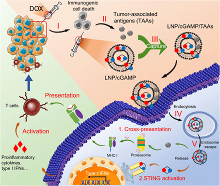
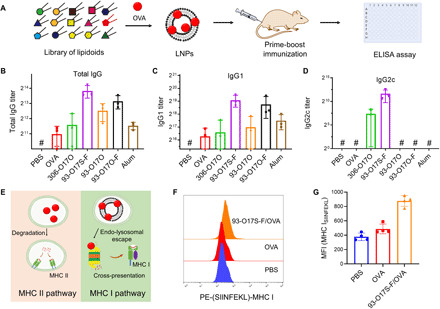
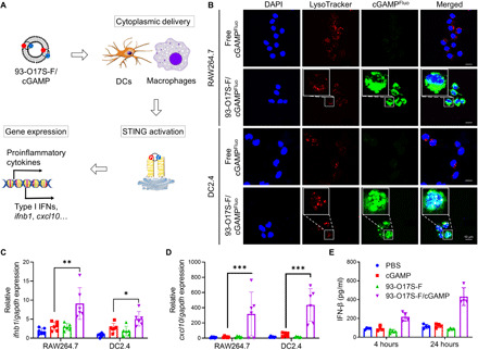
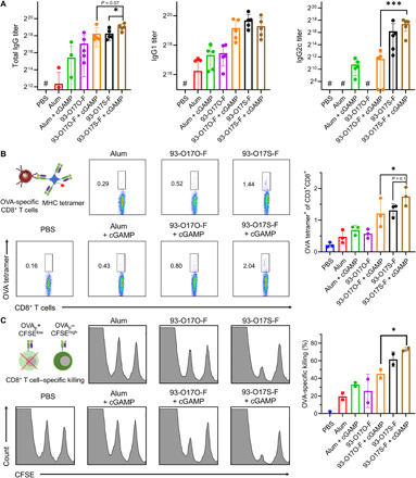
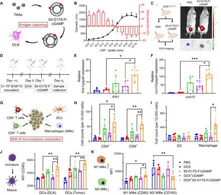
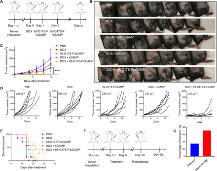
In situ vaccination is a promising strategy for cancer immunotherapy owing to its convenience and the ability to induce numerous tumor antigens. However, the advancement of in situ vaccination techniques has been hindered by low cross-presentation of tumor antigens and the immunosuppressive tumor microenvironment. To balance the safety and efficacy of in situ vaccination, we designed a lipidoid nanoparticle (LNP) to achieve simultaneously enhancing cross-presentation and STING activation. From combinatorial library screening, we identified 93-O17S-F, which promotes both the cross-presentation of tumor antigens and the intracellular delivery of cGAMP (STING agonist). Intratumor injection of 93-O17S-F/cGAMP in combination with pretreatment with doxorubicin exhibited excellent antitumor efficacy, with 35% of mice exhibiting total recovery from a primary B16F10 tumor and 71% of mice with a complete recovery from a subsequent challenge, indicating the induction of an immune memory against the tumor. This study provides a promising strategy for in situ cancer vaccination.


Figures

Fig. 1
Fig. 1. The scheme illustration of LNP system–mediated antigen capturing, cross-presentation, and STING activation.
(I) Low dose of DOX-induced immunogenic cancer cell death. (II) TAAs were released after the administration of low dose of DOX. (III) The released TAAs were captured by lipidoid nanoparticle (LNP)/2′5′-3′5′ cyclic guanosine monophosphate-adenosine monophosphate (cGAMP). (IV) The TAAs and cGAMP encapsulated in LNPs were delivered into APCs via endocytosis. (V) The TAAs and cGAMP escaped from endo/lysosomes to cytoplasm for further cross-presentation and STING activation.
Fig. 2
Fig. 2. The adjuvant effect and enhanced cross-presentation of LNP.
(A) The approach for the screening of LNP library by prime-boost route. (B to D) The OVA-specific immunoglobulin G (IgG) (B), IgG1 (C), and IgG2c (D) antibody titers after immunization with OVA-loaded LNPs. n = 3. #The titer was lower than the minimal dilution. (E) The enhanced cytoplasmic delivery of antigens by LNPs up-regulated the cross-presentation. (F) Typical flow cytometry data of the expression of SIINFEKL–MHC I complex on DC2.4 cells after incubation of different formulation of OVA. (G) The MFI of labeled SIINFEKL–MHC I complex calculated by flow cytometry. n = 4.
Fig. 3
Fig. 3. Enhanced STING activation by cytoplasmic delivery of cGAMP in vitro.
(A) The activation of STING pathway by cytoplasmic delivery of cGAMP using 93-O17S-F. (B) Subcellular distribution of cGAMPFluo and lysosome in RAW264.7 and DC2.4 cells after incubation of free cGAMPFluo or 93-O17S-F/cGAMPFluo for 4 hours. (C and D) Relative expressions of ifnb1 (C) and cxcl10 (D) genes in RAW264.7 and DC2.4 cells after incubation of 93-O17S-F/cGAMP for 4 hours. n = 6, *P ≤ 0.05, **P ≤ 0.01, and ***P ≤ 0.001. (E) The concentration of IFN-β in the medium of DC2.4 cells after incubation of 93-O17S/cGAMP for 4 and 24 hours. n = 4.
Fig. 4
Fig. 4. Enhanced humoral and cellular immune response by codelivery of cGAMP.
(A) The OVA-specific IgG, IgG1, and IgG2c antibody titers after immunization. #The titer was lower than the minimal dilution. n = 5, *P ≤ 0.05, ***P ≤ 0.001. (B) The representative flow images and the quantitated percentages of OVA-peptide (OVAp)–specific CD8+ T cells in spleen of the vaccinated mice. n = 3, *P ≤ 0.05. (C) The representative flow images and the quantitated percentages of OVA-specific killing by CD8+ T cells in spleen of the vaccinated mice. n = 2, *P ≤ 0.05.
Fig. 5
Fig. 5. LNP enhances STING activation and shifts immunocellular composition of the tumor microenvironment in vivo.
(A) Capture of the tumor antigens by 93-O17S-F. (B) The diameters and zeta potentials of 93-O17S-F and tumor lysate complex at different weight ratio. (C) Enhanced delivery of OVAAlexa647 to DLNs after being captured by 93-O17S-F in vivo. Photo credit: J.J. Chen, Tufts University. (D) The route of the in vivo STING activation experiments. (E and F) The relative expression of ifnb1 and cxcl10 genes in B16F10 tumors after the administration of 93-O17S-F/cGAMP for 6 hours. n = 6, *P ≤ 0.05 and ***P ≤ 0.001. (G) The activation of STING pathway recruited the immune cells to tumor sites. (H) The cell numbers of CD4+ and CD8+ T cells at tumor sites after the administration of 93-O17S-F/cGAMP for 48 hours. n = 5, *P ≤ 0.05 and **P ≤ 0.01. (I) The cell numbers of dendritic cells (DCs) and macrophages at tumor sites after the administration of 93-O17S-F/cGAMP for 48 hours. n = 5, *P ≤ 0.05. (J) The MFI of CD80 expressed on CD11c+MHC II+ DCs at DLNs and tumor sites. n = 5, **P ≤ 0.01. (K) The polarization of macrophages at tumor site determined by the MFI of CD80 and CD163 among CD11b+F4/80+ cells. n = 5, *P ≤ 0.05 and **P ≤ 0.01.
Fig. 6
Fig. 6. Antitumor therapeutic effect of 93-O17S-F/cGAMP.
(A) The route of in situ vaccination by 93-O17S/cGAMP. (B) The photographs of B16F10 allograft model tumors at day 6. Photo credit: J.J. Chen, Tufts University. (C) The tumor volumes of B16F10 allograft model tumors after the treatment with different formulations. n = 7, ***P ≤ 0.001. (D) The individual tumor volumes after the treatment with different formulations. (E) The survival rates of mice bearing B16F10 allograft model tumors. (F) The route of tumor rechallenge assay. (G) The percentage of total recovery of primary and rechallenged tumor inoculated mice. For primary tumor, n = 20. For rechallenge, n = 7.

References

    1. Currie G. A., Eighty years of immunotherapy: A review of immunological methods used for the treatment of human cancer. Br. J. Cancer 26, 141–153 (1972). - PMC - PubMed
    1. Kantoff P. W., Higano C. S., Shore N. D., Berger E. R., Small E. J., Penson D. F., Redfern C. H., Ferrari A. C., Dreicer R., Sims R. B., Xu Y., Frohlich M. W., Schellhammer P. F.; IMPACT Study Investigators , Sipuleucel-T immunotherapy for castration-resistant prostate cancer. N. Engl. J. Med. 363, 411–422 (2010). - PubMed
    1. Melero I., Gaudernack G., Gerritsen W., Huber C., Parmiani G., Scholl S., Thatcher N., Wagstaff J., Zielinski C., Faulkner I., Mellstedt H., Therapeutic vaccines for cancer: An overview of clinical trials. Nat. Rev. Clin. Oncol. 11, 509–524 (2014). - PubMed
    1. Melief C. J. M., van Hall T., Arens R., Ossendorp F., van der Burg S. H., Therapeutic cancer vaccines. J. Clin. Invest. 125, 3401–3412 (2015). - PMC - PubMed
    1. Martins F., Sofiya L., Sykiotis G. P., Lamine F., Maillard M., Fraga M., Shabafrouz K., Ribi C., Cairoli A., Guex-Crosier Y., Kuntzer T., Michielin O., Peters S., Coukos G., Spertini F., Thompson J. A., Obeid M., Adverse effects of immune-checkpoint inhibitors: Epidemiology, management and surveillance. Nat. Rev. Clin. Oncol. 16, 563–580 (2019). - PubMed

Publication types