. 2021 Jul;30(7):1397-1407.
doi: 10.1158/1055-9965.EPI-21-0055. Epub 2021 May 5.

Tumor-Associated Stromal Cellular Density as a Predictor of Recurrence and Mortality in Breast Cancer: Results from Ethnically Diverse Study Populations

Affiliations

Tumor-Associated Stromal Cellular Density as a Predictor of Recurrence and Mortality in Breast Cancer: Results from Ethnically Diverse Study Populations

Mustapha Abubakar et al. Cancer Epidemiol Biomarkers Prev. 2021 Jul.

Abstract

Purpose: Tumor-associated stroma is comprised of fibroblasts, tumor-infiltrating lymphocytes (TIL), macrophages, endothelial cells, and other cells that interactively influence tumor progression through inflammation and wound repair. Although gene-expression signatures reflecting wound repair predict breast cancer survival, it is unclear whether combined density of tumor-associated stromal cells, a morphologic proxy for inflammation and wound repair signatures on routine hematoxylin and eosin (H&E)-stained sections, is of prognostic relevance.

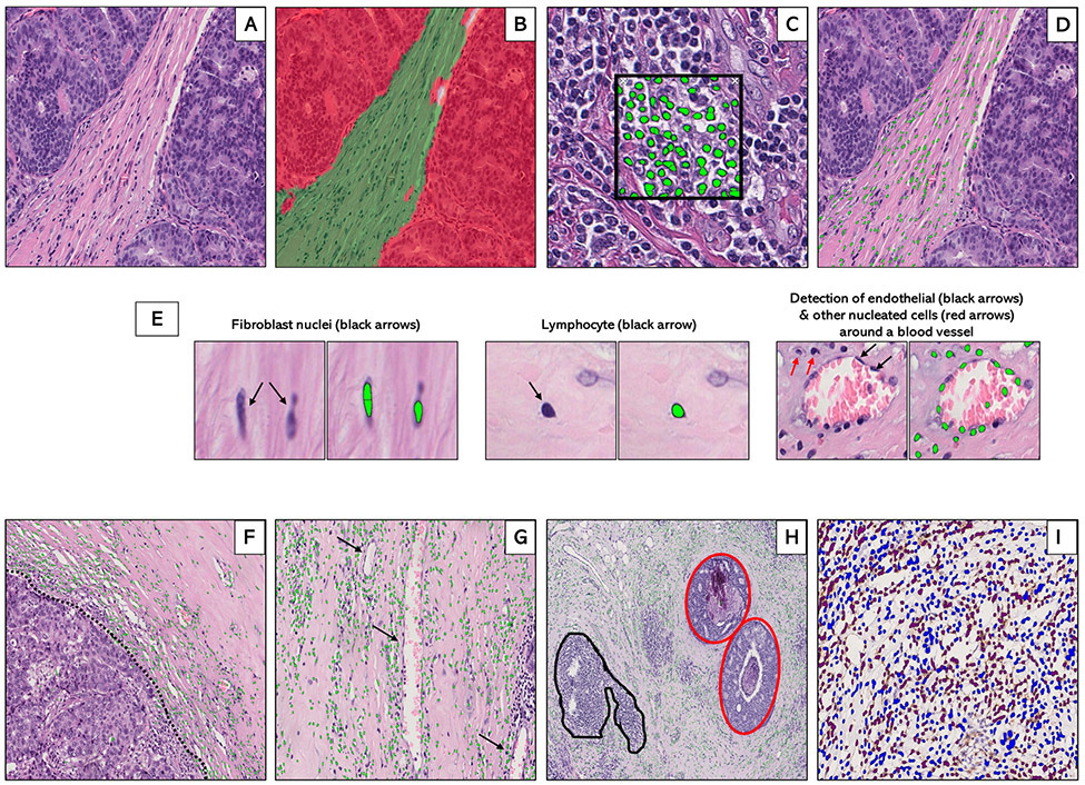
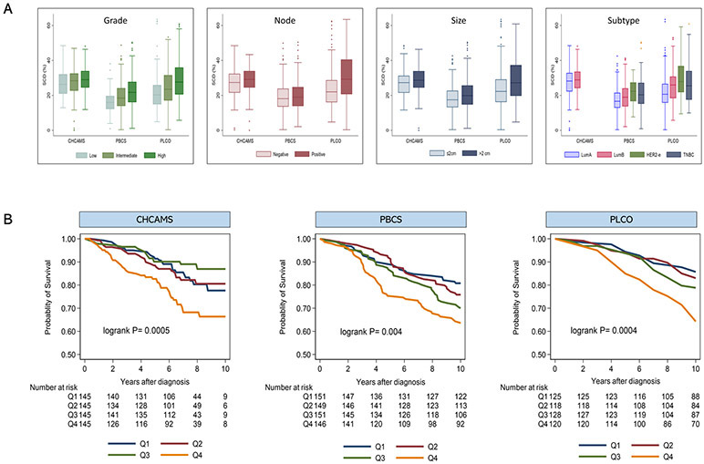
Methods: By applying machine learning to digitized H&E-stained sections for 2,084 breast cancer patients from China (n = 596; 24-55 years), Poland (n = 810; 31-75 years), and the United States (n = 678; 55-78 years), we characterized tumor-associated stromal cellular density (SCD) as the percentage of tumor-stroma that is occupied by nucleated cells. Hazard ratios (HR) and 95% confidence intervals (CI) for associations between SCD and clinical outcomes [recurrence (China) and mortality (Poland and the United States)] were estimated using Cox proportional hazard regression, adjusted for clinical variables.

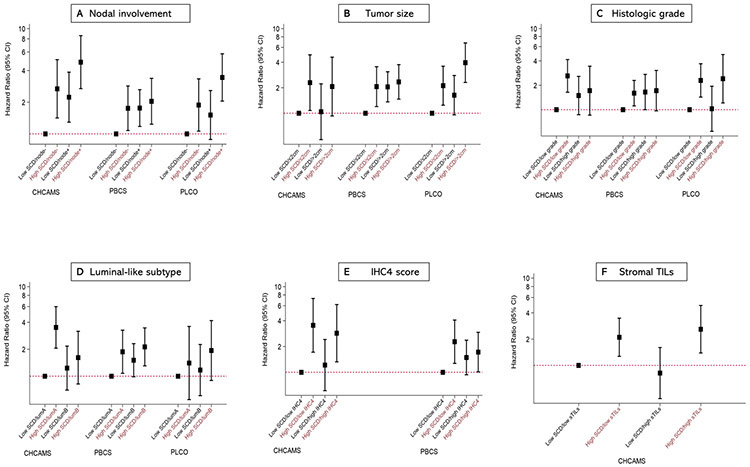
Results: SCD was independently predictive of poor clinical outcomes in hormone receptor-positive (luminal) tumors from China [multivariable HR (95% CI)fourth(Q4) vs. first(Q1) quartile = 1.86 (1.06-3.26); P trend = 0.03], Poland [HR (95% CI)Q4 vs. Q1 = 1.80 (1.12-2.89); P trend = 0.01], and the United States [HR (95% CI)Q4 vs. Q1 = 2.42 (1.33-4.42); P trend = 0.002]. In general, SCD provided more prognostic information than most classic clinicopathologic factors, including grade, size, PR, HER2, IHC4, and TILs, predicting clinical outcomes irrespective of menopausal or lymph nodal status. SCD was not predictive of outcomes in hormone receptor-negative tumors.

Conclusions: Our findings support the independent prognostic value of tumor-associated SCD among ethnically diverse luminal breast cancer patients.

Impact: Assessment of tumor-associated SCD on standard H&E could help refine prognostic assessment and therapeutic decision making in luminal breast cancer.


Conflict of interest statement

Disclosures of Potential Conflicts of Interest:

The authors declare no conflicts of interest

Figures

Figure 1.
Figure 1.
Supervised machine learning for tissue segmentation and cell detection in breast cancer hematoxylin and eosin (H&E)-stained whole tissue sections. Machine-learning algorithms were applied to digitized hematoxylin and eosin (H&E)-stained sections (A) to generate data on stromal cellular density. First, an optimized tissue classification script was used to segment the tumor into epithelial (red) and stromal (green) areas (B). Next, a cell detection script was trained to segment and detect cells (green dots) based on color deconvolution as well as nuclear characteristics (C). Tissue classification and cell detection scripts were combined to enable cell detection to be limited to the stromal compartment (D). Examples of individual stromal cells, including fibroblasts, lymphocytes, and endothelial cells are also shown (E). In general, the machine counted stromal cells (green dots) at the tumor-stroma border (F: dotted black line), around blood vessels (G: black arrows) and in the stroma surrounding foci of ductal carcinoma in-situ (H: inset, red) while excluding tertiary lymphoid structures and lymphoid aggregates (H: inset, black). For a subset of patients with immunohistochemical staining on CD3+ and CD8+ T-cells, optimized scripts were used to quantify the densities of IHC-positive (brown) and IHC-negative (blue) cells within the stroma (I).
Figure 2.
Figure 2.
Tumor-associated stromal cellular density (SCD) in relation to clinicopathological characteristics and breast cancer clinical outcomes. (A) Distribution of SCD by grade, lymph nodal involvement, tumor size, and subtype according to study population. (B) Kaplan-Meier survival curves for the associations between strata (Q1-Q4) of SCD and clinical outcomes (10-year disease-free survival (DFS) and 10-year overall survival (OS)) among patients with luminal breast cancer from three independent study populations, including 580 Chinese patients from CHCAMS (DFS); 597 Polish patients from PBCS (OS); and 492 US patients from the PLCO study (OS). Patients from CHCAMS were premenopausal women, aged 24-55 years, with luminal (HR+) breast cancer that were diagnosed between 2008-2012. Patients from PBCS were women aged 31-75 years, unselected for hormone receptor-status, diagnosed between 2000-2003. PLCO patients were postmenopausal women, aged 55-87 years, unselected for hormone receptor-status, that were participating in the PLCO trial (1993-2001).
Figure 3.
Figure 3.
Joint associations of tumor associated stromal cellular density (SCD) and tumor characteristics with clinical outcomes among luminal breast cancer patients. Hazards ratios (HR) and 95% confidence intervals for the joint associations between SCD and lymph nodal involvement (A), tumor size (B), histologic grade (C), luminal-like subtype (D), IHC4 score (E), and stromal tumor infiltration lymphocytes, sTILs (F) and clinical outcomes (10-year disease-free survival (DFS) and 10-year overall survival (OS)) among hormone receptor-positive breast cancer patients from three independent study populations including CHCAMS (China; DFS), PBCS (Poland; OS) and PLCO (United States; OS). Hazard ratios and corresponding estimates were obtained from multivariable Cox proportional hazard regression models accounting for standard clinical factors, including age, lymph nodal involvement, tumor size, histologic grade, subtype, and systemic therapy, as well as total tissue area. Each primary model exempted the variable in the joint SCD-clinical factor classification.

References

    1. Natrajan R, Sailem H, Mardakheh FK, Arias Garcia M, Tape CJ, Dowsett M, et al. Microenvironmental Heterogeneity Parallels Breast Cancer Progression: A Histology–Genomic Integration Analysis. PLOS Medicine. 2016;13(2):e1001961. - PMC - PubMed
    1. Heindl A, Sestak I, Naidoo K, Cuzick J, Dowsett M, Yuan Y. Relevance of Spatial Heterogeneity of Immune Infiltration for Predicting Risk of Recurrence After Endocrine Therapy of ER+ Breast Cancer. JNCI: Journal of the National Cancer Institute. 2017;110(2):166–75. - PMC - PubMed
    1. Beck AH, Sangoi AR, Leung S, Marinelli RJ, Nielsen TO, van de Vijver MJ, et al. Systematic Analysis of Breast Cancer Morphology Uncovers Stromal Features Associated with Survival. Science Translational Medicine. 2011;3(108):108ra13–ra13. - PubMed
    1. Yuan Y, Failmezger H, Rueda OM, Ali HR, Gräf S, Chin S-F, et al. Quantitative Image Analysis of Cellular Heterogeneity in Breast Tumors Complements Genomic Profiling. Science Translational Medicine. 2012;4(157):157ra43–ra43. - PubMed
    1. Rohan TE, Xue X, Lin H-M, D’Alfonso TM, Ginter PS, Oktay MH, et al. Tumor Microenvironment of Metastasis and Risk of Distant Metastasis of Breast Cancer. JNCI Journal of the National Cancer Institute. 2014;106(8):dju136. - PMC - PubMed

Publication types

MeSH terms