. 2021 May 5;11(1):9616.
doi: 10.1038/s41598-021-89032-7.

Long pentraxin PTX3 is upregulated systemically and centrally after experimental neurotrauma, but its depletion leaves unaltered sensorimotor deficits or histopathology

Affiliations

Long pentraxin PTX3 is upregulated systemically and centrally after experimental neurotrauma, but its depletion leaves unaltered sensorimotor deficits or histopathology

Marco Oggioni et al. Sci Rep. .

Abstract

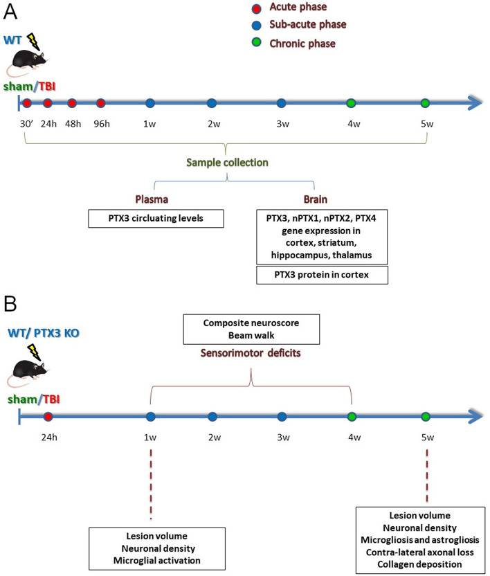
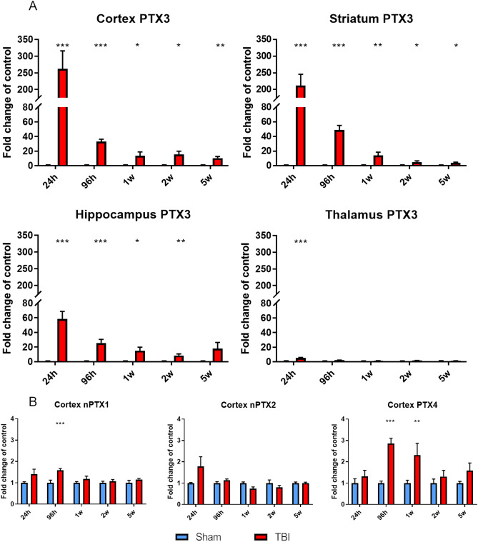
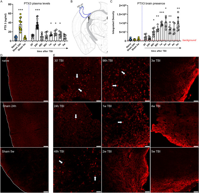
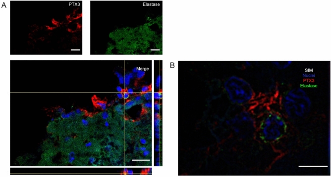
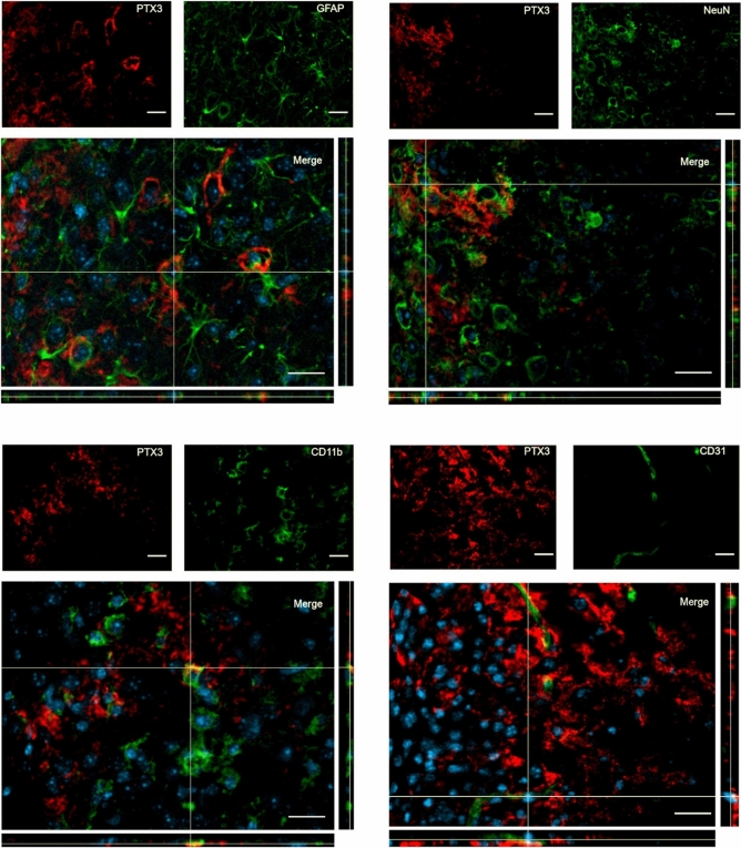
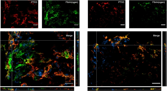
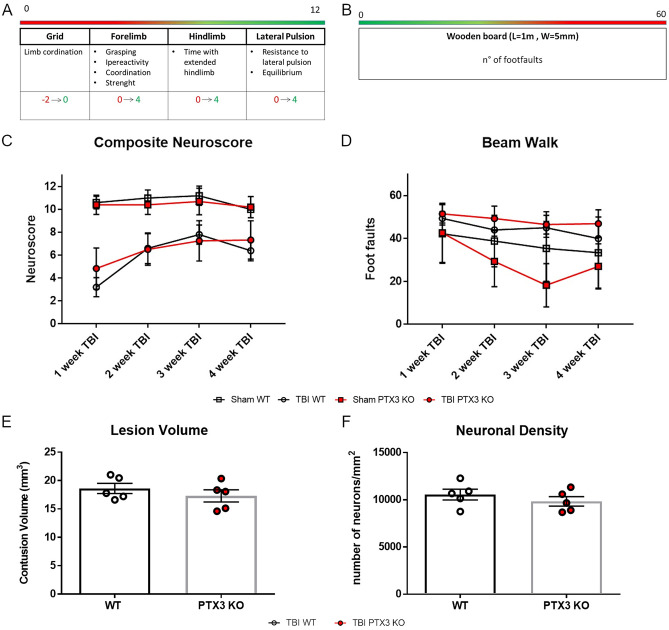
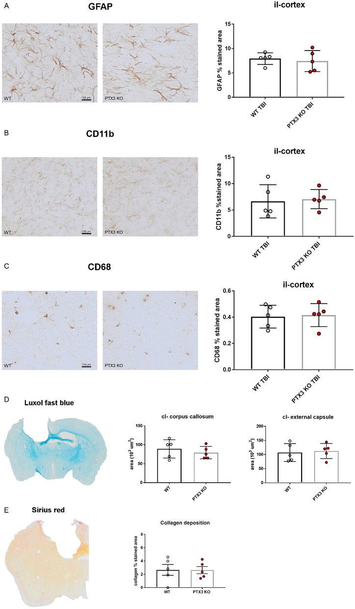
Long pentraxin PTX3, a pattern recognition molecule involved in innate immune responses, is upregulated by pro-inflammatory stimuli, contributors to secondary damage in traumatic brain injury (TBI). We analyzed PTX3 involvement in mice subjected to controlled cortical impact, a clinically relevant TBI mouse model. We measured PTX3 mRNA and protein in the brain and its circulating levels at different time point post-injury, and assessed behavioral deficits and brain damage progression in PTX3 KO mice. PTX3 circulating levels significantly increased 1-3 weeks after injury. In the brain, PTX3 mRNA was upregulated in different brain areas starting from 24 h and up to 5 weeks post-injury. PTX3 protein significantly increased in the brain cortex up to 3 weeks post-injury. Immunohistochemical analysis showed that, 48 h after TBI, PTX3 was localized in proximity of neutrophils, likely on neutrophils extracellular traps (NETs), while 1- and 2- weeks post-injury PTX3 co-localized with fibrin deposits. Genetic depletion of PTX3 did not affect sensorimotor deficits up to 5 weeks post-injury. At this time-point lesion volume and neuronal count, axonal damage, collagen deposition, astrogliosis, microglia activation and phagocytosis were not different in KO compared to WT mice. Members of the long pentraxin family, neuronal pentraxin 1 (nPTX1) and pentraxin 4 (PTX4) were also over-expressed in the traumatized brain, but not neuronal pentraxin 2 (nPTX2) or short pentraxins C-reactive protein (CRP) and serum amyloid P-component (SAP). The long-lasting pattern of activation of PTX3 in brain and blood supports its specific involvement in TBI. The lack of a clear-cut phenotype in PTX3 KO mice may depend on the different roles of this protein, possibly involved in inflammation early after injury and in repair processes later on, suggesting distinct functions in acute phases versus sub-acute or chronic phases. Brain long pentraxins, such as PTX4-shown here to be overexpressed in the brain after TBI-may compensate for PTX3 absence.


Conflict of interest statement

The authors declare no competing interests.

Figures

Figure 1
Figure 1
Experimental Plan. (A) WT mice underwent TBI or sham operation. TBI mice were sacrificed at different time points after surgery and plasma, whole brain or brain areas including cortex, striatum, thalamus and hippocampus were collected. PTX3 plasmatic levels were measured by ELISAs (naive; sham: 24 h, 5w; TBI: 30′, 24 h, 48 h, 96 h, 1w, 2w, 3w, 5w). PTX3 presence (sham: 5w; TBI: 30′, 24 h, 48 h, 96 h, 1w, 2w, 3w, 4w, 5w) and co-localization with neutrophils (Elastase; 48 h), neurons (NeuN; 1w), astrocytes (GFAP; 1w), microglia (CD11b; 1w), endothelial cells (CD31; 1w) and fibrin(ogen) (1w,2w) was studied by immunofluorescence assay on perfused brains. nPTX1, nPTX2, PTX3, PTX4 gene expression analysis was done on snap frozen brain areas by RT-qPCR (sham: 24 h, 96 h, 1w, 2w, 5w; TBI: 24 h, 96 h, 1w, 2w, 5w). (B) WT or PTX3 KO underwent CCI or sham operation. Sensorimotor deficits were assessed by composite neuroscore and beam walk tests on a weekly basis for four weeks after TBI. Brains from both strains were harvested for histopathological analysis: lesion volume and neuronal density with cresyl violet staining (1w; 5w); collagen presence with sirius red staining (5w); contra-lateral white matter loss with luxol fast blue staining (5w); astrogliosis (GFAP; 5w), microgliosis (CD11b/CD68; 5w) and shape descriptors microglia (CD11b; 1w) with immunohistochemistry.
Figure 2
Figure 2
PTX3, nPTX1, nPTX2 and PTX4 mRNA expression in lesioned brain areas. (A) PTX3 was upregulated in the ipsilateral cortex, striatum, hippocampus and thalamus early (24 h) and up to 2w (hippocampus) or 5w (cortex and striatum) after TBI compared to sham mice. (B) nPTX1 and PTX4 cortical expression were significantly increased 96 h after TBI and up to 1w (PTX4) compared to sham, while nPTX2 expression was uneffected. Data is presented as mean ± SEM, n = 6–8. For PTX3 in thalamus and nPTX1 and PTX4 in cortex computations assume that all rows are sampled from populations with the same scatter SD. Multiple t-test followed by Holm-Sidak post hoc test, *adjusted p < 0.05; **p < 0.01; ***p < 0.001 vs sham.
Figure 3
Figure 3
PTX3 protein presence in plasma and in cortex up to 5 weeks after TBI. (A) At 24 h plasmatic PTX3 levels increased in both sham and TBI mice. PTX3 levels were significantly higher than naive from week 1 to week 3. Data is presented as mean ± SEM, n = 4 (TBI 96 h), n = 5 (naive, TBI: 48 h, 1w, 2w, 5w), n = 6 (TBI 30′, 3w), n = 16 (sham 24 h), n = 7 (sham 5w), n = 16 (TBI 24 h). Unequal variances per Bartlett’s test, t-test with Welch correction, *p < 0.05, ***p < 0.001, vs naive. (B) PTX3 presence in cortex was evaluated by immunofluorescence and quantified in a ROI placed in the il-cortex within the first 350 μm from the edge of the contusion (B). (C) PTX3 increased starting from 48 h and reached its maximum at 2 weeks after TBI. Data is presented as mean ± SEM, n = 5–6. Unequal variances per Bartlett’s test, one-way ANOVA with Welch correction, *p < 0.05, **p < 0.01, ***p < 0.001, vs naive. (D) Representative confocal microscopy images showing that PTX3 was not present in naïve or sham mice (sacrificed at 24 h and 5w). Tracings indicate the cortex edge, scale bars = 50 µm. (E) Representative confocal microscopy images showing PTX3 (red) presence next to cell-like shape (arrows) up to 1 week after injury, while it was located mainly extracellularly at longer time points (from 2 to 5w after TBI). Scale bars = 50 µm.
Figure 4
Figure 4
Confocal analysis of PTX3 localization with neutrophils in the contused cortex 48 h after TBI. (A) PTX3 (red) was localized in proximity of neutrophils (Elastase, green). Scale bar = 20 µm. (B) High magnification obtained using SIM showing PTX3 with branches surrounding a neutrophil, likely indicating its presence on NETs. Scale bar = 5 µm.
Figure 5
Figure 5
Confocal analysis of PTX3 presence in the contused cortex 1 week after TBI. PTX3 (red) was present next to but did not co-localize with astrocytes (GFAP, green), neurons (NeuN, green), microglia (CD11b, green) and endothelial cells (CD31, green). Nuclei are in blue. Images are representative of at least two independent experiments. Scale bar = 20 µm.
Figure 6
Figure 6
PTX3 co-localization with fibrin(ogen) in the contused cortex 1 and 2 weeks after TBI. PTX3 (red) co-localized with fibrin(ogen) (green) at 1w (left) and 2w (right) after TBI. Nuclei are in blue. Images are representative of at least two independent experiments. Scale bar = 20 µm.
Figure 7
Figure 7
PTX3 depletion did not affect TBI outcome. (A, B) Sensorimotor function was assessed over 4 weeks after sham injury or TBI using Composite neuroscore (A, worst score = 0) and Beam walk (B, worst score = 60) tests in WT and PTX3 KO mice. (C, D) No difference was observed in post-traumatic sensorimotor deficits between the two strains. Data is presented as mean ± SEM, n = 5 (Sham/TBI WT), n = 10 (Sham PTX3 KO), n = 12 (TBI PTX3 KO). Two-way ANOVA for repeated measures followed by Bonferroni post hoc test = ns. Five weeks after TBI, neither the lesion volume (E) nor the neuronal density (F, neurons per mm2) in the lesioned cortex varied between WT and PTX3 KO mice. Data is presented as mean ± SEM, n = 5 (TBI WT/PTX3 KO). Unpaired t-test = ns (E).
Figure 8
Figure 8
PTX3 depletion did not modify brain inflammatory response at 5 weeks after TBI. Astrocytes (A, GFAP), brain myeloid cells (B, CD11b; C, CD68) and collagen deposition (E, by sirius red) were quantified in the quantified in the ipsilateral (il)-cortex within the first 350 μm from the edge of the contusion showing no difference between WT and PTX3 KO. Axonal loss in contralateral (cl)-corpus callosum and external capsule (D, by luxol fast blue) did not differ between the two strains. Data is presented as mean ± SEM, n = 5 (TBI WT/PTX3 KO). Unpaired t-test = ns.

References

    1. Maas AIR, Menon DK, Adelson PD, Andelic N, Bell MJ, Belli A, et al. Traumatic brain injury: integrated approaches to improve prevention, clinical care, and research. Lancet Neurol. 2017;16:987–1048. doi: 10.1016/S1474-4422(17)30371-X. - DOI - PubMed
    1. Needham EJ, Helmy A, Zanier ER, Jones JL, Coles AJ, Menon DK. The immunological response to traumatic brain injury. J Neuroimmunol. 2019;332:112–125. doi: 10.1016/j.jneuroim.2019.04.005. - DOI - PubMed
    1. Porte R, Davoudian S, Asgari F, Parente R, Mantovani A, Garlanda C, et al. The long pentraxin PTX3 as a humoral innate immunity functional player and biomarker of infections and sepsis. Front Immunol. 2019;10:794. doi: 10.3389/fimmu.2019.00794. - DOI - PMC - PubMed
    1. Bottazzi B, Garlanda C, Teixeira MM. Editorial: the role of pentraxins: from inflammation, tissue repair and immunity to biomarkers. Front Immunol [Internet]. 2019 [cited 2020 Jul 8];10. Available from: https://www.frontiersin.org/articles/10.3389/fimmu.2019.02817/full - PMC - PubMed
    1. de Oliveira THC, Souza DG, Teixeira MM, Amaral FA. Tissue dependent role of PTX3 during ischemia-reperfusion injury. Front. Immunol. 2019;10:1461. doi: 10.3389/fimmu.2019.01461. - DOI - PMC - PubMed

Publication types