Review
. 2021 Jun;28(6):1757-1772.
doi: 10.1038/s41418-021-00785-0. Epub 2021 May 5.

MLKL in cancer: more than a necroptosis regulator

Affiliations
Review

MLKL in cancer: more than a necroptosis regulator

Sofie Martens et al. Cell Death Differ. 2021 Jun.

Abstract

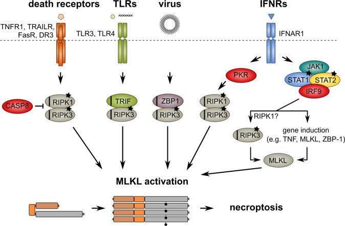
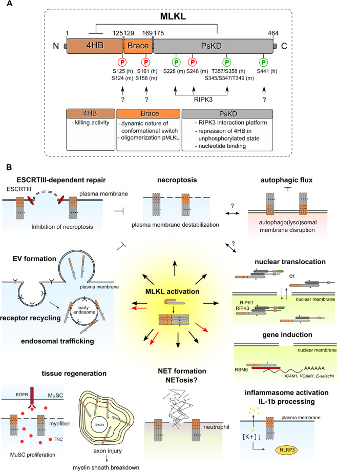
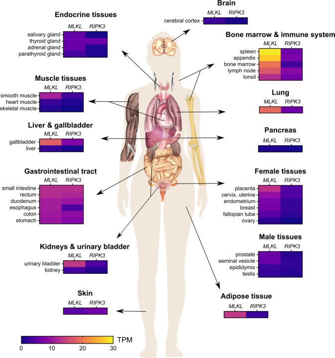
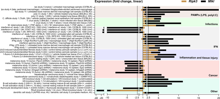
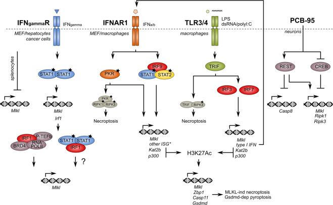
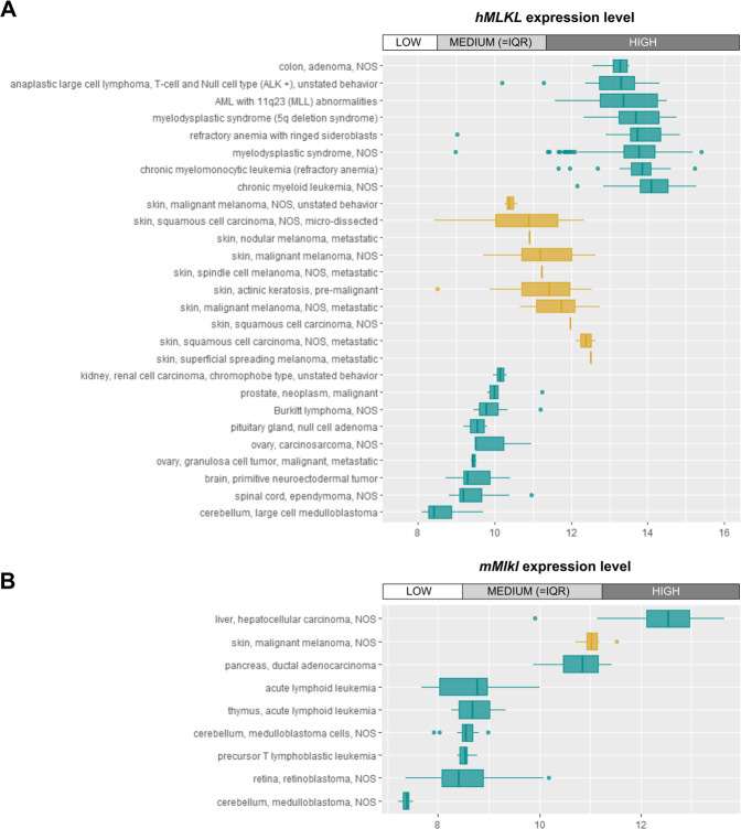
Mixed lineage kinase domain-like protein (MLKL) emerged as executioner of necroptosis, a RIPK3-dependent form of regulated necrosis. Cell death evasion is one of the hallmarks of cancer. Besides apoptosis, some cancers suppress necroptosis-associated mechanisms by for example epigenetic silencing of RIPK3 expression. Conversely, necroptosis-elicited inflammation by cancer cells can fuel tumor growth. Recently, necroptosis-independent functions of MLKL were unraveled in receptor internalization, ligand-receptor degradation, endosomal trafficking, extracellular vesicle formation, autophagy, nuclear functions, axon repair, neutrophil extracellular trap (NET) formation, and inflammasome regulation. Little is known about the precise role of MLKL in cancer and whether some of these functions are involved in cancer development and metastasis. Here, we discuss current knowledge and controversies on MLKL, its structure, necroptosis-independent functions, expression, mutations, and its potential role as a pro- or anti-cancerous factor. Analysis of MLKL expression patterns reveals that MLKL is upregulated by type I/II interferon, conditions of inflammation, and tissue injury. Overall, MLKL may affect cancer development and metastasis through necroptosis-dependent and -independent functions.


Conflict of interest statement

The authors declare no competing interests.

Figures

Fig. 1
Fig. 1. Multiple stimuli lead to necroptosis.
RIPK3-dependent necroptosis can be initiated by engagement of RIPK1, TRIF, or ZBP-1 through their respective RHIM domains (indicated as black stripe) upon activation by phosphorylation (asterisks). Various stimuli induce necroptosis by different pathways that all converge to the executioner of necroptosis, the pseudokinase MLKL. RIPK3-dependent phosphorylation (indicated as black dot on MLKL molecule) induces a conformational switch in MLKL and oligomerization that finally results in necroptosis. Necroptosis occurs especially under conditions of caspase-8 inhibition or ablation. See the legend to Fig. 2 for MLKL molecular domains.
Fig. 2
Fig. 2. Structural domains of MLKL and their functions.
A The functions of each structural domain of MLKL are indicated. The N-terminal 4-helical bundle domain (4HB) shows homology with the N-terminal HeLo-like domain (HELL) of the fungal protein HELLP according to Hidden Markov Models [128]. The PsKD resembles a bi-lobal protein kinase domain, which binds ATP without hydrolyzing it, thus rendering the pseudokinase domain catalytically inactive [91]. Upon RIPK3-dependent phosphorylation (S345) of MLKL, the self-inhibitory pseudokinase domain (PsKD) of MLKL is released from the 4HB, thereby activating MLKL (pMLKLS345) to induce necroptosis. The brace region contributes to the dynamic nature of the 4HB domain, the conformational change induced after activating phosphorylation of the PsKD and the oligomerization of activated MLKL. Phosphorylations that tune cell death activity of MLKL include inhibitory phosphorylation and activating phosphorylation (indicated in red and green respectively). Impact of the phosphorylation of MLKL on its cell death-independent functions is not known yet. h human site, m murine site. B Next to necroptosis execution, MLKL is also involved in ESCRT (Endosomal Sorting Complexes Required for Transport)-dependent repair of the plasma membrane (which restricts necroptotic cell death), release of extracellular vesicles, endosomal trafficking and receptor recycling, myelin sheath membrane breakdown and axon regeneration after injury, muscle stem cell (MuSC) proliferation after muscle injury, NET formation, inflammasome activation, possible nuclear functions including regulation of endothelial cell adhesion molecules such as ICAM1, VCAM1 and E-selectin through interaction with RNA-binding motif protein 6 (RBM6) and stabilization of mRNA, and inhibition of autophagic flux. TNC: tenascin-C. Black dot: phosphorylation of MLKL. Black arrows: processes that require RIPK3-dependent phosphorylation of MLKL, while red arrows: processes that require RIPK3-independent phosphorylation of MLKL.
Fig. 3
Fig. 3. RIPK3 and MLKL genes are differentially expressed in tissues during homeostasis.
RIPK3 and MLKL mRNA expression data in human tissues, generated by the human protein atlas project (HPA) (www.proteinatlas.org). Bone marrow and immune system, lung, adipose tissue and gallbladder exhibit the highest MLKL gene expression, while the pancreas, brain, skin, and some muscle tissues reveal low MLKL mRNA levels. Overall, RIPK3 mRNA expression levels are lower than MLKL. Both RIPK3 and MLKL have low RNA expression in brain and pancreas. Additionally, RIPK3 mRNA levels are the highest in the gastrointestinal tract and the skin (5–10 TPM), while MLKL mRNA levels are highest in bone marrow and immune system (20–30 TPM). TPM transcripts per million.
Fig. 4
Fig. 4. Ripk3 and Mlkl are differentially expressed in tissues during homeostasis and during perturbations.
Overview of all perturbations tested in mouse that result in at least a 5-fold change in mMlkl mRNA levels both up- or downregulation. Data were collected from Genevestigator based on the ‘affymetrix mouse genome 430 2.0 array’ platform. The 2.5-fold change, a fold change considered to be low, is indicated by dotted line. Mlkl mRNA expression is highly inducible in mouse models of infection, inflammation, tissue injury, and cancer. Signals that activate innate immune response including pathogen-associated molecular patterns (PAMPs) such as lipopolysaccharide (LPS, a component of Gram-negative bacteria that activates both TLR4 and inflammasome), infections with parasites, viruses, and bacteria, wound healing as well as chemical-induced inflammation strongly induce Mlkl mRNA expression in various cell types and tissues. Also, IFN signaling induces Mlkl mRNA expression. T. cruzi infection, that activates expression of IFN-stimulated genes through type I IFN receptor (IFNAR1) signaling upregulates Mlkl mRNA [70]. Secondly, IFN-γ-treated BMDMs have increased Mlkl mRNA levels that are even further augmented when treated in combination with LPS, indicating that interferon and TLR/inflammasome signaling operate in synergy to regulate Mlkl mRNA expression. Finally, IFN-regulated transcription factors such as signal transducer and activator of transcription 1 (STAT1), STAT2, and IFN-regulatory factor 9 (IRF9) seem to mediate the induction of Mlkl by IFNα, as respective KOs result in reduced Mlkl mRNA levels. Mice that develop nonalcoholic steatohepatitis (NASH; Gnmt KO mice), have a higher risk to develop hepatocellular carcinoma (HCC). Not only does NASH upregulate Mlkl mRNA expression in the liver, but also HCC further increases its level (up to 7-fold change).
Fig. 5
Fig. 5. Transcriptional regulation of MLKL/Mlkl.
IFN signaling induces transcription of MLKL by different mechanisms, including direct binding of STAT1 to the promoter of MLKL and IRF-regulated expression of acetyltransferases that can activate transcription of MLKL by acetylation of its promoter. Also CREB can directly bind to the MLKL promoter, thereby repressing MLKL expression. Finally, REST can (in)directly activate MLKL transcription. * = another interferon-stimulated gene that might be involved in necroptosis signaling. Irf1/3/7/9 interferon regulatory factor 1/3/7/9, Gsdmd gasdermin D, Casp 8/11 caspase-8/11, Zbp1 Z-DNA binding protein 1, STAT1/2 signal transducer and activator of transcription 1/2, PKR protein kinase R, ISG interferon-stimulated gene, H3K27Ac histon 3 lysin 27 acetylation, p300 histone acetyltransferase p300, Kat2b lysine acetyltransferase 2b, TRIF TIR-domain-containing adaptor-protein inducing IFN-β, PCB-95 polychlorinated biphenyl-95, LPS lipopolysaccharide, IFNgammaR interferon gamma receptor, IFNAR1 interferon alpha receptor, TLR3/4 toll-like receptor 3/4, REST RE1-silencing transcription factor, CREB cAMP responsive element binding protein, BRD4 bromodomain 4 protein, RNA-POLII RNA polymerase II, P-TEFb positive transcription elongation factor.
Fig. 6
Fig. 6. High variation in MLKL/Mlkl expression between different types of cancer.
MLKL/Mlkl mRNA expression data of a small selection of human (A) and mouse (B) cancer types. Cancers displaying high levels of MLKL/Mlkl mRNA include colon adenoma and hematopoietic cancers, while brain tumors display very low levels. This expression pattern may be reflecting different MLKL/Mlkl mRNA expression in these tissues (Fig. 2). Although MLKL/Mlkl mRNA level is low in skin tissue, different types of skin cancer have variable MLKL/Mlkl mRNA levels ranging from medium to high (orange). Data were collected from Genevestigator using ‘affymetrix human genome U133 Plus 2.0 Array’ (A) and ‘affymetrix mouse genome 430 2.0 Array’ (B) respectively as platform. Expression levels are indicated according to ‘LOW’, ‘MEDIUM’, and ‘HIGH’, referring to the expression value range determined by looking at all expression values of all genes over all samples for the platform used. LOW = first quartile range, MEDIUM = interquartile range (IQR), and HIGH = fourth quartile.

References

    1. Choi ME, Price DR, Ryter SW, Choi AMK. Necroptosis: a crucial pathogenic mediator of human disease. JCI Insight. 2019;4:e128834. - PMC - PubMed
    1. Sun L, Wang H, Wang Z, He S, Chen S, Liao D, et al. Mixed lineage kinase domain-like protein mediates necrosis signaling downstream of RIP3 kinase. Cell. 2012;148:213–27. - PubMed
    1. Zhao J, Jitkaew S, Cai Z, Choksi S, Li Q, Luo J, et al. Mixed lineage kinase domain-like is a key receptor interacting protein 3 downstream component of TNF-induced necrosis. Proc Natl Acad Sci USA. 2012;109:5322–7. - PMC - PubMed
    1. Pasparakis M, Vandenabeele P. Necroptosis and its role in inflammation. Nature. 2015;517:311–20. - PubMed
    1. Delanghe T, Dondelinger Y, Bertrand MJM. RIPK1 kinase-dependent death: a symphony of phosphorylation events. Trends Cell Biol. 2020;30:189–200. - PubMed

Publication types