Review
. 2021 Jul;21(7):461-475.
doi: 10.1038/s41568-021-00355-z. Epub 2021 May 5.

Leukaemia: a model metastatic disease

Affiliations
Review

Leukaemia: a model metastatic disease

Andrew E Whiteley et al. Nat Rev Cancer. 2021 Jul.

Abstract

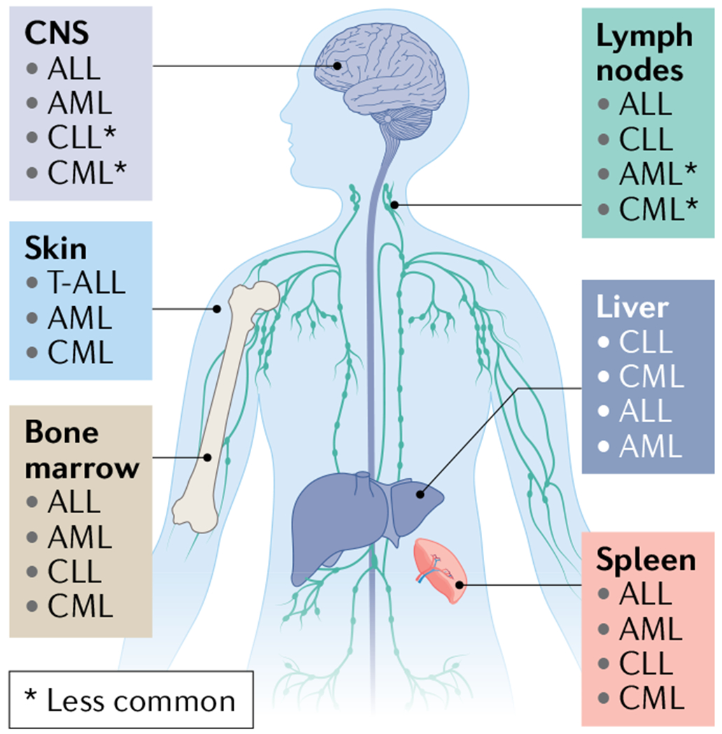
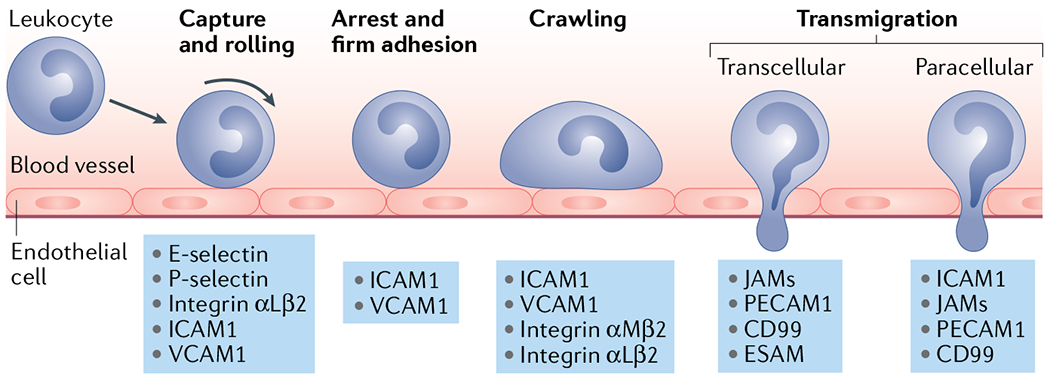
In contrast to solid cancers, which often require genetic modifications and complex cellular reprogramming for effective metastatic dissemination, leukaemic cells uniquely possess the innate ability for migration and invasion. Dedifferentiated, malignant leukocytes retain the benign leukocytes' capacity for cell motility and survival in the circulation, while acquiring the potential for rapid and uncontrolled cell division. For these reasons, leukaemias, although not traditionally considered as metastatic diseases, are in fact models of highly efficient metastatic spread. Accordingly, they are often aggressive and challenging diseases to treat. In this Perspective, we discuss the key molecular processes that facilitate metastasis in a variety of leukaemic subtypes, the clinical significance of leukaemic invasion into specific tissues and the current pipeline of treatments targeting leukaemia metastasis.


Figures

Fig. 1 |
Fig. 1 |. Primary metastatic profiles of leukaemia metastasis.
Acute lymphoblastic leukaemia (ALL) cells are typically initially found in the peripheral vascular system and the haematopoietic organs, including the bone marrow (BM), lymphatic system and spleen; however, central nervous system (CNS) involvement occurs in 5–10% of adult patients at diagnosis and can occur in up to 30–50% of patients in the absence of CNS-directed therapy. WHO classifies ALL and lymphoblastic lymphoma as a spectrum disorder and distinguishes these diseases according to clinical presentation favouring BM (involvement of >20% of blasts) or lymphatic tissue involvement, respectively. Acute myeloid leukaemia (AML) primarily develops and spreads within the BM and less frequently involves other haematopoietic organs. It may invade non-haematopoietic organs including the gingiva, skin, muscle and CNS, although uncommonly. Chronic lymphocytic leukaemia/small lymphocytic lymphoma (CLL/SLL) is notable for its highly stereotyped clinical evolution: at the earliest stages, bloodstream lymphocytosis is the sole clinical finding, followed over time by the development of lymph node enlargement, then splenomegaly and in advanced stages, progressive BM involvement. CLL and SLL are considered the same entity, but with differing disease presentation of bloodstream (CLL) or nodal involvement (SLL),. Chronic myeloid leukaemia (CML) primarily involves the BM, peripheral circulation and spleen. In rare cases including during evolution to blast crisis, CML expansion can result in infiltration into other less common sites, including the lymph nodes, liver, skin or CNS. T-ALL, T cell ALL.
Fig. 2 |
Fig. 2 |. The leukaemia bone marrow microenvironment.
Leukaemia cell homing is mediated by the chemokine stromal cell-derived factor 1 (SDF1),– and adhesion molecules such as the integrin VLA-4,– and E-selectin expressed by the vascular endothelium of fenestrated, sinusoidal bone marrow vessels. In this niche, growth factors, cytokines, extracellular matrix (ECM) components as well as stromal cells such as NG2+ cells, pericytes and colony stimulating factor 1 receptor-positive (CSF1R+) monocytes all serve to promote leukaemia cell growth and survival. As leukaemia cells colonize the bone marrow (BM), they deplete resident haematopoietic stem and progenitor cells (HSPCs) through the secretion of stem cell factor (SCF), creating a malignant niche that disrupts normal haematopoiesis. Leukaemia cells may also migrate to pro-dormancy endosteal niches where osteopontin (OPN), transforming growth factor-β (TGFβ) and hypoxic conditions support a quiescent state, thus protecting them from chemotherapy. Niche factors such as granulocyte colony-stimulating factor (G-CSF) can mobilize leukaemia cells from the metastatic niche into circulation where they can seed distant sites. Matrix-degrading enzymes such as elastase, matrix metalloproteinase 2 (MMP2) and MMP9 (REFS,) can facilitate the extravasation process. Finally, immune cells such as natural killer (NK) cells may limit the survival of leukaemia cells in the BM,. PSGL1, P-selectin glycoprotein ligand 1; VCAM1, vascular cell adhesion molecule 1; VEGF, vascular endothelial growth factor.
Fig. 3 |
Fig. 3 |. The leukaemia splenic microenvironment.
The spleen is a site of leukaemic progression primarily in chronic leukaemias and contains specific microenvironmental niches that can actively promote leukaemia cell homing and support disease expansion. a | As disease progresses, the white pulp expands, leading to a hypertrophic phenotype seen in many patients,. b | Leukaemia cells enter the spleen through large, fenestrated sinusoidal vessels in the red pulp. This is mediated by stromal cell-derived factor 1 (SDF1)–CXCR4 (REFS,) and CCL19–CCR7 (REF.) expression in the splenic sinusoidal niche. Ly6C+ leukaemia-associated macrophages (LAMs) may also promote leukaemia cell migration to the spleen through the secretion of the CCL8 and CCL9 chemokines. Leukaemia proliferation and survival in the spleen is supported by various stromal components such as hyaluronan, monocyte-derived nurse-like cells and helper B cells. For example, the reciprocal interaction between CD84 expressed by both leukaemia cells and stromal cells can promote the survival of either. TGFβ; transforming growth factor-β.
Fig. 4 |
Fig. 4 |. Routes of leukaemia central nervous system invasion.
Leukaemia cell invasion of the central nervous system (CNS) mostly relates to acute leukaemias. Leukaemia cells have been shown to invade the CNS by breaching the blood–brain barrier of parenchymal or leptomeningeal vessels (part a), crossing the blood–cerebrospinal fluid (CSF) barrier of the choroid plexus (part b) or travelling along the abluminal surface of emissary blood vessels that exit the bone marrow through fenestrations in the bone and transition into leptomeningeal vessels (part c). The choroid plexus is a secretory tissue in the brain that is responsible for producing CSF. It contains fenestrated vessels and a monolayer of ependymal cells with tight junctions, ion pumps and transporters that filter out many cells, ions and proteins to produce CSF. The meninges comprise three membranous layers–the dura, the arachnoid and the pia mater–that form a continuous physical barrier surrounding the brain parenchyma and spinal cord. The dura mater is the outermost layer that is adjacent to the calvarium (skull) or vertebral bone. The subsequent arachnoid and pia mater are connected and form the leptomeninges. Between these two layers that form the leptomeninges is the subarachnoid space, which is filled with an acellular CSF that physically cushions the brain and spinal cord.
None

References

    1. Lapidot T et al. A cell initiating human acute myeloid leukaemia after transplantation into SCID mice. Nature 367, 645–648 (1994). - PubMed
    1. Miles LA et al. Single-cell mutation analysis of clonal evolution in myeloid malignancies. Nature 587, 477–482 (2020). - PMC - PubMed
    1. Vetrie D, Helgason GV & Copland M The leukaemia stem cell: similarities, differences and clinical prospects in CML and AML. Nat. Rev. Cancer 20, 158–173 (2020). - PubMed
    1. Jin L, Hope KJ, Zhai Q, Smadja-Joffe F & Dick JE Targeting of CD44 eradicates human acute myeloid leukemic stem cells. Nat. Med 12, 1167–1174 (2006). - PubMed
    1. Krause DS, Lazarides K, Lewis JB, Von Andrian UH & Van Etten RA Selectins and their ligands are required for homing and engraftment of BCR-ABL1+ leukemic stem cells in the bone marrow niche. Blood 123, 1361–1371 (2014). - PMC - PubMed