. 2021 Apr 28:27:191-205.
eCollection 2021.

Differences in activation of intracellular signaling in primary human retinal endothelial cells between isoforms of VEGFA 165

Affiliations

Differences in activation of intracellular signaling in primary human retinal endothelial cells between isoforms of VEGFA 165

Wendelin Dailey et al. Mol Vis. .

Abstract

Purpose: There are reports that a b-isoform of vascular endothelial growth factor-A 165 (VEGFA165b) is predominant in normal human vitreous, switching to the a-isoform (VEGFA165a) in the vitreous of some diseased eyes. Although these isoforms appear to have a different ability to activate the VEGF receptor 2 (VEGFR2) in various endothelial cells, the nature of their ability to activate intracellular signaling pathways is not fully characterized, especially in retinal endothelial cells. We determined their activation potential for two key intracellular signaling pathways (MAPK, AKT) over complete dose-response curves and compared potential effects on the expression of several VEGFA165 target genes in primary human retinal microvascular endothelial cells (HRMECs).

Methods: To determine full dose-response curves for the activation of MAPK (ERK1/2), AKT, and VEGFR2, direct in-cell western assays were developed using primary HRMECs. Potential differences in dose-response effects on gene expression markers related to endothelial cell and leukocyte adhesion (ICAM1, VCAM1, and SELE) and tight junctions (CLDN5 and OCLN) were tested with quantitative PCR.

Results: Activation dose-response analysis revealed much stronger activation of MAPK, AKT, and VEGFR2 by the a-isoform at lower doses. MAPK activation in primary HRMECs displayed a sigmoidal dose-response to a range of VEGFA 165 a concentrations spanning 10-250 pM, which shifted higher into the 100-5,000 pM range with VEGFA 165 b. Similar maximum activation of MAPK was achieved by both isoforms at high concentrations. Maximum activation of AKT by VEGFA 165 b was only half of the maximum activation from VEGFA 165 a. At a lower intermediate dose, where VEGFA 165 a activated intracellular signaling stronger than VEGFA 165 b, the changes in VEGFA target gene expression were generally greater with VEGFA 165 a.

Conclusions: In primary HRMECs, VEGFA 165 a could maximally activate MAPK and AKT at lower concentrations where VEGFA 165 b had relatively little effect. The timing for maximum activation of MAPK was similar for the isoforms, which is different from that reported for non-retinal endothelial cells. Although differences in VEGFA 165 a and VEGFA 165 b are limited to the sequence of their six C-terminal six amino acids, this results in a large difference in their ability to activate at least two key intracellular signaling pathways and VEGF-target gene expression in primary human retinal endothelial cells.


Figures

Figure 1
Figure 1
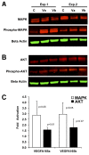
MAPK and AKT are activated in HRMECs by VEGFA165a and VEGFA165b. A: Immunoblots of total MAPK and activated MAPK (phospho-MAPK) 10 min after treatment with VEGFA165a (Va), VEGFA165b (Vb), or media control (C). Two representative experiments are shown. B: Immunoblots of total AKT and activated AKT (phospho-AKT) 10 min after treatment with VEGFA165a (Va), VEGFA165b (Vb), or media control (C). Two representative experiments are shown. C: Fold activation of MAPK and AKT from three immunoblotting experiments. P values are shown, t test relative to media controls (onefold).
Figure 2
Figure 2
VEGFA165a strongly activates MAPK in primary HRMECs at doses where VEGFA165b has little effect. Activation of MAPK was measured with in situ immunofluorescence with a phosphospecific antibody (phospho-Thr202/Tyr204) for p44/42. A: Activation of MAPK at three time points after the addition of 1,000 pM VEGFA165a or VEGFA165b. Bar shows standard deviation. (t test, ***p<0.001 relative to untreated control, n=8 biologic replicates.) B: Dose–response curves for activation of MAPK. The median effective dose (ED50) was 73 pM for VEGFA165a and 1,015 pM for VEGFA165b. Bars indicate the 95% confidence interval for data fit to the four-parameter log-logistic function. n=4 biologic replicates per dose.
Figure 3
Figure 3
VEGFA165b is a weaker activator of AKT in primary HRMECs compared to VEGFA165a. A: Activation of AKT was measured with in situ immunofluorescence with a phosphospecific antibody for Ser473-AKT at five time points after the addition of 20 ng/ml (1,050 pM) VEGFA165a or VEGFA165b. Bar shows standard deviation. (t test, ***p<0.001 relative to untreated control, n=4 biologic replicates.) B: Dose–response curves for activation of AKT. The median effective dose (ED50) value for AKT activation by VEGFA165a was 53 pM compared to 126 pM for VEGFA165b. Bars indicate the 95% confidence interval for data fit to the four-parameter log-logistic function. n=4 biologic replicates per dose.
Figure 4
Figure 4
Differences in the activation of intracellular signaling between VEGFA165a and VEGFA165b in primary HRMECs begin at the receptor VEGFR2. VEGFA165a was a stronger activator of VEGFR2 compared to VEGFA165b. The median effective dose (ED50) for activation by VEGFA165a was 254 pM compared to 1,192 pM for VEGFA165b. Bars indicate the 95% confidence interval for data fit to the four-parameter log-logistic function. n=4 biologic replicates per dose.
Figure 5
Figure 5
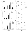
VEGFA165a has a stronger effect on leukocyte–endothelial cell adhesion gene expression in primary HRMECs than VEGFA165b. Relative gene expression was measured with quantitative PCR (qPCR). A: Expression of the ICAM1 gene at 1, 3, 6, and 24 h after treatment with VEGFA165. Confluent human retinal microvascular endothelial cells (HRMECs) were treated with a saturating high dose of VEGFA165a or VEGFA165b (100 ng/ml, 5,300 pM). B: Expression of the ICAM1 gene comparing VEGFA165a and VEGFA165b at low, intermediate, and high doses measured after 6 h. C: Expression of the SELE gene at 1, 3, 6, and 24 h after treatment with VEGFA165. D: Expression of the SELE gene comparing VEGFA165a and VEGFA165b at low, intermediate, and high doses measured after 6 h. E: Expression of the VCAM1 gene at 1, 3, 6, and 24 h after treatment with VEGFA165. F: Expression of the VCAM1 gene comparing VEGFA165a and VEGFA165b at low, intermediate, and high doses measured after 6 h. (Triplicate assays, error bars show standard deviation. ANOVA (ANOVA): compared to media control *p<0.05, **p<0.01; compared to VEGFA165a p<0.05, ††p<0.01).
Figure 6
Figure 6
VEGFA165a has a stronger effect on the expression of tight-junction-protein genes in primary HRMECs than VEGFA165b. Relative gene expression measured with quantitative PCR (qPCR). A: Expression of the CLDN5 gene at 1, 3, 6, and 24 h after treatment with VEGFA165. Confluent human retinal microvascular endothelial cells (HRMECs) were treated with a saturating high dose of VEGFA165a or VEGFA165b (100 ng/ml, 5,300 pM). B: Expression of the CLDN5 gene comparing VEGFA165a and VEGFA165b at low, intermediate, and high doses measured after 6 h. C: Expression of the OCLN gene at 1, 3, 6, and 24 h after treatment with VEGFA165. D: Expression of the OCLN gene comparing VEGFA165a and VEGFA165b at low, intermediate, and high doses measured after 6 h. (Triplicate assays, error bars show standard deviation. ANOVA (ANOVA): compared to media control *p<0.05, **p<0.01; compared to VEGFA165a †† p<0.01).

References

    1. Aiello LP, Avery RL, Arrigg PG, Keyt BA, Jampel HD, Shah ST, Pasquale LR, Thieme H, Iwamoto MA, Park JE. Vascular endothelial growth factor in ocular fluid of patients with diabetic retinopathy and other retinal disorders. N Engl J Med. 1994;331:1480–7. - PubMed
    1. Miller JW, Adamis AP, Aiello LP. Vascular endothelial growth factor in ocular neovascularization and proliferative diabetic retinopathy. Diabetes Metab Rev. 1997;13:37–50. - PubMed
    1. Robbins SG, Conaway JR, Ford BL, Roberto KA, Penn JS. Detection of vascular endothelial growth factor (VEGF) protein in vascular and non-vascular cells of the normal and oxygen-injured rat retina. Growth Factors. 1997;14:229–41. - PubMed
    1. Amadio M, Govoni S, Pascale A. Targeting VEGF in eye neovascularization: What’s new?: A comprehensive review on current therapies and oligonucleotide-based interventions under development. Pharmacol Res. 2016;103:253–69. - PubMed
    1. Avery RL, Castellarin AA, Steinle NC, Dhoot DS, Pieramici DJ, See R, Couvillion S, Nasir MA, Rabena MD, Le K, Maia M, Visich JE. Systemic pharmacokinetics following intravitreal injections of ranibizumab, bevacizumab or aflibercept in patients with neovascular amd. Br J Ophthalmol. 2014;98:1636–41. - PMC - PubMed

Publication types

MeSH terms

LinkOut - more resources