. 2021 Apr 19:12:632304.
doi: 10.3389/fimmu.2021.632304. eCollection 2021.

A Comparison Between Recombinant Listeria GAPDH Proteins and GAPDH Encoding mRNA Conjugated to Lipids as Cross-Reactive Vaccines for Listeria, Mycobacterium, and Streptococcus

Affiliations

A Comparison Between Recombinant Listeria GAPDH Proteins and GAPDH Encoding mRNA Conjugated to Lipids as Cross-Reactive Vaccines for Listeria, Mycobacterium, and Streptococcus

Hector Teran-Navarro et al. Front Immunol. .

Abstract

Cross-reactive vaccines recognize common molecular patterns in pathogens and are able to confer broad spectrum protection against different infections. Antigens common to pathogenic bacteria that induce broad immune responses, such as the glyceraldehyde-3-phosphate dehydrogenase (GAPDH) of the genera Listeria, Mycobacterium, or Streptococcus, whose sequences present more than 95% homology at the N-terminal GAPDH1-22 peptide, are putative candidates for universal vaccines. Here, we explore vaccine formulations based on dendritic cells (DC) loaded with two molecular forms of Listeria monocytogenes GAPDH (LM-GAPDH), such as mRNA carriers or recombinant proteins, and compare them with the same molecular forms of three other antigens used in experimental vaccines, listeriolysin O of Listeria monocytogeness, Ag85A of Mycobacterium marinum, and pneumolysin of Streptococcus pneumoniae. DC loaded with LM-GAPDH recombinant proteins proved to be the safest and most immunogenic vaccine vectors, followed by mRNA encoding LM-GAPDH conjugated to lipid carriers. In addition, macrophages lacked sufficient safety as vaccines for all LM-GAPDH molecular forms. The ability of DC loaded with LM-GAPDH recombinant proteins to induce non-specific DC activation explains their adjuvant potency and their capacity to trigger strong CD4+ and CD8+ T cell responses explains their high immunogenicity. Moreover, their capacity to confer protection in vaccinated mice against challenges with L. monocytogenes, M. marinum, or S. pneumoniae validated their efficiency as cross-reactive vaccines. Cross-protection appears to involve the induction of high percentages of GAPDH1-22 specific CD4+ and CD8+ T cells stained for intracellular IFN-γ, and significant levels of peptide-specific antibodies in vaccinated mice. We concluded that DC vaccines loaded with L. monocytogenes GAPDH recombinant proteins are cross-reactive vaccines that seem to be valuable tools in adult vaccination against Listeria, Mycobacterium, and Streptococcus taxonomic groups.

Keywords: cross-reactive vaccines; glyceraldehyde-3-phosphate-dehydrogenase; innate immunity; listeriosis; pneumonia; tuberculosis.


Conflict of interest statement

MF was employed by company DIOMUNE S.L. The remaining authors declare that the research was conducted in the absence of any commercial or financial relationships that could be construed as a potential conflict of interest.

Figures
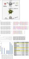
Figure 1
Figure 1
Scheme of the study and selection of the bacterial antigens for the vaccine vectors. (A) Scheme explaining our strategy in this study. First DC are incubated with the different antigen forms: recombinant L. monocytogenes GAPDH proteins or mRNA-LIPO-GAPDH carriers for screening of the suitable ones, causing DC activation with minimal apoptosis induction. DC vaccines loaded with the different antigens are tested for DTH responses as a measurement of T cell responses. DC vaccines with the maximal DTH responses are tested for vaccination experiments followed by bacterial challenge. (B) Multiple alignment of GAPDH sequences of NAD-binding domains of the following bacteria detected at our Health Institution showing more than 95% homology: Listeria monocytogenes (A0A0B8RGN3_LISM), M. tuberculosis (A0A045ITJ4_MYCTX), M. chelonae (A0A0E3TR96_MYCCH) M. marinum (A0A2Z5YDP2_MYCMR), S. agalactiae (Q9ALW2_STRAG), S. pyogenes (G3P_STRPY), and S. pneumoniae (I6L8L9_STREE) protein sequences using CLUSTAL O (1.2.4) multiple sequence alignment. The aligned regions correspond to the InterPro domain IPR020828 that all the proteins have at the beginning of their sequence. The InterPro domain IPR020828 corresponds to the Glyceraldehyde 3-phosphate dehydrogenase, NAD(P) binding domain: https://www.ebi.ac.uk/interpro/entry/InterPro/IPR020828/. The consensus symbols are taken from https://www.ebi.ac.uk/seqdb/confluence/display/JDSAT/Clustal+Omega+FAQ#ClustalOmegaFAQ-Whatdotheconsensussymbolsmeaninthealignment? The symbols meaning is explained in Materials and Methods section Bioinformatics Analyses. Colors on protein alignments correspond to residues according to their physicochemical properties: RED corresponds to Small (small + hydrophobic-including aromatic-Y), BLUE corresponds to acidic, MAGENTA corresponds to basic—H, GREEN corresponds to hydroxyl + sulfhydryl + amine + G and GRAY corresponds to unusual amino/imino acids (see Supplementary Material for complete residues description). (C) Phylogenetic tree of the seven bacteria species compared in this study. The tree data are the following: (Listeria_monocytogenes_GAPDH_NAD-binding:0.09732, ((Mycobacterium_tuberculosis_GAPDH_NAD-binding:0.04475, Mycobacterium_marinum_GAPDH_NAD-binding:0.05140):0.03060, Mycobacterium_chelonae_GAPDH_NAD-binding:0.07196):0.31226):0.14525, Streptococcus_agalactiae_GAPDH_NAD-binding:0.03596, (Streptococcus_pneumonia_GAPDH_NAD-binding:0.02528, Streptococcus_pyogenes_GAPDH_NAD-binding:0.02170):0.01751); (D) Analyses of the clinical cases of bacteria species after the bioinformatic analysis of GAPDH sequences showing 95% homologies in B and detected in the year 2016 at the Hospital U. Marqués de Valdecilla (Microbiology Dpt) from a complete study from 2014 to 2019 (graphic on the left). In the Table (on the right), we show sera from patients (HUMV codes) infected with the bacterial strains of B and examined for anti-LM-GAPDH1−22 antibodies using a peptide ELISA. Sera were collected from patients and storage at −80°C. In the table, we present the antibody titers of patients from a representative 2016 year and with anti-GAPDH-L1 titers higher than 2.0 OD after performing a peptide-specific ELISA. Results are presented as the mean ± SD of OD units in triplicate experiments (P < 0.05). Asterisks and highlighted in yellow correspond to the selected clinical isolates for our further study. Virulence of these clinical isolates into human MoDC (2 × 10⁵/mL) from healthy donors is evaluated in vitro after MoDC infection with 2 × 10⁶ CFU/sample of the clinical isolates detailed in the Table. MoDC were lysed at two different times, at 1 h and at 16 h infection and lysates cultured in agar plates to count CFU. In vitro virulence is expressed as a replication index (RI) of the ration of CFU at 16 h to CFU at 1-h post-infection. Results expressed as RI numbers ± SD of three different experiments. ELISA and virulence assays in vitro using MoDC are performed in triplicate. A Student t-Test is applied for statistical analysis (P ≤ 0.05).
Figure 2
Figure 2
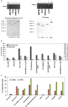
Selection of the antigen forms for the vaccines. (A) Upper geles correspond to preparation of the mRNA antigens from cDNA plasmids encoding for Ag85A antigen of M. marinum (MM), PLY of S. pneumoniae (SP), and GAPDH or LLO of L. monocytogenes (LM) after in vitro transcription. Gels show the linear plasmids (upper left) and mRNA transcripts (upper right). Concentration of the mRNA preparations and qualities are shown in Supplementary Figure 1A. Lower gels correspond to Coomasie stained gels of purification of His-recombinant proteins (lower left) and DC uptake of prepared mRNA from all antigens (PLY of SP, Ag85A of MM, LLO, or GAPDH of LM) conjugated to the lipid carrier, lipofectamine, and after 16 h DC cells are lysed. Lysates were immunoprecipitated with rabbit anti-LLO antibody (DIATEVA), rabbit anti-Mycobacterium antibody (Colorado University), rabbit anti-PLY (a gift from JR de los Toyos, Oviedo, Spain), and rabbit anti-LM-GAPDH1-22 antibody (C. A-D and M.F obtained at CBMSO) (24). All immunoprecitpates were stained with Coomasie blue. (B) DC apoptosis (light gray bars) and DTH responses measured as the footpad swelling (dark gray bars) evaluated after incubation of DC with different antigens: empty DC (DC-CONT), lipofectamine (DC-LIPO), recombinant proteins as PLYrec from SP, LLOrec, and GAPDHrec from LM and antigen Ag85A from MM or mRNA-LIPO complexes of PLY, LLO, GAPDH, or Ag85A. Apoptosis is measured in vitro by flow cytometry and results are expressed as the percentages of annexin-V positive cells ± SD of three different experiments. ANOVA test was applied for flow cytometry results (P ≤ 0.05). DTH responses are measured in vivo after inoculation of right hind footpads of C57BL/6 mice with the different DC vaccines (n = 5 per DC vaccine). Forty-eight hours after inoculation of DC vaccines, DTH responses are evaluated by the swelling of the hind footpads measured with a caliper. Results are expressed as millimeters ± SD of each group of 5 mice. Student t-Test was applied for statistical analysis (P ≤ 0.05). (C) C57BL/6 mice were immunized i.v with 5 × 10³ CFU/mice (HUMV-LM01, HUMV-MM01, or HUMV-SP01) and 7 days later, left hind footpads were inoculated with 1 × 10⁶ DC vaccines (pre-loaded with 5 μg/mL of LLOrec, GAPDHrec, or 50 g/mL of mRNA-LIPO-GAPDH or mRNA-LIPO-LLO, or 1 × 10⁶ CFU of LM, MM or SP, LIPO incubated DC, or saline incubated DC). Popliteal lymph nodes are isolated from mice legs and after homogenization, T cells sub-populations are analyzed by flow cytometry. Percentages of CD4+ (green bars) or CD8+ T cells (red bars) are shown. Results are expressed as the percentages of positive cells ± SD of three different experiments. Student t-Test was applied for statistical analysis (P < 0.05).
Figure 3
Figure 3
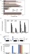
Adjuvant and vaccine abilities of DC vaccine vectors loaded with recombinant or mRNA-LIPO antigens. (A) Flow cytometry analysis of DC surface markers after incubation with recombinant proteins LLOrec or GAPDHrec or mRNA-LIPO complexes: mRNA-LIPO-LLO, mRNA-LIPO-GAPDH, bacteria MM (HUMV-MM01), SP (HUMV-SP01), or LM (HUMV-LM01) or two adjuvants, LPS or DIO-1. Results show the percentages of CD11c+, MHC-II+, CD40+, or CD86+ positive cells. Results are the mean of three different experiments ± SD. Student t-Test was applied for statistical analysis (P ≤ 0.05). (B) Cytokine levels released to the supernatants of DC and measured with a multiparametric CBA kit (BD Biosciences). Results are expressed as pg/mL of each cytokine ± SD of triplicate samples. ANOVA test was applied to the cytokine's concentrations according to the manufacturer recommendation (P ≤ 0.05). (C) Scheme of vaccination model and sample collection to analyze immune responses and protection: spleens and sera. (D) Vaccination of C57BL/6 mice with a single dose of DC vaccines. Seven days later, each group of vaccinated mice are divided in 3 sets and challenged i.v with 104 CFU/mice of hypervirulent strains of HUMV-LM01, HUMV-MM01, or HUMV-SP01. Next, after 14 days mice are bled, sacrificed and spleens collected. Vaccination results expressed percentages of protection as the mean ± SD of triplicates. Percentages are calculated as the number of CFU/mL counted in spleen homogenates of NV mice (saline) divided by CFU/mL of each set of vaccinated mice. Results are expressed as the mean ± SD of triplicates. Student t-Test was applied for statistical analysis (P ≤ 0.05). CFU of non-vaccinated mice are the following: saline LM (HUMV-LM01) 2.75 × 10⁵ CFU/mL, DC-CONT LM (HUMV-LM01) 2.60 × 10⁵ CFU/mL, saline MM (HUMV-MM01) 1 × 10⁵ CFU/mL, DC-CONT MM (HUMV-MM01) 0.9 × 10⁵ CFU/mL, saline SP (HUMV-SP01) 2.5 × 10⁵ CFU/mL, DC-CONT SP (HUMV-SP01) 2.49 × 10⁵ CFU/mL.

Similar articles

Cited by

References

    1. Mora M, Telford JL. Genome-based approaches to vaccine development. J Mol Med. (2010) 88:143–7. 10.1007/s00109-009-0574-9 - DOI - PubMed
    1. Calderon-Gonzalez R, Tobes R, Pareja E, Frande-Cabanes E, Alaez-Alvarez L, Petrovsky N, et al. . Identification and characterisation of T-cell epitopes for incorporation into dendritic cell-delivered Listeria vaccines. J Immunol Methods. (2015) 424:111–9. 10.1016/j.jim.2015.05.009 - DOI - PMC - PubMed
    1. Stephenson KE, Neubauer GH, Reimer U, Pawlowski N, Knaute T, Zerweck J, et al. . Quantification of epitope diversity of HIV-1-specific binding antibodies by peptidemicroarrays for global HIV-1 vaccine development. J Immunol Methods. (2015) 416:105–23. 10.1016/j.jim.2014.11.006 - DOI - PMC - PubMed
    1. Martínez L, Milanič M, Malaina I, Álvarez C, Pérez MB, M de la Fuente I. Weighted lambda superstrings applied to vaccine design. PLoS ONE. (2019) 14:e0211714. 10.1371/journal.pone.0211714 - DOI - PMC - PubMed
    1. Sanchez-Ramon S, Conejero L, Netea MG, Sancho D, Palomares O, Subiza JL. Trained immunity based-vaccines: a new paradigm for the development of broad-spectrum anti-infectious formulations. Front Immunol. (2018) 9:2936. 10.3389/fimmu.2018.02936 - DOI - PMC - PubMed

Publication types

MeSH terms