. 2021 Apr 17:2021:6629204.
doi: 10.1155/2021/6629204. eCollection 2021.

Chemerin-9 Attenuates Experimental Abdominal Aortic Aneurysm Formation in ApoE-/- Mice

Affiliations

Chemerin-9 Attenuates Experimental Abdominal Aortic Aneurysm Formation in ApoE-/- Mice

Shuxiao Chen et al. J Oncol. .

Abstract

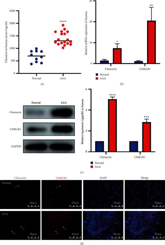
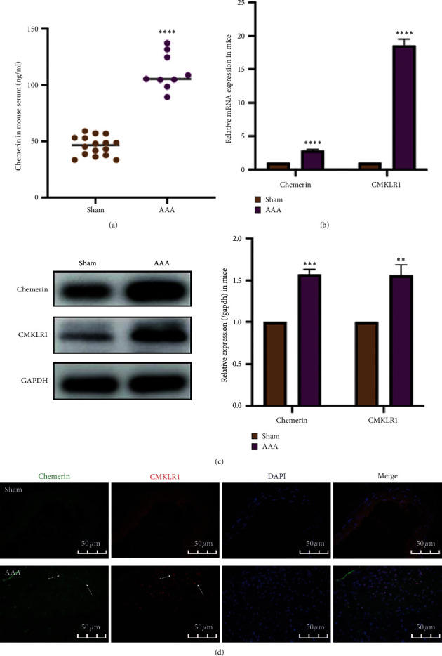
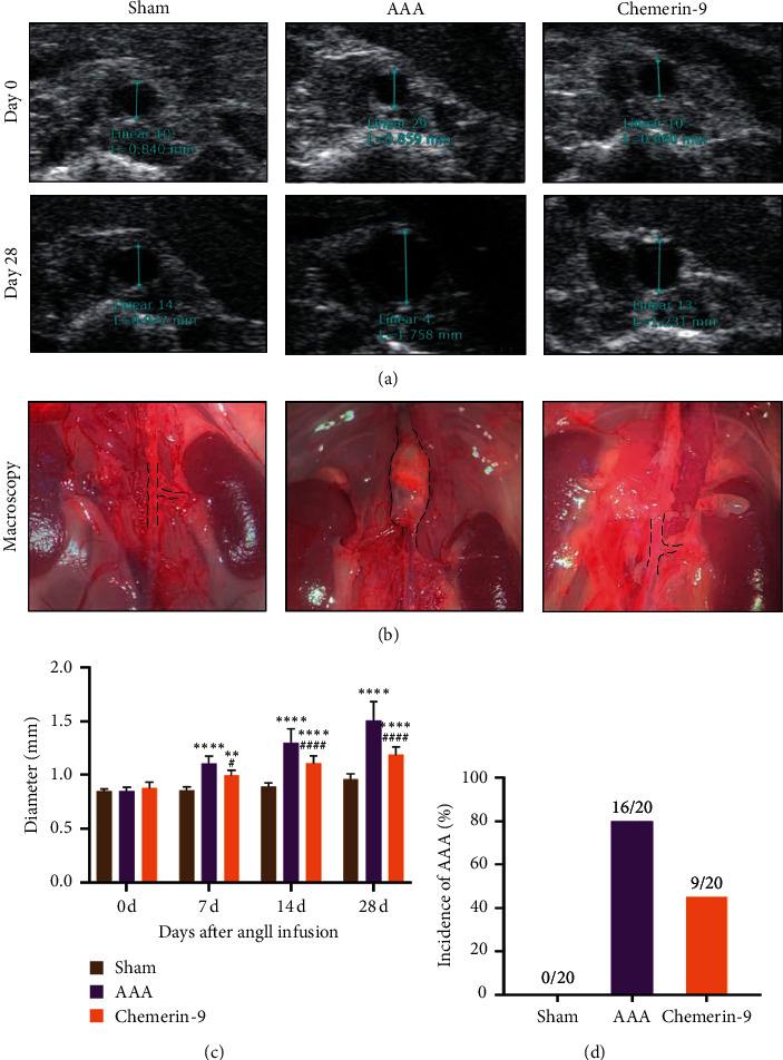
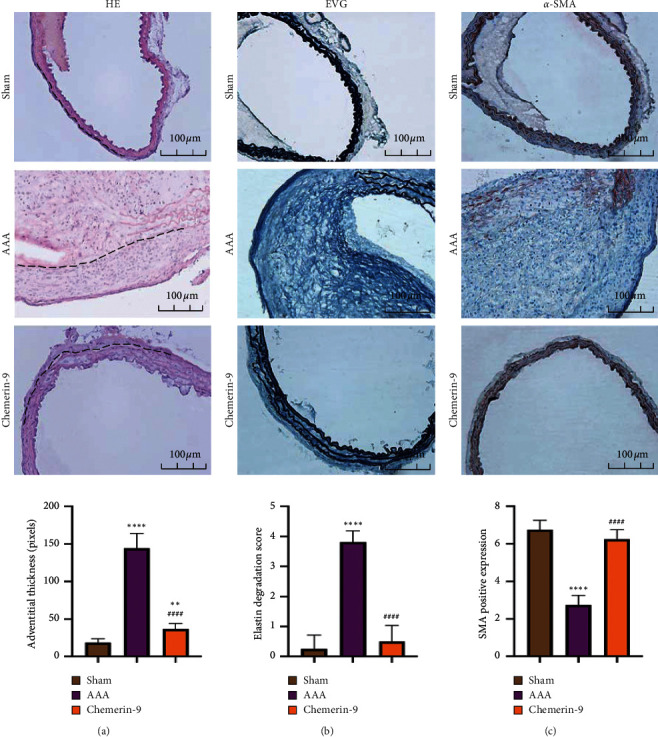
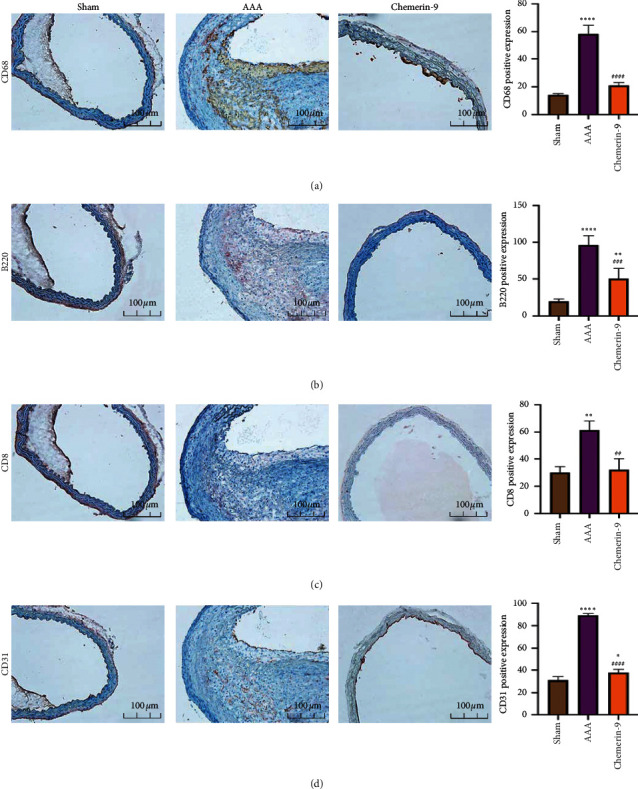
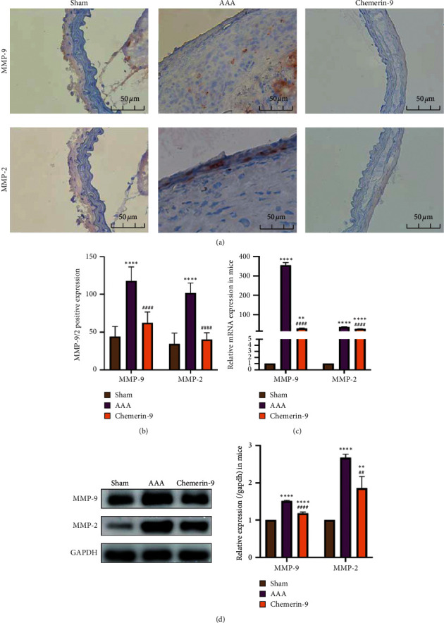
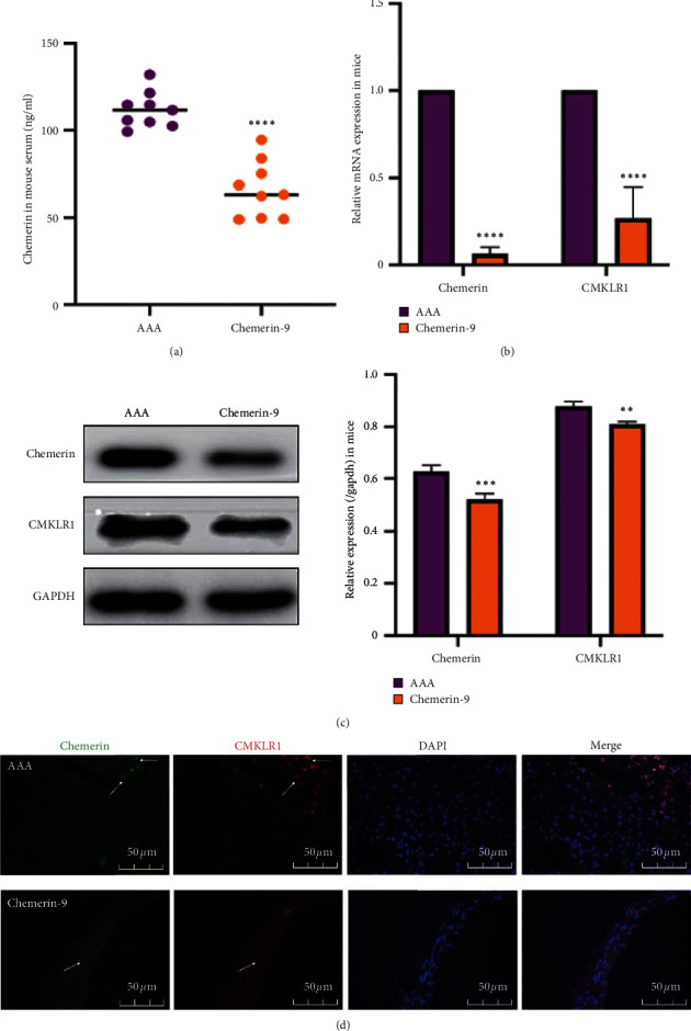
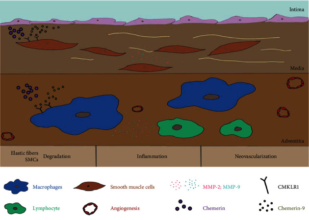
Chronic inflammation plays an essential role in the pathogenesis of abdominal aortic aneurysm (AAA), a progressive segmental abdominal aortic dilation. Chemerin, a multifunctional adipocytokine, is mainly generated in the liver and adipose tissue. The combination of chemerin and chemokine-like receptor 1 (CMKLR1) has been demonstrated to promote the progression of atherosclerosis, arthritis diseases, and Crohn's disease. However, chemerin-9 acts as an analog of chemerin to exert an anti-inflammatory effect by binding to CMKLR1. Here, we first demonstrated that AAA exhibited higher levels of chemerin and CMKLR1 expression compared with the normal aortic tissues. Hence, we hypothesized that the chemerin/CMKLR1 axis might be involved in AAA progression. Moreover, we found that chemerin-9 treatment markedly suppressed inflammatory cell infiltration, neovascularization, and matrix metalloproteinase (MMP) expression, while increasing the elastic fibers and smooth muscle cells (SMCs) in Ang II-induced AAA in ApoE-/- mice. This demonstrated that chemerin-9 could inhibit AAA formation. Collectively, our findings indicate a potential mechanism underlying AAA progression and suggest that chemerin-9 can be used therapeutically.


Conflict of interest statement

All the authors declare that they have no conflicts of interest related to this paper.

Figures

Figure 1
Figure 1
The expression of chemerin and CMKLR1 in humans. (a) Changes in circulating chemerin levels in humans. (b) Relative mRNA expression of chemerin and CMKLR1 in the human aortas. (c) Representative western blot images (left) and semiquantitative analysis (right) of protein expression of chemerin and CMKLR1 in the human aortas. (d) Double immunofluorescence staining for chemerin and CMKLR1 in human aortas. The data are shown as mean ± SD. P < 0.05, ∗∗P < 0.01, ∗∗∗P < 0.001, ∗∗∗∗P < 0.0001 vs. the normal group.
Figure 2
Figure 2
The expression of chemerin and CMKLR1 in mice. (a) Changes in circulating chemerin levels in mice (n = 16/Sham group; n = 9/AAA group). (b) Relative mRNA expression of chemerin and CMKLR1 in the mice aortas (n = 6/group). (c) Representative western blot images (left) and semiquantitative analysis (right) of protein expression of chemerin and CMKLR1 in the mice aortas (n = 6/group). (d) Double immunofluorescence staining for chemerin and CMKLR1 in the mice aortas. The data are shown as mean ± SD. ∗∗P < 0.01, ∗∗∗P < 0.001, ∗∗∗∗P < 0.0001 vs. the Sham group.
Figure 3
Figure 3
Effects of chemerin-9 on abdominal aortic diameter in ApoE−/− mice. (a) Representative ultrasonography images on the 0 and 28 days after pump implantation (n = 20/group). (b) Representative microscopical images of the abdominal aorta in mice on the 28th day. (c) The maximal diameters of each group on the 0, 7, 14, and 28 days after pump implantation (n = 20/group). (d) The AAA incidence of each group 28 days after pump implantation (n = 20/group). The data are shown as mean ± SD. ∗∗P < 0.01, ∗∗∗∗P < 0.0001 vs. the Sham group; #P < 0.05, ####P < 0.0001 vs. the AAA group.
Figure 4
Figure 4
Effects of chemerin-9 on the morphological changes of the abdominal aorta in ApoE−/− mice. (a) Histopathological analysis of representative abdominal aortas of the Sham group, AAA group and chemerin-9 group using HE staining (upper) and quantification (lower) of adventitia thickness (n = 6/group). The adventitial area is shown with black dotted lines. (b) Representative EVG staining images (upper) of elastin and semiquantitative analysis (lower) of elastic laminae deficiency in the medial layer of the aneurysm (n = 6/group). (c) Representative IHC staining images (upper) of SMCs and semiquantitative analysis (lower) of depleted medial smooth muscle in three groups (n = 6/group). The data are shown as mean ± SD. ∗∗P < 0.01; ∗∗∗∗P < 0.0001 vs. the Sham group; ####P < 0.0001 vs. the AAA group.
Figure 5
Figure 5
Effects of chemerin-9 on inflammatory cell infiltration and neoangiogenesis in the aortic wall. (a-d) Representative IHC staining images (left) and semiquantitative analysis (right) of CD68, B220, CD8, and CD31 in 3 groups (n = 6/group). The data are shown as mean ± SD. P < 0.05; ∗∗P < 0.01; ∗∗∗∗P < 0.0001 vs. the Sham group; ##P < 0.01; ###P < 0.001; ####P < 0.0001 vs. the AAA group.
Figure 6
Figure 6
Effects of chemerin-9 on matrix metalloproteinase (MMP)-2 and MMP-9 expression in ApoE−/− mice. (a) Representative IHC staining images of MMP-9 and MMP-2 in 3 groups. (b) Semiquantitative analysis of positive expression of MMP-9 and MMP-2 in 3 groups (n = 6/group). (c) Relative mRNA expression of MMP-9 and MMP-2 in the mice aortas (n = 6/group). (d) Representative western blot images (left) and semiquantitative analysis (right) of protein expression of MMP-9 and MMP-2 in 3 groups (n = 6/group). The data are shown as mean ± SD. ∗∗P < 0.01, ∗∗∗∗P < 0.0001 vs. the Sham group. ##P < 0.01, ####P < 0.0001 vs. the AAA group.
Figure 7
Figure 7
Effects of chemerin-9 on the levels of chemerin and CMKLR1 in ApoE−/− mice. (a) Changes in circulating chemerin levels in mice (n = 9/group). (b) Relative mRNA expression of chemerin and CMKLR1 in the aortas of mice (n = 6/group). (c) Representative western blot images (left) and semiquantitative analysis (right) of protein expression of chemerin and CMKLR1 in 3 groups (n = 6/group). (d) Double immunofluorescence staining for chemerin and CMKLR1 in mice. The data are shown as mean ± SD. ∗∗P < 0.01, ∗∗∗P < 0.001, ∗∗∗∗P < 0.0001 vs. the AAA group.
Figure 8
Figure 8
The potential mechanism of chemerin-9 on AAA.

References

    1. Sakalihasan N., Michel J. B., Katsargyris A., et al. Abdominal aortic aneurysms. Nature Reviews Disease Primers. 2018;4(1):p. 34. doi: 10.1038/s41572-018-0030-7. - DOI - PubMed
    1. Kuivaniemi H., Ryer E. J., Elmore J. R., Tromp G. Understanding the pathogenesis of abdominal aortic aneurysms. Expert Review of Cardiovascular Therapy. 2015;13(9):975–987. doi: 10.1586/14779072.2015.1074861. - DOI - PMC - PubMed
    1. Guirguis-Blake J. M., Beil T. L., Senger C. A., Coppola E. L. Primary care screening for abdominal aortic aneurysm. Journal of the American Medical Association. 2019;322(22):2219–2238. doi: 10.1001/jama.2019.17021. - DOI - PubMed
    1. Jana S., Chute M., Hu M., et al. ADAM (a disintegrin and metalloproteinase) 15 deficiency exacerbates Ang II (angiotensin II)–Induced aortic remodeling leading to abdominal aortic aneurysm. Arteriosclerosis, Thrombosis, and Vascular Biology. 2020 - PMC - PubMed
    1. Go A. S., Mozaffarian D., Roger V. L., et al. Heart disease and stroke statistics-2013 update. Circulation. 2013;127(1):e6–e245. doi: 10.1161/cir.0b013e31828124ad. - DOI - PMC - PubMed

LinkOut - more resources