Review
. 2021 Jun;254(3):321-341.
doi: 10.1007/s00232-021-00179-w. Epub 2021 May 5.

Recent Insights into the Structure and Function of Mycobacterial Membrane Proteins Facilitated by Cryo-EM

Affiliations
Review

Recent Insights into the Structure and Function of Mycobacterial Membrane Proteins Facilitated by Cryo-EM

Ameya D Bendre et al. J Membr Biol. 2021 Jun.

Abstract

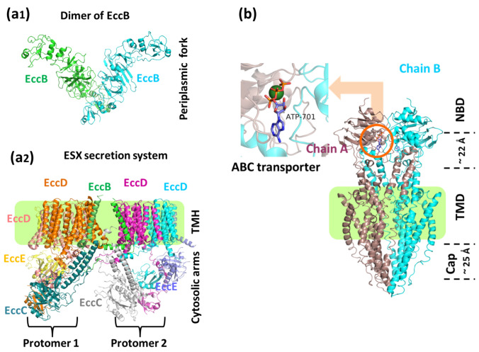
Mycobacterium tuberculosis (Mtb) is one of the deadliest pathogens encountered by humanity. Over the decades, its characteristic membrane organization and composition have been understood. However, there is still limited structural information and mechanistic understanding of the constituent membrane proteins critical for drug discovery pipelines. Recent advances in single-particle cryo-electron microscopy and cryo-electron tomography have provided the much-needed impetus towards structure determination of several vital Mtb membrane proteins whose structures were inaccessible via X-ray crystallography and NMR. Important insights into membrane composition and organization have been gained via a combination of electron tomography and biochemical and biophysical assays. In addition, till the time of writing this review, 75 new structures of various Mtb proteins have been reported via single-particle cryo-EM. The information obtained from these structures has improved our understanding of the mechanisms of action of these proteins and the physiological pathways they are associated with. These structures have opened avenues for structure-based drug design and vaccine discovery programs that might help achieve global-TB control. This review describes the structural features of selected membrane proteins (type VII secretion systems, Rv1819c, Arabinosyltransferase, Fatty Acid Synthase, F-type ATP synthase, respiratory supercomplex, ClpP1P2 protease, ClpB disaggregase and SAM riboswitch), their involvement in physiological pathways, and possible use as a drug target. Tuberculosis is a deadly disease caused by Mycobacterium tuberculosis. The Cryo-EM and tomography have simplified the understanding of the mycobacterial membrane organization. Some proteins are located in the plasma membrane; some span the entire envelope, while some, like MspA, are located in the mycomembrane. Cryo-EM has made the study of such membrane proteins feasible.

Keywords: Cryo-EM; Drug discovery; Membrane protein; Mycobacterium; Protein structure.


Conflict of interest statement

PJP is co-founder and shareholder of www.cryosol-world.com.

Figures

Fig. 1
Fig. 1
a1 Structure of EccB dimer from the ESX-3 core complex showing fork architecture at the periplasm (PDB: 6SGY). a2 The ESX-3 core complex structure formed by EccC, EccD, and EccE subunits is arranged laterally. The pale green box is the trans-membrane region. The translocon pore is formed by the EccD subunit (PDB: 6SGW). b The structure of ABC transporter Rv1819c. In the inset, the binding site for the nucleotide (PDB: 6TQE) is shown. The substrate enters from the cap end of the transporter. The conversion of ATP to ADP leads to the import of the incoming substrate from the NBD end (Color figure online)
Fig. 2
Fig. 2
a Mycobacterial Arabinosyltransferase complex. The complex is composed of two chains, each of EmbB and AcpM. On binding of DPA (red-green spheres), resting-state complex (PDB: 7BX8) is activated (PDB: 7BWR). The arabinosyltransferase is inhibited by ethambutol (Etmb: red) binding in the active site in EmbB (7BVC). b The barrel-shaped MsFAS is a hexamer with one monomer chain shown in cyan (PDB: 6GJC). Each chain is made up of 6 subunits, namely—acetyltransferase (AT), enoyl reductase (ER), dehydratase (DH), malonyl transacylase (MPT), ketoacyl reductase (KR), and ketoacyl synthase (KS) (Color figure online)
Fig. 3
Fig. 3
The complete F-type ATPase complex. a F0 is the membrane-bound region that mainly consists of a C-ring. The F1 is the peripheral region where ATP is synthesized (PDB: 7JG5). b SF0 regions with and without BDQ (dark red) bound to it. c BDQ binds in two sites. The leading site lies between ‘a’ and ‘c’ subunits, while the lagging site lies only on the ‘c’ subunit (Color figure online)
Fig. 4
Fig. 4
Structure of respiratory supercomplex (PDB: 6HWH). a1 The supercomplex is composed of two subcomplexes and some unidentified peptides (grey). Complex IV (blue) consists of CtaC, CtaD, CtaE, and CtaF units. Complex III is composed of QcrA, QcrB, and QcrC domains. Open QcrC is in orange, while closed conformation is in red. a2 The electron from heme bL is transferred to heme bH rather than to FeS. The cardiolipins (CDL) are anchored to the two Arg residues near the menaquinone binding site in QcrB and the helix of QcrC that embeds in the membrane. The heme cofactors are shown in purple and the cardiolipin in green. b The MtClpP1P2 complex (PDB: 6VGK). The heptamer ClpP1 (cyan) ring is placed below the heptameric ClpP2 (maroon) ring, but each monomer of ClpP1 interacts with its ClpP2 counterpart. c The ClpB is a hexamer of six protomers. From the top view, the core of the free ClpB appears like a tunnel (PDB: 6ED3). On the binding of nucleotides (ADP: blue spheres, ATPγS brown spheres) and the substrate—casein (red), the size of the tunnel shrinks due to the movement of protomers (e.g., P1), and the overall structure becomes more compact with the movement of each monomer subunit (PDB: 6DJU) (Color figure online)
Fig. 5
Fig. 5
a The overall architecture of tmRNA and SmpB in the 70S bound state—the trans-translation. The SmpB is a small protein (green) that helps in the stabilization of tmRNA at A and P-tRNA binding sites (PDB: 5ZEY). b The open clamp structure of the Mtb RNAP bound to Fidaxomicin (in cyan), which stabilizes the clamp in a fully open position without DNA in the active site cleft (PDB: 6BZO). The Fidaxomicin binds in the narrow gap between the clamp and the RNAP, as shown in the inset. c Overlapping structure of AdnAB helicase-nuclease before (PDB: 6PPR) and after (PDB: 6PPU) cleavage DNA-complex. Pre-cleavage DNA is shown in orange, while post-cleavage is in red. d PafE-doubly capped (PDB: 6BGL), and PafE-singly capped (PDB: 6BGO) 20S core particle in Mtb. The GQYL motif (yellow spheres) can be seen in the doubly capped CP complex. This motif enables PafE to mount on the α ring, thus allowing the substrate to enter (Color figure online)

References

    1. Abdallah AM, Verboom T, Hannes F, et al. A specific secretion system mediates PPE41 transport in pathogenic mycobacteria. Mol Microbiol. 2006 doi: 10.1111/j.1365-2958.2006.05409.x. - DOI - PubMed
    1. Acharya KR, Lloyd MD. The advantages and limitations of protein crystal structures. Trends Pharmacol Sci. 2005;26:10–14. - PubMed
    1. Al-Amoudi A, Chang J-J, Leforestier A, et al. Cryo-electron microscopy of vitreous sections. EMBO J. 2004;23:3583–3588. - PMC - PubMed
    1. Alderwick LJ, Harrison J, Lloyd GS, Birch HL. The mycobacterial cell wall—peptidoglycan and arabinogalactan. Cold Spring Harb Perspect Med. 2015 doi: 10.1101/cshperspect.a021113. - DOI - PMC - PubMed
    1. Alhuwaider AAH, Dougan DA. AAA+ machines of protein destruction in mycobacteria. Front Mol Biosci. 2017;4:49. - PMC - PubMed

Publication types

MeSH terms

Substances

LinkOut - more resources