Review
. 2022 Jan 12;43(1):160-197.
doi: 10.1210/endrev/bnab014.

How Protein Methylation Regulates Steroid Receptor Function

Affiliations
Review

How Protein Methylation Regulates Steroid Receptor Function

Lucie Malbeteau et al. Endocr Rev. .

Abstract

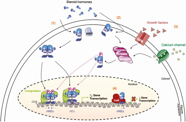
Steroid receptors (SRs) are members of the nuclear hormonal receptor family, many of which are transcription factors regulated by ligand binding. SRs regulate various human physiological functions essential for maintenance of vital biological pathways, including development, reproduction, and metabolic homeostasis. In addition, aberrant expression of SRs or dysregulation of their signaling has been observed in a wide variety of pathologies. SR activity is tightly and finely controlled by post-translational modifications (PTMs) targeting the receptors and/or their coregulators. Whereas major attention has been focused on phosphorylation, growing evidence shows that methylation is also an important regulator of SRs. Interestingly, the protein methyltransferases depositing methyl marks are involved in many functions, from development to adult life. They have also been associated with pathologies such as inflammation, as well as cardiovascular and neuronal disorders, and cancer. This article provides an overview of SR methylation/demethylation events, along with their functional effects and biological consequences. An in-depth understanding of the landscape of these methylation events could provide new information on SR regulation in physiology, as well as promising perspectives for the development of new therapeutic strategies, illustrated by the specific inhibitors of protein methyltransferases that are currently available.

Keywords: AR; ERα; GR; PR; coregulators; lysine methyltransferases; methylation; protein arginine methyltransferases; protein demethylases; steroid receptors.


Figures

Graphical Abstract
Graphical Abstract
Figure 1.
Figure 1.
Common structural organization of steroid receptors. (A) Schematic representation of steroid receptors (SRs) structure. The amino-terminal domain (NTD) is variable in size and composition and contains a ligand-independent transactivation domain (AF-1). The DNA binding domain (DBD) is the most conserved region, in which 2 zinc fingers maintain the core of the domain and bind to DNA. A less-conserved hinge region is present between the DBD and the ligand binding domain (LBD) and contains a nuclear localization signal (NLS). The ligand associates with the receptor through the LBD, which also contains a ligand-dependent transactivation domain (AF-2). The functions associated with the F domain are still not clearly understood. (B) The members of the steroid receptor (ie, ERα, PR, GR, and AR) subgroup share a deeply conserved structure of functional domains with some specificities. The main biological roles of these SRs, and the associated pathological disorders when SR signaling are dysregulated, are pointed out on the right.
Figure 2.
Figure 2.
Steroid receptor signaling pathways. The steroid hormone enters into the cell by passive diffusion through the plasma membrane and binds with high affinity to its specific receptor. (1) Classical steroid hormone nuclear signaling. The ligand–receptor complex undergoes conformational changes triggering its dissociation from the chaperone heat-shock protein (HSP), receptor dimerization, and its translocation into the nuclear compartment. Inside the nucleus, the ligand-associated SR binds to specific DNA sequences that serve as enhancer or silencer elements, recruits coregulators and enzyme-modifying chromatin complexes to locally perturb the chromatin organization and regulate assembly or disassembly of an active transcription complex. SR-dependent multiprotein complexes target selective hormone-response elements (HREs) on target promoters, or indirectly interact with chromatin through transcription factors (TFs) on their response elements (REs). This could affect the level of growth factor receptors (GFRs), calcium signaling actors, or cellular proliferation effectors, among many other cellular pathways regulated by SR target genes. (2) Nongenomic signaling. The steroid ligand binds to SRs located at the plasma membrane or in the cytoplasm and triggers rapid post-translational modifications, often dependent on activating kinase cascades (MAPK, PI3K, Src), that in turn result in the transcriptional activation of the receptor. Conversely, SR genomic effects can regulate rapid nongenomic events, highlighting a potent crosstalk dependent on ligand-bound SRs. (3) Nonclassical steroid hormone nuclear signaling. Apart from the binding of the specific steroid hormone, SRs can also be indirectly activated by growth factors, leading the recruitment and the activity of cytoplasmic phosphorylation cascades, the same involved in the classical signaling, namely MAPK and PI3K/Akt kinases. (4) Unliganded-receptor nuclear signaling. More recent data revealed that unliganded forms of SRs play critical roles on chromatin and deeply take part in gene repression of a subset of target genes after recruitment of corepressors (CoR).
Figure 3.
Figure 3.
Process of protein methylation. Lysine residues are methylated by lysine methyltransferases (KMTs, green arrow) to generate, mono- (Kme1), di- (Kme2), or tri-methyllysines (Kme3). (A) KMTs use the methyl donor S-Adenosylmethionine (AdoMet) to add methyl (-CH3) groups on targets and produce S-adenosylhomocysteine (AdoHcy) in addition to methyllysines. This process is highly dynamic and can be reversed by lysine demethylases (KDMs, red arrow). (B) Arginine methylation is catalyzed by a family of 9 PRMTs, divided into 3 subgroups (type I, II, or III, green arrows). All use the methyl donor AdoMet to add methyl (-CH3) groups on targets and produce AdoHcy in addition to methylarginines. PRMTs that promote monomethylation (MMA), symmetric dimethylation (sDMA), or asymmetric dimethylation (aDMA) lead to the production of monomethylarginine, asymmetric dimethylarginine, or symmetric dimethylarginine respectively. JMJD6 is currently the only enzyme identified with an arginine demethylase activity (red arrow). PRMTs on which we focus in this article are highlighted in bold.
Figure 4.
Figure 4.
Biological consequences of SR methylation. All the methylation events targeting the steroid receptors on arginine (R) and lysine (K) residues and reported at this time are represented for (A) ERα, (B) PR, (C) AR, and (D) GR. When identified, the protein methyltransferases involved are noted in black and the demethylases in brown. The methylation events leading to repressive functions are represented in red and the activating functions are in green. For ERα, we enlarged the hinge domain as it is the main region modified by methylation. When decrypted and reported, the biological consequences of the methylation event on the physiology/pathology have been indicated (in green for activating functions, red for repressive functions and blue when no effect). NTD, N-terminal domain; DBD, DNA-binding domain; h, hinge; LBD, ligand binding domain; NLS, nuclear localization signal; NES, nuclear export signal; BC, breast cancer; PC, prostate cancer.
Figure 5.
Figure 5.
Indirect methylation events regulating SR signaling. Here, we highlight 2 examples, in (A) GR and in (B) ERα, of indirect methylation events (ie, not directly on SRs), regulating the transcriptional activity of these 2 receptors. This concerns the methylation of histone tails on chromatin and/or the methylation of coregulators. When identified, the targeted lysines (K) or arginines (R) and the methyltransferases are noted in black and the demethylases in brown. The methylation events leading to repressive functions are represented with red lines and the activating functions with green arrows. Me, methylation; GRE, GR response elements; ERE, estrogen response elements; H3, histone H3; H4, histone H4; CoA, coactivators; Dex, dexamethasone; E2, estrogens; BC, breast cancer.

References

    1. Baschant U, Lane NE, Tuckermann J. The multiple facets of glucocorticoid action in rheumatoid arthritis. Nat Rev Rheumatol. 2012;8(11):645-655. - PubMed
    1. Jensen EV, Jacobson HI, Walf AA, Frye CA. Estrogen action: a historic perspective on the implications of considering alternative approaches. Physiol Behav. 2010;99(2):151-162. - PMC - PubMed
    1. Ashburner M. Sequential gene activation by ecdysone in polytene chromosomes of Drosophila melanogaster. II. The effects of inhibitors of protein synthesis. Dev Biol. 1974;39(1):141-157. - PubMed
    1. Islam MS, Afrin S, Jones SI, Segars J. Selective progesterone receptor modulators-mechanisms and therapeutic utility. Endocr Rev. 2020;41(5):643-694. - PMC - PubMed
    1. Nicolaides NC, Galata Z, Kino T, Chrousos GP, Charmandari E. The human glucocorticoid receptor: molecular basis of biologic function. Steroids. 2010;75(1):1-12. - PMC - PubMed

Publication types

MeSH terms

Substances