. 2021 May 5;33(3):511-530.
doi: 10.1093/plcell/koaa060.

Distinct identities of leaf phloem cells revealed by single cell transcriptomics

Affiliations

Distinct identities of leaf phloem cells revealed by single cell transcriptomics

Ji-Yun Kim et al. Plant Cell. .

Abstract

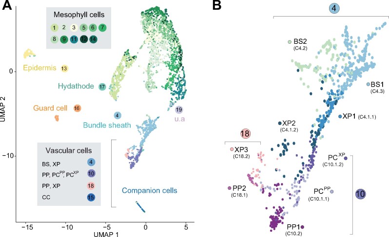
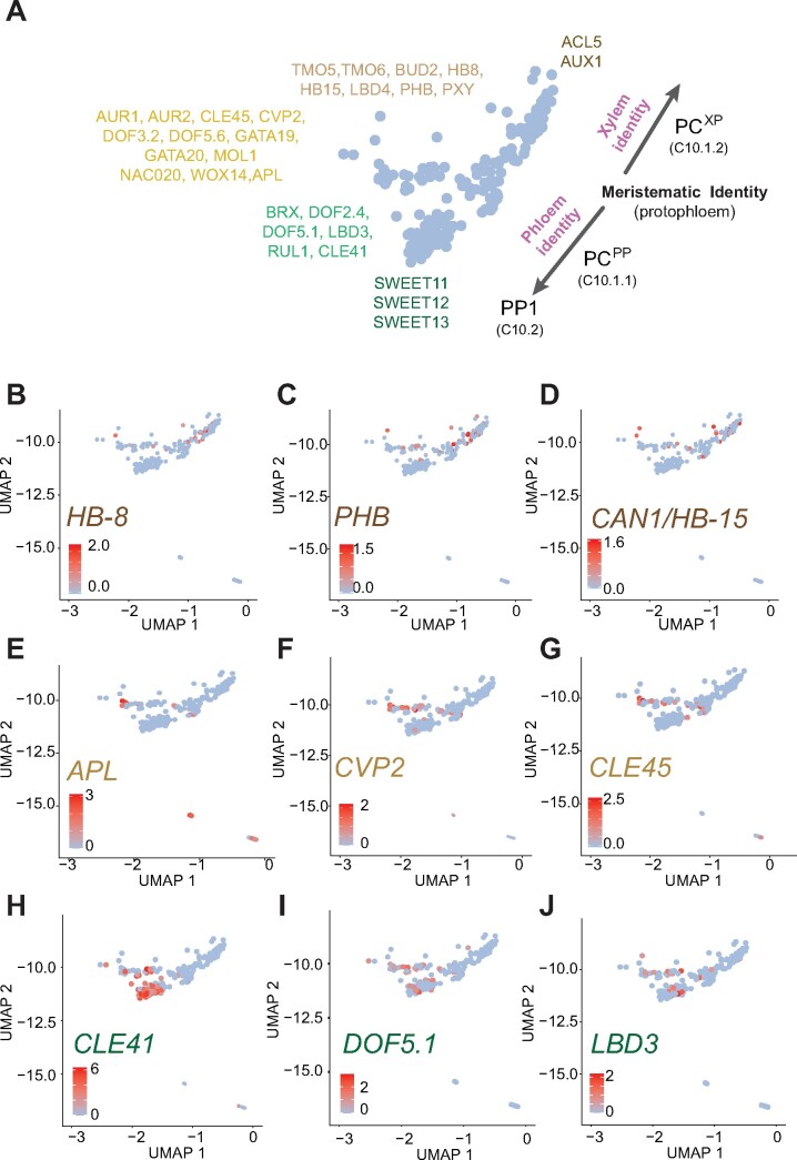
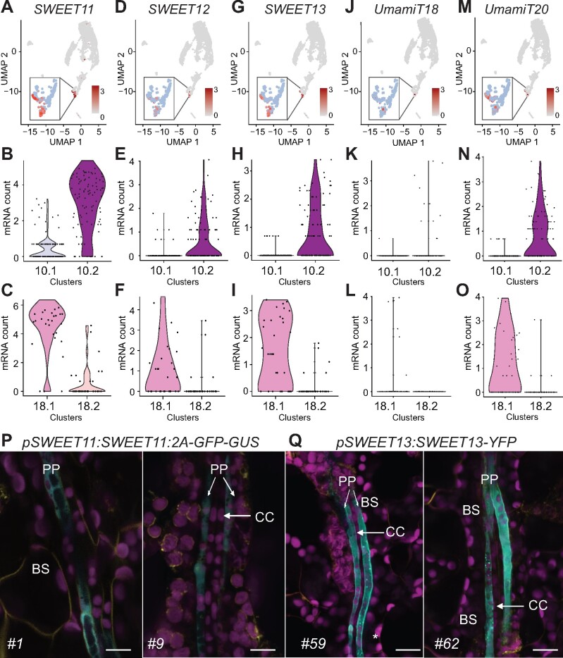
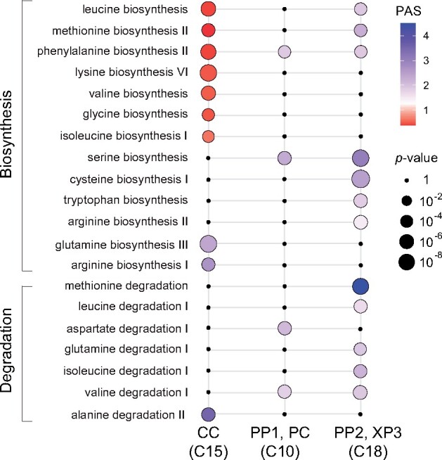
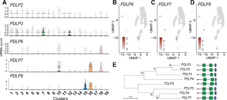
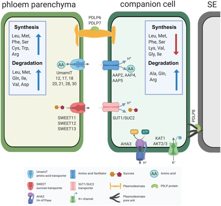
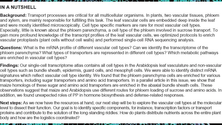
The leaf vasculature plays a key role in solute translocation. Veins consist of at least seven distinct cell types, with specific roles in transport, metabolism, and signaling. Little is known about leaf vascular cells, in particular the phloem parenchyma (PP). PP effluxes sucrose into the apoplasm as a basis for phloem loading, yet PP has been characterized only microscopically. Here, we enriched vascular cells from Arabidopsis leaves to generate a single-cell transcriptome atlas of leaf vasculature. We identified at least 19 cell clusters, encompassing epidermis, guard cells, hydathodes, mesophyll, and all vascular cell types, and used metabolic pathway analysis to define their roles. Clusters comprising PP cells were enriched for transporters, including SWEET11 and SWEET12 sucrose and UmamiT amino acid efflux carriers. We provide evidence that PP development occurs independently from ALTERED PHLOEM DEVELOPMENT, a transcription factor required for phloem differentiation. PP cells have a unique pattern of amino acid metabolism activity distinct from companion cells (CCs), explaining differential distribution/metabolism of amino acids in veins. The kinship relation of the vascular clusters is strikingly similar to the vein morphology, except for a clear separation of CC from the other vascular cells including PP. In summary, our single-cell RNA-sequencing analysis provides a wide range of information into the leaf vasculature and the role and relationship of the leaf cell types.


Figures

Figure 1
Figure 1
Strategies to enrich vascular protoplasts. (A–C) Box plot representation of the RT-qPCR analysis results showing the relative transcript level of the marker for the PP, SWEET11, and the marker for XP, GLR3;6. UBQ10 was used as an internal control for normalization. The abaxial epidermis was removed from leaves harvested from 6-weeks-old plants grown under short day conditions and was treated as indicated. The data shown are from a single experiment with three technical replicates (mean ± se, n = 3). (A) Leaves from which the abaxial epidermis had been stripped were either cut on each side of the main vein before enzymatic digestion of the cell wall (wounded, W) or directly placed in the digesting enzyme solution (nonwounded, NW). (B) Leaves from which the abaxial epidermis had been stripped were cut on each side of the main vein and incubated in the enzyme solution containing 0.4 M or 0.6 M mannitol. (C) Protoplasts isolated from leaf sample prepared as in (B) were incubated in an enzyme solution composed of cell wall degrading enzymes; cellulase Onozuka R-10, macerozyme R-10 obtained from Yakult (Tokyo), and Duchefa (Haarlem). (D) GFP (Green fluorescent protein) fluorescence (cyan) marking PP cells in leaf protoplasts. Protoplasts were isolated from 6-week-old pAtSWEET11:AtSWEET11-GFP plants expressing a PP cell-specific marker. Magenta, chlorophyll autofluorescence. Scale bar: 10 µm. (E) GFP fluorescence (cyan) marking procambium cells in leaf protoplasts. Protoplasts were isolated from 6-week-old Q0990 plants expressing a procambium cell-specific marker. Magenta, chlorophyll autofluorescence. Scale bar: 20 µm.
Figure 2
Figure 2
Assignment of cellular identity to clusters. (A) UMAP dimensional reduction projection of 5,230 Arabidopsis leaf cells. Cells were grouped into 19 distinct clusters using Seurat (Butler et al., 2018). The cluster number is shown and colored based on the colors assigned to each cell type (u.a.—unassigned, i.e. cluster could not be assigned to a known cell type). Each dot indicates individual cells colored according to the cell type assigned. (B) Magnification of C4, C18, and C10 subclusters. Different colors indicate distinct cell identities. BS, bundle sheath; PC, procambium; PP, phloem parenchyma; PCXP, procambium; XP, xylem parenchyma cells with features relating to xylem differentiation. PCPP, procambium cells with features relating to phloem differentiation.
Figure 3
Figure 3
mRNA levels of marker genes in clusters used to assign cell types. Violin plots showing transcript enrichment of known cell type-specific marker genes across clusters. Clusters are indicated on the x-axis. The name of the cell type assigned to each cluster is indicated on the right side of the violin plots.
Figure 4
Figure 4
Identification of the procambium cell cluster with distinct procambium cell identities. (A) Schematics representing subpopulations of Cluster 10. Genes enriched in the subpopulations are indicated. (B–D) UMAP showing enrichment of transcripts of genes related to xylem differentiation. (E–G) UMAP showing enrichment of transcripts of genes related to maintenance of protophloem pluripotency and differentiation. (H–J) UMAP showing the distribution transcripts related to phloem differentiation.
Figure 5
Figure 5
Three SWEET sucrose transporters and UmamiT amino acid transporters mark the PP cluster. (A–O) UMAP and violin plots of C10 and C18 subclusters showing enrichment of SWEET11 (A–C), SWEET12 (D–F), SWEET13 (G–I), UmamiT18/SIAR1 (J–L), and UmamiT20 (M–O) transcripts in PP clusters. Subcluster 10.1 corresponds to PC, 18.2 to XP3, and 10.2 and 18.1 to PP. Inset show magnification of C10 and C18. (P and Q) Confocal microscopy images of pSWEET11:SWEET11-2A-GFP-GUS (P) and pSWEET13:SWEET13-YFP (Q) leaf showing GFP (P) or YFP (Q) fluorescence specific to PP. Magenta, chlorophyll autofluorescence. Yellow, FM4-64FX. Cyan GFP fluorescence (P) or YFP fluorescence (Q). Scale bars: 10 µm. BS, bundle sheath; CC, companion cell; PP, phloem parenchyma cells are marked. Numbers on the bottom left indicate independent transgenic lines.
Figure 6
Figure 6
Reporter gene analysis of pbZIP9:GFP-GUS plants. (A and B) GUS-stained transgenic plants expressing the transcriptional pbZIP9:GFP-GUS reporter construct show GUS activity in the leaf vasculature. Four-week-old plants grown in LD conditions were used for GUS histochemistry. A magnified image of the seventh leaf is shown in b. Scale bars: 1 mm (A) and 0.5 mm (B). (C and D) Confocal microscopy images of two independent pbZIP9:GFP-GUS reporter lines showing GFP fluorescence specific for PP. Magenta, chlorophyll autofluorescence. Yellow, FM4-64FX, Cyan, GFP fluorescence. BS, bundle sheath; CC, companion cell; PP, phloem parenchyma cells are marked. Scale bar: 10 µm.
Figure 7
Figure 7
Distinct PP and CC marker gene expression in the apl mutant. (A) Morphology of apl mutant (right) grown on LD conditions for 2 weeks. WT plant grown under the same condition is shown on the left side. Scale bar: 1 cm. (B) RT-qPCR analysis of CC-marker gene (SUC2) and PP- marker genes (SWEET11 and bZIP9). Segregating seeds from heterozygous parents were plated on MS for 2 weeks. The first and second leaves from plants homozygous for the APL mutation (apl) and heterozygous for the mutation or WT (APL) were collected for RNA extraction and RT-qPCR. Three independent replicates showed similar results and a representative experiment with three technical replicates are shown (mean ± se, n = 3).
Figure 8
Figure 8
Amino acid biosynthesis and degradation pathways are differentially represented in the CC, PP, PC, and XP3 cells. Metabolic pathway activities of amino acid biosynthesis and degradation pathways in Clusters 15, 10, and 18. Statistical significance is represented as differences in dot size. Statistically insignificant values are shown as black dots (random permutation test, P >0.05). Colors represent the PAS; a score 1 (violet) indicates a higher activity. Activities were compared between all clusters in the scRNA-seq data set.
Figure 9
Figure 9
Transcript enrichment of PDLP genes in vascular cell types. (A) Violin plot showing transcript enrichment of PDLPs. (B–D) UMAP plot showing the enrichment of PDLP6 (B), PDLP7 (C), and PDLP8 (D) transcripts in the PP clusters (B and C), guard cell cluster (C and D), and CC cluster (D). (E) Phylogenetic analysis of PDLPs in Arabidopsis. The phylogenetic tree was generated with the maximum likelihood method implemented in PhyML (Lemoine et al., 2019). Percent support values from 1,000 bootstrap samples are shown. Protein motifs predictions are based on the SMART database (http://smart.embl-heidelberg.de). DUF26 domains, transmembrane region, and signal peptide are shown in green, blue, and red, respectively.
Figure 10
Figure 10
Hypothetical phloem loading process in an Arabidopsis leaf. Sucrose produced during photosynthesis in the mesophyll cells is transported across the bundle sheath to the PP. Biosynthesis and catabolism of multiple amino acids is highly active in the PP. Transporters present in the PP secrete sucrose (SWEET11, 12, 13) or amino acids (UmamiT11, 12, 17, 18, 20, 21, 28, 30) into the apoplasm. H+/sucrose cotransporters import sucrose (SUT1/SUC2) and amino acids (AAP2, 4, 5) into the SE/CC. The H+ gradient required for the active import of sucrose and amino acids into the SE/CC is provided by plasma membrane H+-ATPases. The membrane potential is maintained by the potassium channels (KAT1 and AKT2/3). Symplasmic transport is mediated by PDLP6 and PDLP7 in the PD in PP cells and PDLP8 enriched in the PD-pore unit of the CC. Note that the schematic is based on transcript levels and the distribution could differ at the protein level. Schematics was made in © BioRender—https://biorender.com.
None

Similar articles

Cited by

References

    1. Arun Chinnappa KS, Nguyen TTS, Hou J, Wu Y, McCurdy DW (2013) Phloem parenchyma transfer cells in Arabidopsis - an experimental system to identify transcriptional regulators of wall ingrowth formation. Front Plant Sci 4:102. - PMC - PubMed
    1. Atkins CA (2000) Biochemical aspects of assimilate transfers along the phloem path: N-solutes in lupins. Austr J Plant Physiol 27: 531–537
    1. Baima S, Possenti M, Matteucci A, Wisman E, Altamura MM, Ruberti I, Morelli G (2001) The Arabidopsis ATHB-8 HD-zip protein acts as a differentiation-promoting transcription factor of the vascular meristems. Plant Physiol 126: 643–655 - PMC - PubMed
    1. van Bel AJE, Knoblauch M (2000) Sieve element and companion cell: the story of the comatose patient and the hyperactive nurse. Funct Plant Biol 27: 477–487
    1. Bezrutczyk M, Zöllner NR, Kruse CPS, Hartwig T, Lautwein T, Köhrer K, Frommer WB, Kim J-Y (2021) Evidence for phloem loading via the abaxial bundle sheath cells in maize leaves. The Plant Cell 33: 531--547 - PMC - PubMed

Publication types

MeSH terms