. 2021 May 5;33(3):531-547.
doi: 10.1093/plcell/koaa055.

Evidence for phloem loading via the abaxial bundle sheath cells in maize leaves

Affiliations

Evidence for phloem loading via the abaxial bundle sheath cells in maize leaves

Margaret Bezrutczyk et al. Plant Cell. .

Abstract

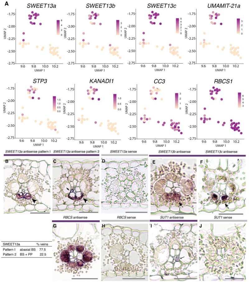
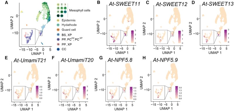
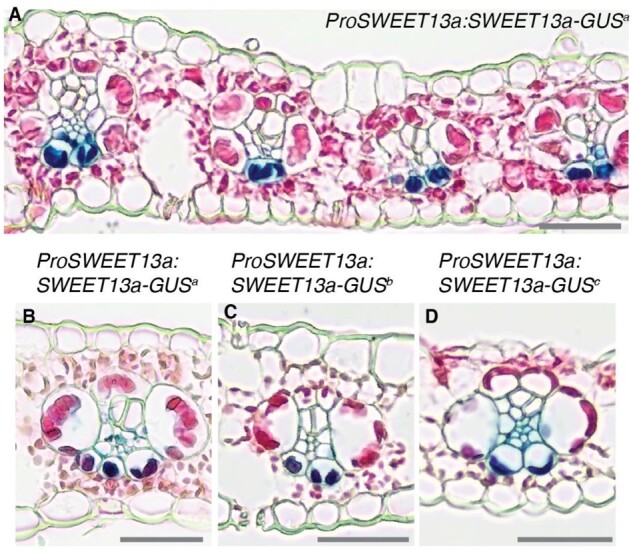
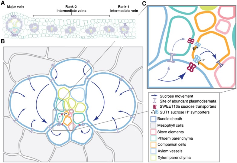
Leaves are asymmetric, with different functions for adaxial and abaxial tissue. The bundle sheath (BS) of C3 barley (Hordeum vulgare) is dorsoventrally differentiated into three types of cells: adaxial structural, lateral S-type, and abaxial L-type BS cells. Based on plasmodesmatal connections between S-type cells and mestome sheath (parenchymatous cell layer below bundle sheath), S-type cells likely transfer assimilates toward the phloem. Here, we used single-cell RNA sequencing to investigate BS differentiation in C4 maize (Zea mays L.) plants. Abaxial BS (abBS) cells of rank-2 intermediate veins specifically expressed three SWEET sucrose uniporters (SWEET13a, b, and c) and UmamiT amino acid efflux transporters. SWEET13a, b, c mRNAs were also detected in the phloem parenchyma (PP). We show that maize has acquired a mechanism for phloem loading in which abBS cells provide the main route for apoplasmic sucrose transfer toward the phloem. This putative route predominates in veins responsible for phloem loading (rank-2 intermediate), whereas rank-1 intermediate and major veins export sucrose from the PP adjacent to the sieve element companion cell complex, as in Arabidopsis thaliana. We surmise that abBS identity is subject to dorsoventral patterning and has components of PP identity. These observations provide insights into the unique transport-specific properties of abBS cells and support a modification to the canonical phloem loading pathway in maize.


Figures

Figure 1
Figure 1
Mesophyll and bundle sheath clusters show canonical expression of C4 photosynthesis-related genes. A, Workflow for expression analysis in maize cells. Illustration of protoplasting, 10× chromium gel bead-in-emulsion (GEM) partitioning and cDNA synthesis, RNA sequencing, and data analysis. B, UMAP plot showing a two-dimensional representation of cell relationships from scRNA-seq data set A in multidimensional space. Bundle sheath cells separate into two subclusters at higher resolution (inset). The upper and lower clusters were later determined to correspond to abaxial and adaxial BS cells (Figure 2), and are therefore named abBS and adBS (C) Violin plots showing distribution of normalized mRNA counts of marker genes for cells in each cluster. Genes listed are known to be differentially expressed in MS and BS cells in C4 maize (PEP1, MDH6, CA, ME1, RBCS1) or are genes that identify unique clusters (NAAT1, TAAT). D, Violin plots showing normalized mRNA levels both of genes differentially expressed between abBS and BSad subclusters (SWEET13a, SWEET13b, and SWEET13c, CC3 (cystatin3) and of genes highly expressed in both clusters (ME1, RBCS1). Gene symbols are shown in Figure 1, C and D, and corresponding gene IDs from the most complete current B73 genome annotation available on the Ensembl genome browser (B73 RefGen_v4), as well as full names, are provided in Supplemental Table S2.
Figure 2
Figure 2
The abaxial BS cluster contains high levels of mRNAs encoding transport proteins. A, Feature plots show normalized levels of mRNAs for maize genes differentially expressed between the two clusters of bundle sheath cells from data set A plotted in UMAP space. B–F, In situ hybridizations of SWEET13a, SWEET13b, and SWEET13c to localize their mRNAs. Rank-2 intermediate veins from sections hybridized with antisense probes for SWEET13a, SWEET13b, and SWEET13c showed that the mRNA localization of three SWEET13 genes was largely limited to abaxial bundle sheath cells. B and C, SWEET13a mRNA localization was predominantly in abBSCs in the majority of veins (77.5%) and to PP in a subset of veins (22.5%; n = 824). D, No staining was visible after hybridization with the SWEET13a sense probe (negative control). E and F, SWEET13b and SWEET13c probes showed staining predominantly in abBSCs. G, RBCS1 antisense probe hybridized sections showed staining in all BS cells. H, No staining was detected in sections hybridized with an RBCS1 sense probe. I, SUT1 mRNA was localized to a vascular cell which is likely a companion cell in rank-2 intermediate veins (arrow). J, No staining was detected after hybridization with a SUT1 sense probe (negative control). See Supplemental Figure S7 for intermediate rank-1 and major veins. Bars = 100 µm.
Figure 3
Figure 3
Arabidopsis homologs of genes enriched in maize abBS cells are expressed in Arabidopsis PP. Maize transporters showing mRNA enrichment in abBSCs are homologous to many Arabidopsis transporters enriched in Arabidopsis PP cells. A UMAP plot showing two-dimensional representation of cell relationships in multidimensional space for single-cell sequencing of Arabidopsis (At) leaf cells. Clusters are indicated by colors in the key to the right of the UMAP plot. Feature plots show normalized levels of mRNA transcripts for Arabidopsis transport proteins homologous to abBS transport proteins. B–D At-SWEET11 (AT3G48740), At-SWEET12 (AT5G23660), and At-SWEET13 (AT5G50800) are homologous to Zm-SWEET13a, Zm-SWEET13b, and Zm-SWEET13c. E, At-UmamiT21 (AT5G64700) is homologous to Zm-UmamiT21a. F, At-UmamiT20 (AT4G08290) is homologous to Zm-UmamiT20a (Supplemental Figure S6). G and H, At-NPF5.8 (AT5G14940)_and At-NPF5.9 (AT3G01350) are homologous to Zm-NRT1.
Figure 4
Figure 4
SWEET13a is localized to abaxial bundle sheath cells of rank-2 veins. GUS histochemistry in leaves of maize lines transformed with the translational fusion construct ProSWEET13a:SWEET13a-GUS. A, The dark blue chlorobromoindigo precipitate indicative of GUS activity was detected in abaxial bundle sheath cells of maize plants transformed with ProSWEET13a:SWEET13a-GUSplus. B–D, Three independent transformation events (a, b, and c = ProSWEET13a:SWEET13a-GUSa, b, and c) resulted in similar expression patterns in rank-2 intermediate veins, rank-1 intermediate veins, and major veins (for rank-1 and major veins see Supplemental Figure 7). B, Line “a,” (C) Line “b,” and (D) Line “c.” Sections were counterstained with eosin-Y; bars = 100 µm.
Figure 5
Figure 5
Abaxial BS transcripts are co-regulated during the sink–source transition. A, Tissues selected for qRT-PCR: a V2-stage seedling (upper) with source and sink tissue highlighted (lower). The base of leaf 3 is still in the whorl and is net sink tissue (Li et al., 2010). B, 18S-normalized mRNA levels of SWEET13a, UmamiT21a, AAP45 (encoding proteins transport proteins enriched in abBS cells) and SUT1 in source (leaf tip) and sink (leaf base) tissue. Values are average of three technical (qRT-PCR) replicates of three pools of two plants; error bars represent sem. *Students two-tailed paired t test values are shown. Independent repeats confirmed the data. C, ProSWEET13a:SWEET13a-GUSplus transformed B73 seedling segments after a 12–48 h incubation in GUS staining solution. V2 leaf 3 tip (12 h), (D) leaf 3 sheath (48 h), (E) stem cross section 1 cm above soil (48 h), and (F) cross-section across root tip (48 h). Of these, only the tip of leaf 3 (source) showed chlorobromoindigo precipitate indicative of GUS activity due to expression of the SWEET13a–GUS fusion protein. Bars: 100 µm.
Figure 6
Figure 6
Phloem loading occurs via the abBS in maize. A, Arrangement and relative numbers of major veins, rank-1 intermediate veins, and rank-2 intermediate veins in a mature maize leaf. Note that rank-1 intermediate veins are distinguished from rank-2 by the presence of hypodermal sclerenchyma. B, A rank-2 intermediate vein surrounded by bundle sheath (blue outline) and mesophyll (gray) cells. Sucrose movement down its concentration gradient is indicated by blue arrows. C, The inset shows details of sucrose movement either from bundle sheath cells into the apoplasm via SWEET13 transporters or to PP (teal) via plasmodesmata, where sucrose is then exported to the apoplasmic space by SWEETs. Sucrose in the apoplasm is taken up by SUT1 into the sieve element (orange, pink) complex of the companion cells for long distance transport.
None

Similar articles

Cited by

References

    1. Ashburner M, Ball CA, Blake JA, Botstein D, Butler H, Cherry JM, Davis AP, Dolinski K, Dwight SS, Eppig JT et al. (2000) Gene ontology: tool for the unification of biology. The Gene Ontology Consortium. Nat Genet 25: 25–2910.1038/75556 - DOI - PMC - PubMed
    1. Baker RF, Leach KA, Boyer NR, Sawyers MJ, Benitez-Alfonso Y, Skopelitis T, Luo A, Sylvester A, Jackson D, Braun DM (2016) Sucrose transporter ZmSut1 expression and localization uncover new insights into sucrose phloem loading. Plant Physiol 172: 1876–1898 - PMC - PubMed
    1. Bezrutczyk M, Hartwig T, Horschman M, Char SN, Yang J, Yang B, Frommer WB, Sosso D (2018) Impaired phloem loading in zmsweet13a,b,c sucrose transporter triple knock-out mutants in Zea mays. New Phytol 218: 594–603 - PubMed
    1. Bosabalidis AM, Evert RF, Russin WA (1994) Ontogeny of the vascular bundles and contiguous tissues in the maize leaf blade. Am J Bot 81: 745–752
    1. Bürkle L, Hibberd JM, Quick WP, Kühn C, Hirner B, Frommer WB (1998) The H+-sucrose cotransporter NtSUT1 is essential for sugar export from tobacco leaves. Plant Physiol 118: 59–68 - PMC - PubMed

Publication types