. 2021 Jun;35(6):e23791.
doi: 10.1002/jcla.23791. Epub 2021 May 6.

Identification of seven tumor-educated platelets RNAs for cancer diagnosis

Affiliations

Identification of seven tumor-educated platelets RNAs for cancer diagnosis

Xinxin Ge et al. J Clin Lab Anal. 2021 Jun.

Abstract

Background: Tumor-educated platelets (TEPs) may enable blood-based cancer diagnosis. This study aimed to identify diagnostic TEPs genes involved in carcinogenesis.

Materials and methods: The TEPs differentially expressed genes (DEGs) between healthy samples and early/advanced cancer samples were obtained using bioinformatics. Gene ontology (GO) analysis and Kyoto encyclopedia of genes and genomes (KEGG) pathway enrichment analysis were used to identify the pathways and functional annotation of TEPs DEGs. Protein-protein interaction of these TEPs DEGs was analyzed based on the STRING database and visualized by Cytoscape software. The correlation analysis and diagnostic analysis were performed to evaluate the diagnostic value of TEPs mRNAs expression for early/advanced cancers. Quantitative real-time PCR (qRT-PCR) was applied to validate the role of DEGs in cancers.

Results: TEPs mRNAs were mostly involved in protein binding, extracellular matrix, and cellular protein metabolic process. RSL24D1 was negatively correlated to early-stage cancers compared to healthy controls and may be potentially used for early cancer diagnosis. In addition, HPSE, IFI27, LGALS3BP, CRYM, HBD, COL6A3, LAMB2, and IFITM3 showed an upward trend in the expression from early to advanced cancer stages. Moreover, ARL2, FCGR2A, and KLHDC8B were positively associated with advanced, metastatic cancers compared to healthy controls. Among the 12 selected DEGs, the expression of 7 DEGs, including RSL24D1, IFI27, CRYM, HBD, IFITM3, FCGR2A, and KLHDC8B, were verified by the qRT-PCR method.

Conclusion: This study suggests that the 7-gene TEPs liquid-biopsy biomarkers may be used for cancer diagnosis and monitoring.

Keywords: bioinformatics analysis; diagnosis; mRNA; tumor educated platelets.


Conflict of interest statement

The authors declare no conflict of interest.

Figures
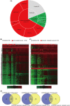
FIGURE 1
FIGURE 1
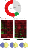
Identification of DEGs in TEPs in localized and metastatic pan‐cancer patients, and platelets from healthy controls, based on the datasets GSE68086. (A) A number of platelet samples of healthy controls and cancer patients with different stages or types of cancer‐based on GSE68086. (B, C) Hierarchical clustering heatmap of DEGs in the expression profiling datasets GSE68086. (B) Heatmap of DEGs in TEPs collected from healthy controls and early, localized cancer patients. (C) Heatmap of DEGs in TEPs collected from healthy controls and advanced metastatic cancer patients. The horizontal axis indicates the sample, and the vertical axis indicates the DEGs. Red represents the up‐regulated DEGs, and green represents the down‐regulated DEGs. DEGs, differentially expressed genes. (D) Identification of TEPs mRNAs between localized and metastatic pan‐cancer. Left, commonly altered differential expressed TEPs mRNAs. Middle, identification of up‐regulated differential expressed TEPs mRNAs. Right, identification of down‐regulated differential expressed TEPs mRNAs
FIGURE 2
FIGURE 2
mRNA profiles of TEPs from localized and metastatic NSCLC cancer patients, and platelets from healthy controls, based on the datasets GSE89843. (A) Number of platelet samples of healthy controls and NSCLC cancer patients at different stages. (B, C) Hierarchical clustering heatmap of DEGs in the expression profiling datasets GSE89843. (B) Heatmap of DEGs in TEPs collected from healthy controls and early, localized NSCLC cancer patients. (C) Heatmap of DEGs in TEPs collected from healthy controls and metastatic NSCLC cancer patients. The horizontal axis indicates the sample, and the vertical axis indicates the DEGs. Red represents the up‐regulated DEGs, and green represents the down‐regulated DEGs. (D) Identification of TEPs mRNAs between localized and metastatic NSCLC cancer. Left, commonly altered differential expressed TEPs mRNAs. Middle, identification of up‐regulated differential expressed TEPs mRNAs. Right, identification of down‐regulated differential expressed TEPs mRNAs
FIGURE 3
FIGURE 3
Analysis of the spliced RNA repertoire of TEPs from pan‐cancer patients at different stages. (A, B) Identification of DEGs in the four datasets (GSE68086 early pan‐cancer and metastatic pan‐cancer, GSE89843 early NSCLC cancer, and metastatic NSCLC cancer) via Venn diagrams software. (A) Up‐regulated differential expressed TEPs mRNAs. (B) Down‐regulated differential expressed TEPs mRNAs. Different colors represent different datasets. (C, D) GO analyses of the DEGs according to their biological process, cellular component, and molecular function. GO, gene ontology. (E) KEGG pathway enrichment analysis. Dot size represents the number of genes in each KEGG pathway; p‐value: Red < purple < blue. KEGG, Kyoto Encyclopedia of Genes and Genomes. (F) Protein‐protein interaction network of DEGs visualized through String datasets
FIGURE 4
FIGURE 4
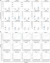
Diagnostic value of TEPs DEGs for early, localized pan‐cancer based on the datasets GSE68086. (A) Correlation analysis between expression levels of 15 TEPS DEGs and two groups, including healthy control groups and early cancer groups. HC, healthy control; EC, early cancer; The 15 TEPS DEGs are PLXNB3, SAMD14, ALAS2, C4orf48, PARP10, EHBP1L1, DYSF, SSBP4, LRRC75A, CD69, RSL24D1, ZNF667, PPT1, IARS, and HERC3, respectively. (B) ROC analysis of sensitivity and specificity of the above 15 TEPS DEGs signature in predicting the diagnosis of early pan‐cancer patients
FIGURE 5
FIGURE 5
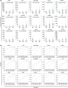
Identification of 15 TEPS DEGs signatures for pan‐cancer diagnosis based on the datasets GSE68086. (A) Correlation analysis between expression levels of 15 TEPS DEGs and three groups, including healthy control groups, early cancer groups, and advanced cancer groups. HC, healthy control; EC, early cancer; AC, advanced cancer. The 15 TEPS DEGs are HPSE, IFI27, LGALS3BP, CRYM, WASF1, HBD, COL6A3, PRSS50, LAMB2, LTF, TPM2, TYMP, NELL2, SLC38A1, and IFITM3, respectively. (B) ROC analysis of sensitivity and specificity of the above 15 TEPS DEGs signature in predicting the diagnosis of pan‐cancer patients
FIGURE 6
FIGURE 6
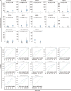
Diagnostic value of 13 TEPs DEGs for advanced, metastatic pan‐cancer based on the datasets GSE68086. (A) Correlation analysis between expression levels of 13 TEPS DEGs and two groups, including healthy control groups and advanced cancer groups. HC, healthy control; AC, advanced cancer. The 13 TEPS DEGs are FCGR2A, KLHDC8B, DEFA3, IGFBP2, MAOB, ZNF346, ARL2, MMP1, KLHL35, CA1, RP11‐525A16.4, CTD‐2509G16.2, and MS4A1, respectively. (B) ROC analysis of sensitivity and specificity of the above 15 TEPS DEGs signature for predicting advanced pan‐cancer
FIGURE 7
FIGURE 7
Relative expression level of 12 selected DEGs. Relative expression level of KLHDC8B, FCGR2A, ARL2, IFITM3, LAMB2, COL6A3, HBD, CRYM, LGALS3BP, IFI27, HPSE and RSL24D1. NSCLC: non‐small cell lung carcinoma; CRC: colorectal cancer. *p < 0.05, **p < 0.01, and ***p < 0.001

Similar articles

Cited by

References

    1. Siegel RL, Miller KD, Jemal A. Cancer statistics, 2020. CA Cancer J Clin. 2020;70(1):7‐30. - PubMed
    1. Vaidyanathan R, Soon RH, Zhang P, Jiang K, Lim CT. Cancer diagnosis: from tumor to liquid biopsy and beyond. Lab Chip. 2018;19(1):11‐34. - PubMed
    1. San Lucas FA, Allenson K, Bernard V,, et al. Minimally invasive genomic and transcriptomic profiling of visceral cancers by next‐generation sequencing of circulating exosomes. Ann Oncol. 2016;27(4):635‐641. - PMC - PubMed
    1. Pos O, Biró O, Szemes T, Nagy B. Circulating cell‐free nucleic acids: characteristics and applications. Eur J Hum Genet. 2018;26(7):937‐945. - PMC - PubMed
    1. Krebs MG, Metcalf RL, Carter L, Brady G, Blackhall FH, Dive C. Molecular analysis of circulating tumour cells‐biology and biomarkers. Nat Rev Clin Oncol. 2014;11(3):129‐144. - PubMed

MeSH terms