. 2021 May 6;16(5):e0251184.
doi: 10.1371/journal.pone.0251184. eCollection 2021.

Genetic analysis of the Drosophila ESCRT-III complex protein, VPS24, reveals a novel function in lysosome homeostasis

Affiliations

Genetic analysis of the Drosophila ESCRT-III complex protein, VPS24, reveals a novel function in lysosome homeostasis

Jonathan R Florian et al. PLoS One. .

Abstract

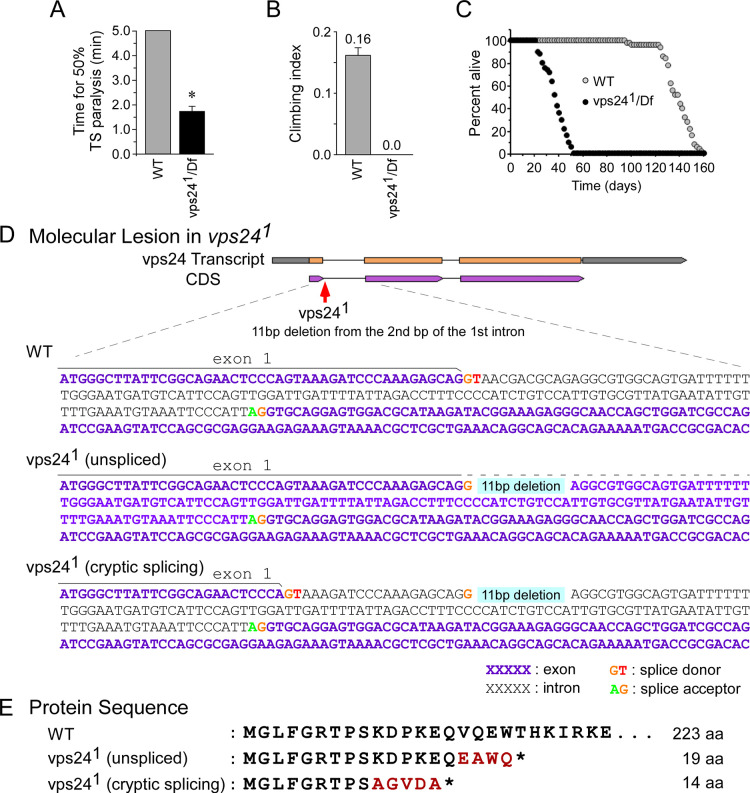
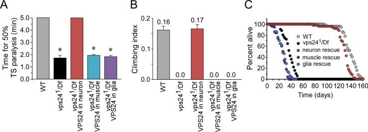
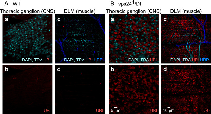
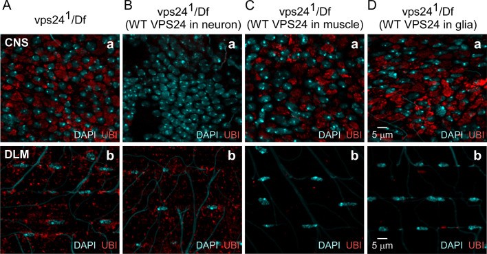
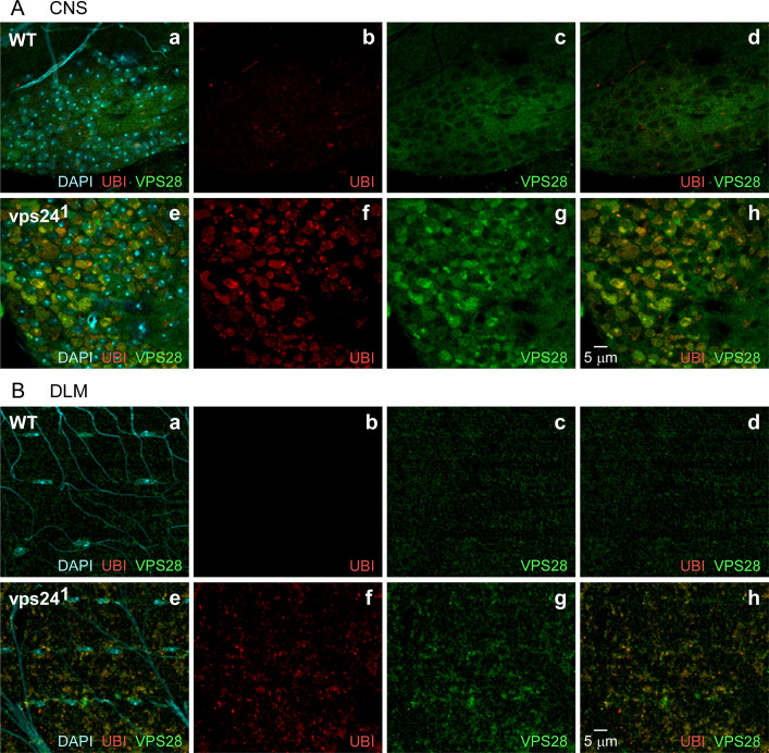
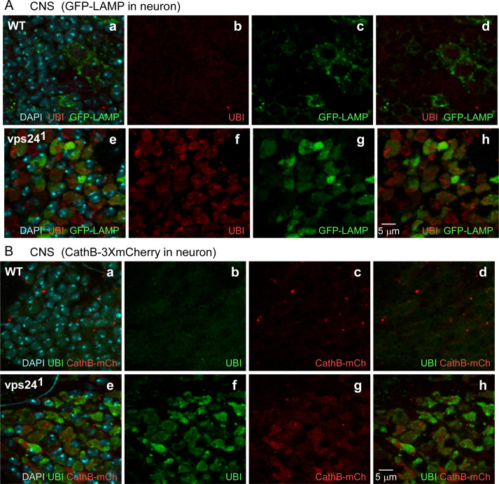
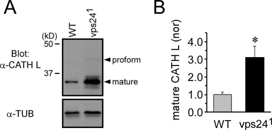
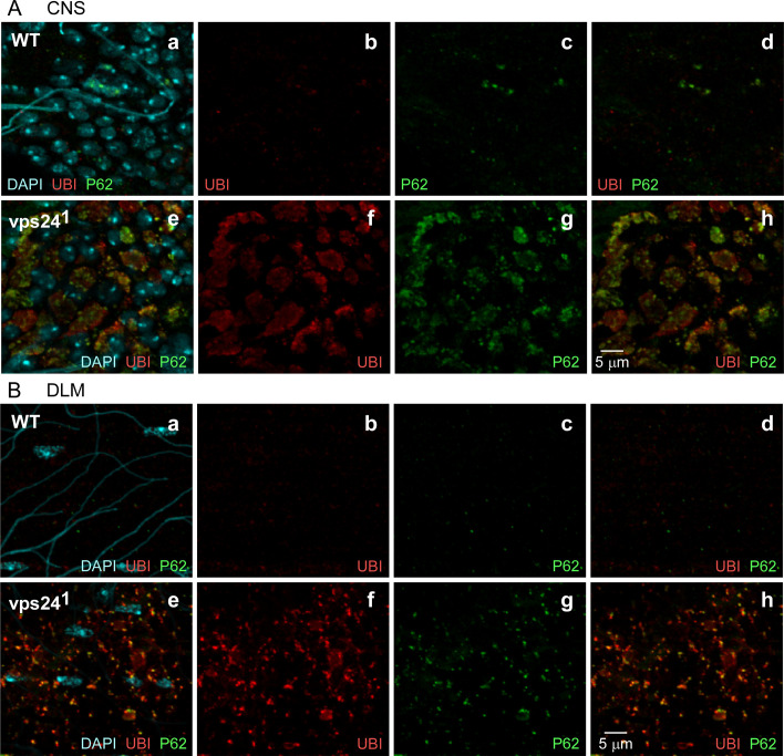
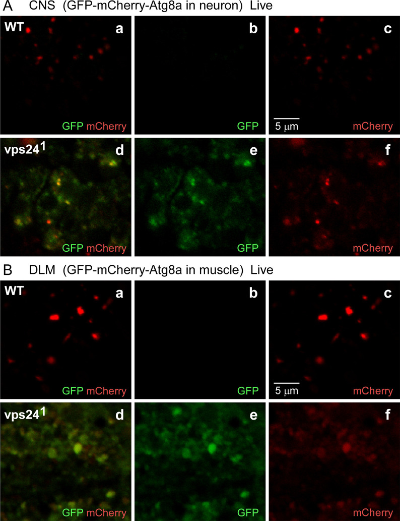
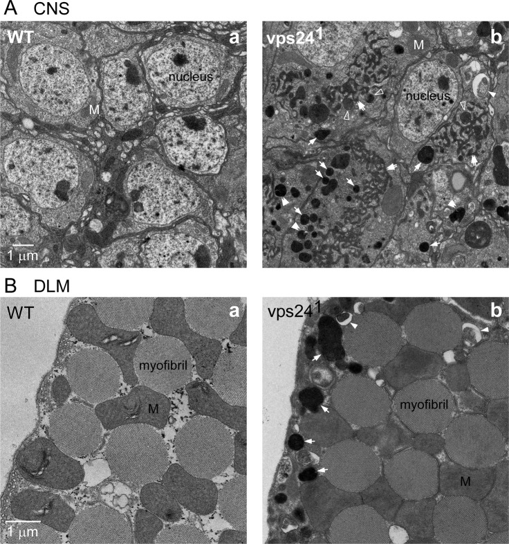
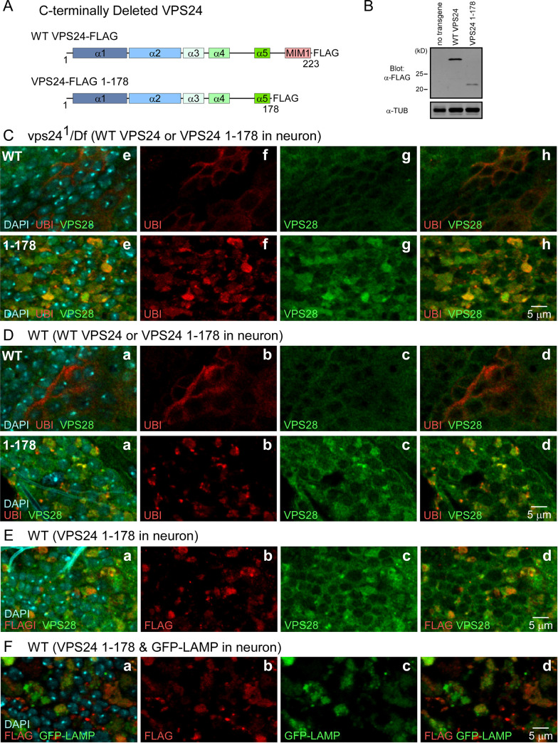
The ESCRT pathway is evolutionarily conserved across eukaryotes and plays key roles in a variety of membrane remodeling processes. A new Drosophila mutant recovered in our forward genetic screens for synaptic transmission mutants mapped to the vps24 gene encoding a subunit of the ESCRT-III complex. Molecular characterization indicated a loss of VPS24 function, however the mutant is viable and thus loss of VPS24 may be studied in a developed multicellular organism. The mutant exhibits deficits in locomotion and lifespan and, notably, these phenotypes are rescued by neuronal expression of wild-type VPS24. At the cellular level, neuronal and muscle cells exhibit marked expansion of a ubiquitin-positive lysosomal compartment, as well as accumulation of autophagic intermediates, and these phenotypes are rescued cell-autonomously. Moreover, VPS24 expression in glia suppressed the mutant phenotype in muscle, indicating a cell-nonautonomous function for VPS24 in protective intercellular signaling. Ultrastructural analysis of neurons and muscle indicated marked accumulation of the lysosomal compartment in the vps24 mutant. In the neuronal cell body, this included characteristic lysosomal structures associated with an expansive membrane compartment with a striking tubular network morphology. These findings further define the in vivo roles of VPS24 and the ESCRT pathway in lysosome homeostasis and their potential contributions to neurodegenerative diseases characterized by defective ESCRT or lysosome function.


Conflict of interest statement

The authors have declared that no competing interests exist.

Figures

Fig 1
Fig 1. A new mutant of vps24, vps241.
(A-C) The vps24 mutant exhibits locomotor and lifespan deficits. (A) vps241 /Df(3R)Exel6140 flies (vps241 /Df) exhibited rapid temperature sensitive (TS) paralysis at 38°C, whereas wild-type flies (WT) did not. Tests were truncated at 5 min if 50% TS paralysis had not occurred. (B) Climbing tests, carried out at a room temperature (RT) of 22–24°C, indicated no detectable climbing ability in the vps24 mutant. The climbing tests were truncated at 2 min if 50% climbing had not occurred and zero was given for the climbing index (see Materials and Methods). (C) Loss of the VPS24 function reduced lifespan with respect to WT. 7d old female flies raised at 20°C were examined in these and the following behavioral studies. Here and in subsequent figures, data points represent the mean ± SEM and asterisks mark significant differences from control values (P = 0.05). (D, E) vps241 mutation. (D) In vps241, an 11 bp deletion occurs at the beginning of the 1st intron and removes its splice donor signal. This disrupts splicing of the 1st intron in two ways: complete failure of splicing to remove the first intron (unspliced) or use of a cryptic splice donor site contained within the WT Exon 1 (see text). The broken line above the vps241 (unspliced) sequence represents continuation of Exon 1. (E) Each type of aberrant vps24 transcript (unspliced or cryptically spliced) encodes a drastically truncated polypeptide composed of the first 15 or 9 amino acids of VPS24 (full length is 223) followed by several amino acids of non-VPS24 protein sequence (in red). *, stop codon.
Fig 2
Fig 2. Neuronal expression of the wild-type VPS24 protein rescues the vps241 TS paralytic phenotype as well as locomotor and lifespan deficits.
(A) The TS paralytic phenotype of the vps24 mutant was rescued by expression of the wild-type EGFP-VPS24 in neurons but not in muscle or glia. The GAL4 drivers for neuronal, muscle and glial expression were Appl-GAL4, Mhc-GAL4 and Repo-Gal4 respectively. (B) Climbing tests, carried out at a room temperature (RT) of 22–24°C, indicated no detectable climbing ability in the vps24 mutant. The climbing tests were truncated at 2 min if 50% climbing had not occurred and zero was given for the climbing index. (C) Loss of the VPS24 function reduced lifespan with respect to WT when raised at a permissive temperature of 20°C. These mutant phenotypes were rescued by expression of wild-type EGFP-VPS24 in neurons but not in muscle or glia. The same GAL4 drivers were used for subsequent cell-type specific expression studies.
Fig 3
Fig 3. Ubiquitin-positive compartments accumulate in neurons and muscle of the vps24 mutant.
Confocal immunofluorescence images from wild-type (WT) (A) or vps241/Df(3R)Exel6140 (vps241/Df) mutant flies (B). Ubiquitin (UBI) staining indicates that the vps24 mutant exhibits a marked increase in ubiquitin-positive compartments in CNS (a, b) and muscle (c, d) with respect to WT. In contrast to neurons and muscle, ubiquitin positive structures are not prominent in glia of the vps24 mutant (see S4C and S4D Fig). DAPI labeling of nuclei and autofluorescence from trachea (TRA) appear in the same channel. Anti-HRP labels the neuronal plasma membrane.
Fig 4
Fig 4. Cell-autonomous rescue and -nonautonomous suppression of ubiquitin-positive compartment accumulation in the vps24 mutant.
Confocal immunofluorescence images of CNS neurons (a) and DLM (b) of the vps24 mutant (A), or the mutant rescued by neuronal (B), muscle (C) or glial (D) expression of the wild-type EGFP-VPS24 protein. Neuronal or muscle expression of wild-type EGFP-VPS24 produces cell-autonomous rescue of the vps24 mutant phenotype in the respective cell type. Notably, glial expression of wild-type VPS24 produces cell-nonautonomous suppression of the vps24 phenotype in muscle but not neurons.
Fig 5
Fig 5. An ESCRT-I component, VPS28, associates with ubiquitin-positive compartments.
Confocal immunofluorescence images of CNS neurons (A) and DLMs (B) from WT (a-d) or the vps24 mutant (e-h). Endogenous VPS28 was detected using an anti-VPS28 antibody. In both neurons and muscle, VPS28 is highly colocalized with ubiquitin-positive compartments and also exhibits a broader diffuse pattern. Only the diffuse VPS28 pattern was observed in neurons lacking ubiquitinated protein deposits. Thus ESCRT-I and III complexes may cooperate in clearance of ubiquitin-positive compartments.
Fig 6
Fig 6. Expansion of lysosomal compartments in neurons of the vps24 mutant.
Confocal immunofluorescence and native GFP or mCherry fluorescence images of CNS neurons from WT (a-d) or vps24 mutant (e-h) flies exhibiting neuronal expression of the lysosomal markers, GFP-LAMP (A) or Cathepsin-3xmCherry (CathB-mCh) (B). Neurons of the vps24 mutant exhibited markedly enlarged ubiquitin-positive lysosomal compartments.
Fig 7
Fig 7. Normal proteolytic processing of Cathepsin L in the vps24 mutant.
(A) Western blot analysis of endogenous Cathepsin L (CATH L) showing the proform and processed form of CATH L. Whole fly lysate from WT or the vps24 mutant was analyzed. In WT, the proform of CATH L is not visible in the image but is present at a detectable level. (B) Quantification of processed CATH L. The vps24 mutant exhibited significant accumulation of processed CATH L (3.1 ± 0.64 fold increase, n = 4) with respect to WT. However, the ratio of proform to processed form was similar in WT and the mutant [WT: 0.023 ± 0.004 (n = 5), 241/Df: 0.013 ± 0.002 (n = 5), P = 0.062]. Tubulin (TUB) was used as a loading control.
Fig 8
Fig 8. Disruption of autophagy contributes to accumulation of ubiquitin-positive compartments in the vps24 mutant.
Confocal immunofluorescence images of CNS neurons (A) and DLM (B) from WT (a-d) or the vps24 mutant (e-h). Localization of endogenous P62, the autophagy adaptor protein, was used to mark intermediates in autophagy. In both neurons and muscle, the vps24 mutant exhibits accumulation of P62. The distribution of P62 overlaps with but is more punctate and restricted than that of the ubiquitin-positive compartment.
Fig 9
Fig 9. The GFP-mCherry-Atg8a fusion proteins accumulate in a non-acidic compartment in the vps24 mutant.
Confocal live-imaging of the GFP-mCherry-Atg8a fusion protein in CNS neurons (A) and DLM (B) from WT (a-c) or the vps24 mutant (d-f). Neuronal (A) or muscle (B) expression of the tandem-tagged fusion protein was achieved as described in Fig 2. In both neurons and muscle of the vps24 mutant, colocalized GFP and mCherry fluorescence was detected (Ad-f, Bd-f). This indicates that the pH-sensitive GFP tag is not quenched and thus the tandem-tagged fusion protein is in a non-acidic compartment. In WT neurons and muscle (Aa-c, Ba-c), the GFP fluorescence signal is diminished with respect to the mCherry fluorescence.
Fig 10
Fig 10. Ultrastructural analysis of the vps24 mutant.
Transmission electron microscopy images of CNS neurons (A) and DLM (B) from WT (a) or the vps24 mutant (b). The vps24 mutant exhibited accumulation of autolysosomes (arrows) and tubular membrane compartments (double arrows) as well as autophagosome structures (filled arrowheads), whereas these were absent in WT. The electron dense tubular membrane compartment appeared to be continuous with some electron dense spherical autolysosomes (open arrowheads). In mutant muscle, mitochondria appear to be swollen. Double arrowheads, autophagic intermediates closely associated with spherical autolysosome structures (see S10 Fig); M, mitochondrion.
Fig 11
Fig 11. Transgenic expression of a VPS24 mutant lacking the C-terminus disrupts VPS24 function and reveals colocalization of VPS24 with VPS28 at the expanded, ubiquitin-positive lysosomal compartment.
(A) Schematic representations of control (WT VPS24-FLAG) and C-terminal truncation mutant (VPS24-FLAG 1–178) VSP24 proteins carrying a C-terminal FLAG tag. The MIM1 [Microtubule-interacting and transport (MIT)-interacting motif 1] domain present in the VPS24 C-terminal mediates binding interactions with MIT domain proteins such as VPS4. The vps24 mutant form lacks this interaction domain. (B) Western blot analysis of transgene products following neuronal expression of the control or mutant transgene. Lysate was prepared from fly heads and Tubulin (TUB) was used as a loading control. (C-F) Confocal immunofluorescence images of CNS neurons following neuronal transgene expression in a WT or vps24 mutant background. (C) Neuronal expression of WT VPS24, but not VPS24 1–178, rescues the neuronal phenotype of the vps24 mutant. (D) Neuronal expression of VPS24 1–178, but not WT VPS24, in a WT background mimics the neuronal phenotype of the vps24 mutant. (E) Colocalization of VPS24 1–178 and endogenous VPS28 following neuronal transgene expression in a WT background. (F) Colocalization of VPS24 1–178 and the expanded lysosome membrane compartment following neuronal transgene expression in a WT background.

References

    1. Christ L, Raiborg C, Wenzel EM, Campsteijn C, Stenmark H. Cellular Functions and Molecular Mechanisms of the ESCRT Membrane-Scission Machinery. Trends in Biochemical Sciences 2017;42(1):42–56. 10.1016/j.tibs.2016.08.016 - DOI - PubMed
    1. Hurley JH, Hanson PI. Membrane budding and scission by the ESCRT machinery: it’s all in the neck. Nature Reviews Molecular Cell Biology 2010;11(8):556–66. 10.1038/nrm2937 - DOI - PMC - PubMed
    1. McCullough J, Frost A, Sundquist WI. Structures, Functions, and Dynamics of ESCRT-III/Vps4 Membrane Remodeling and Fission Complexes. Annual Review of Cell and Developmental Biology 2018;34:85–109. 10.1146/annurev-cellbio-100616-060600 - DOI - PMC - PubMed
    1. Schoneberg J, Lee IH, Iwasa JH, Hurley JH. Reverse-topology membrane scission by the ESCRT proteins. Nature Reviews Molecular Cell Biology 2017;18(1):5–17. 10.1038/nrm.2016.121 - DOI - PMC - PubMed
    1. Frankel EB, Audhya A. ESCRT-dependent cargo sorting at multivesicular endosomes. Semin Cell Dev Biol 2018;74:4–10. 10.1016/j.semcdb.2017.08.020 - DOI - PMC - PubMed

Publication types

MeSH terms

Substances

LinkOut - more resources