. 2021 Jun 3;28(6):1016-1022.e4.
doi: 10.1016/j.stem.2021.04.031. Epub 2021 May 5.

Naive stem cell blastocyst model captures human embryo lineage segregation

Affiliations

Naive stem cell blastocyst model captures human embryo lineage segregation

Ayaka Yanagida et al. Cell Stem Cell. .

Abstract

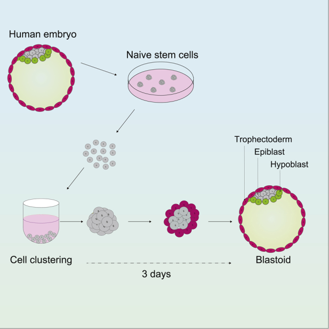
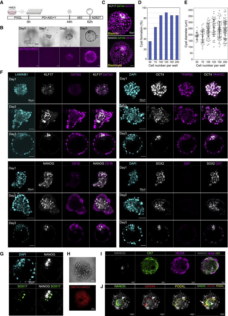
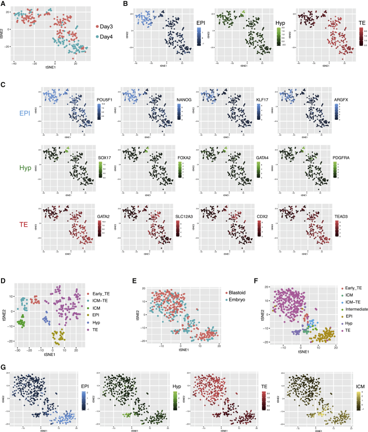
Human naive pluripotent cells can differentiate into extraembryonic trophectoderm and hypoblast. Here we describe a human embryo model (blastoid) generated by self-organization. Brief induction of trophectoderm leads to formation of blastocyst-like structures within 3 days. Blastoids are composed of three tissue layers displaying exclusive lineage markers, mimicking the natural blastocyst. Single-cell transcriptome analyses confirm segregation of trophectoderm, hypoblast, and epiblast with high fidelity to the human embryo. This versatile and scalable system provides a robust experimental model for human embryo research.

Keywords: blastocyst; embryonic stem cells; epiblast; human embryo; hypoblast; lineage segregation; pluripotency; self-organization; trophoblast.


Conflict of interest statement

Declaration of interests G.G. and A.S. are inventors on a patent relating to human naive pluripotent stem cells filed by the University of Cambridge.

Figures

None
Graphical abstract
Figure 1
Figure 1
Formation of model human blastocysts (blastoids) (A) Schematic of procedure. (B) Phase contrast and florescence images showing formation of a GATA3:mKO2 positive cyst. (C) z-projections of a day 3 blastoid and a human late blastocyst (E7) stained for epiblast (KLF17, NANOG), hypoblast (GATA4), and trophectoderm (GATA3) markers. (D) Quantification of cavitated mKO2-positive cyst formation related to cell number seeded. (E) Quantification of diameter of cysts as in (D). Error bars are SD. (F) Immunofluorescence staining for epiblast and trophectoderm markers during blastoid formation. (G) Immunofluorescence staining for hypoblast marker SOX17 in day 3 blastoid. (H) Phase contrast and fluorescence images of GATA3:mKO blastoid outgrowth after 4 days. (I) z-projections of immunofluorescence staining of day 4 outgrowth stained for differentiated trophoblast (CK7) and syncytiotrophoblast (HCGB) markers and NANOG. (J) z-projections of immunofluorescence staining of day 4 outgrowth stained for PODXL, NANOG, GATA4, and DAPI. Arrows point to cavities (see Video S1). Scale bars in all images are 50 μm.
Figure 2
Figure 2
Single-cell transcriptome analysis (A) t-distributed stochastic neighbor embedding (tSNE) of day 3 and day 4 blastoid cells. (B) tSNE plot in (A) showing averaged expression scores of classifier genes (Table S1) enriched in human E6/7 embryo lineages, epiblast (EPI), hypoblast (Hyp), and trophectoderm (TE). (C) Expression (FPKM) of selected lineage markers on tSNE plot in (A). (D) tSNE analysis of human embryos at E5, E6, E7 with lineage assignment by marker profile. ICM_TE denotes ambiguous ICM and TE identity. (E) Integrated tSNE plot for blastoid and embryo cells. (F) tSNE plot in (E) colored to show cell lineage assignments from Figures S1A and S1D. (G) tSNE in (E) showing averaged expression scores of lineage classifiers.

Comment in

References

    1. Bedzhov I., Zernicka-Goetz M. Self-organizing properties of mouse pluripotent cells initiate morphogenesis upon implantation. Cell. 2014;156:1032–1044. - PMC - PubMed
    1. Blakeley P., Fogarty N.M.E., del Valle I., Wamaitha S.E., Hu T.X., Elder K., Snell P., Christie L., Robson P., Niakan K.K. Defining the three cell lineages of the human blastocyst by single-cell RNA-seq. Development. 2015;142:3151–3165. - PMC - PubMed
    1. Boroviak T., Loos R., Lombard P., Okahara J., Behr R., Sasaki E., Nichols J., Smith A., Bertone P. Lineage-Specific Profiling Delineates the Emergence and Progression of Naive Pluripotency in Mammalian Embryogenesis. Dev. Cell. 2015;35:366–382. - PMC - PubMed
    1. Boroviak T., Stirparo G.G., Dietmann S., Hernando-Herraez I., Mohammed H., Reik W., Smith A., Sasaki E., Nichols J., Bertone P. Single cell transcriptome analysis of human, marmoset and mouse embryos reveals common and divergent features of preimplantation development. Development. 2018;145:dev167833. - PMC - PubMed
    1. Bredenkamp N., Stirparo G.G., Nichols J., Smith A., Guo G. The Cell-Surface Marker Sushi Containing Domain 2 Facilitates Establishment of Human Naive Pluripotent Stem Cells. Stem Cell Reports. 2019;12:1212–1222. - PMC - PubMed

Publication types