Review
. 2021 Jan-Jun:296:100745.
doi: 10.1016/j.jbc.2021.100745. Epub 2021 May 3.

50 Years of structural immunology

Affiliations
Review

50 Years of structural immunology

Ian A Wilson et al. J Biol Chem. 2021 Jan-Jun.

Abstract

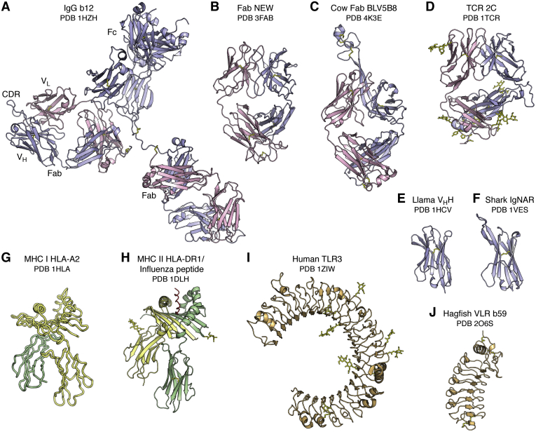
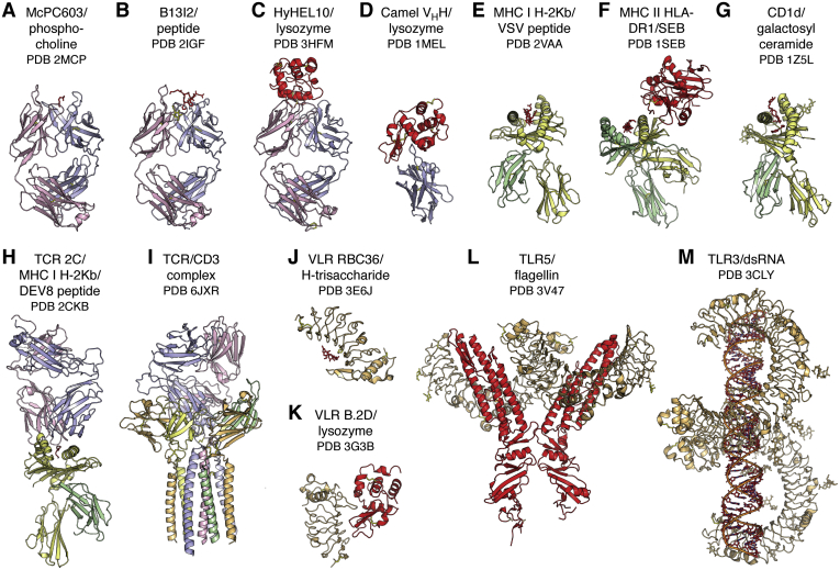
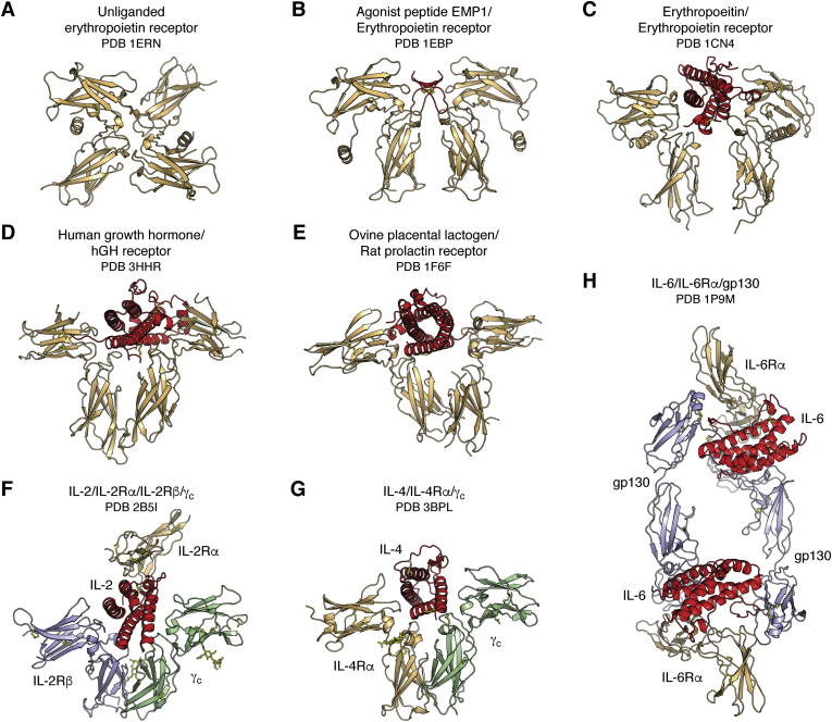
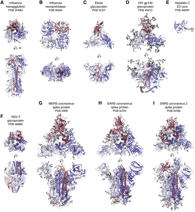
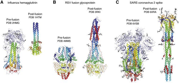
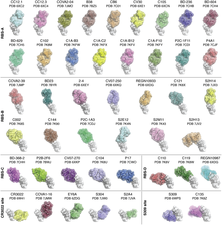
Fifty years ago, the first landmark structures of antibodies heralded the dawn of structural immunology. Momentum then started to build toward understanding how antibodies could recognize the vast universe of potential antigens and how antibody-combining sites could be tailored to engage antigens with high specificity and affinity through recombination of germline genes (V, D, J) and somatic mutation. Equivalent groundbreaking structures in the cellular immune system appeared some 15 to 20 years later and illustrated how processed protein antigens in the form of peptides are presented by MHC molecules to T cell receptors. Structures of antigen receptors in the innate immune system then explained their inherent specificity for particular microbial antigens including lipids, carbohydrates, nucleic acids, small molecules, and specific proteins. These two sides of the immune system act immediately (innate) to particular microbial antigens or evolve (adaptive) to attain high specificity and affinity to a much wider range of antigens. We also include examples of other key receptors in the immune system (cytokine receptors) that regulate immunity and inflammation. Furthermore, these antigen receptors use a limited set of protein folds to accomplish their various immunological roles. The other main players are the antigens themselves. We focus on surface glycoproteins in enveloped viruses including SARS-CoV-2 that enable entry and egress into host cells and are targets for the antibody response. This review covers what we have learned over the past half century about the structural basis of the immune response to microbial pathogens and how that information can be utilized to design vaccines and therapeutics.

Keywords: MHC; T cells; TLR; VLR; antibodies; cellular immunity; humoral immunity; immune recognition; microbial pathogens; viral antigens.


Conflict of interest statement

Conflict of interest The authors declare that they have no conflicts of interest with the contents of this article.

Figures

Figure 1
Figure 1
The diversity of antigen receptors in the immune system. The immunoglobulin fold is utilized as the recognition motif in antibodies (A, B, C, E and F) in the humoral adaptive immune system and T cell receptors (D) in the cellular adaptive immune system. The intact IgG b12 (A) is labeled to illustrate the relative positions of the two Fab and one Fc regions, the VL and VH immunoglobulin domains within one Fab, and the complementarity-determining region containing region of the Fab. The camelid family (E), which includes llamas, and the shark family (F), also have smaller antibodies that contain only a single VH domain (termed VHH or nanobody) instead of the two VH and VL domains in conventional antibodies. The major histocompatibility complex (MHC) fold (G and H) is used to present peptide antigens to T cell receptors for classical MHC I and II, and lipids, glycolipids, specialized peptides, and other antigens in nonclassical MHC-like molecules. Note that, in (G), the unrefined HLA-A2 structure was deposited with only Cα atoms, so a cartoon trace is shown. Toll-like receptors (I) in the innate immune system adopt a Leu-rich repeat fold and recognize specific antigens, including proteins, nucleic acids, lipopolysaccharides, unmethylated CpG, and small molecules. Variable lymphocyte receptors (J) (VLRs) also are composed of Leu-rich repeats and function as the adaptive immune responses in the jawless vertebrates, lampreys, and hagfish. In this and following figures, the immunoglobulin light and heavy chains are colored pink and light blue. The MHC Class I heavy chain is colored yellow and β2 microglobulin chain in green, whereas the MHC Class II α and β chains are colored yellow and green. The TLRs and VLRs are colored in beige. For all figures, carbohydrate and disulfide bonds are colored yellow. The name of the receptor, ligand if any, and Protein Data Bank (PDB) ID are shown below each figure.
Figure 2
Figure 2
Diversity of antigens recognized by antigen receptors in the immune system. The diverse antigen receptors in the immune system can detect, interact with, and respond to the universe of potential antigens, shown here colored in red and identified under the name of its receptor. Antibodies (AD) can recognize virtually any antigen whether large or small, and which can have diverse chemical compositions from small molecules (A) to carbohydrates to lipids to peptides (B) to proteins (C and D) and combinations thereof. Classical major histocompatibility complex (MHC) molecules (E and F) bind processed peptide antigens in their binding groove for presentation to T cell receptors (TCRs). CD1 (G) is related to class I MHC but presents lipid-containing antigens to certain types of T cells and NK cells. TCR signaling is initiated by binding to MHC-peptide complexes (H). The T cell–bound TCRs associate with the multisubunit, membrane-spanning CD3 (I). Variable lymphocyte receptors (J and K) and Toll-like receptors (L and M) can sense specific types of antigens present on microbial pathogens in the innate immune system and regulate inflammatory responses and transcriptional events through antigen-induced signal transduction pathways. Molecules are colored as in (A). In (I), the CD3 complex is colored with ζ chains in blue-white, δ chain in pale green, ε chains in light orange, and γ chain in pale yellow.
Figure 3
Figure 3
Hematopoietic and type I cytokine receptors. These signaling receptors have a self-ligand and can form homodimers (EPOR, AC; hGHR, D; rat prolactin receptor, E), heterodimers (IL-4R, G), heterotrimers (IL-2R, F), and other high-order complexes (IL-6R, H) with their ligands. EPO receptors also exist as inactive preformed dimers (A) on the cell surface and change conformation to an active, signaling-competent state on interaction with their natural ligand (C). Preformed dimers have now been found in other cytokine receptors. Cytokine receptor α, β or gp130, and γ chains are colored beige, blue, and green, respectively, with the bound hormone or agonist colored in red.
Figure 4
Figure 4
Viral antigens on enveloped viruses. Viral glycoproteins embedded in the viral membrane are responsible for entry of viruses into host cells. These envelope proteins contain receptor-binding sites and membrane fusion activities or are involved in progeny release and cell egress (influenza neuraminidase). Many of these viral antigens are homotrimers (e.g., influenza HA (A), Ebola virus GP (C), HIV-1 (D), RSV F (F), MERS (G), and SARS-CoV 1 and 2 spike proteins (H and I)), whereas Influenza NA (B) is a homotetramer. The HepC E1E2 glycoprotein is thought to exist as a heterodimer, but only the structure for the monomeric E2 core has been determined (E). These antigens are usually heavily glycosylated to shield themselves from antibodies in the immune system. In all panels, the individual subunits of each glycoprotein are colored red, white, or blue and top (looking down the trimer or tetramer axis) and side views where the viral cell membrane would be on the bottom are shown for each. Carbohydrate is shown in black.
Figure 5
Figure 5
Spring-loaded conformational rearrangements in viral glycoproteins to attain their fusogenic state. Viral antigens involved in cell entry are metastable and undergo large conformational changes to attain their fusion-active form after engaging their receptor(s). Prefusion (left) and postfusion (right) structures of (A) Influenza hemagglutinin, (B) RSV fusion glycoprotein, and (C) SARS-CoV-2 spike are illustrated with the fusion portions of the proteins colored in a rainbow with blue and red corresponding to the N and C termini. Molecules are oriented so that the viral membrane would be on the bottom and target cell membrane on the top.
Figure 6
Figure 6
Antibody recognition of the RBD of SARS-CoV-2. A large number of neutralizing antibody structures to SARS-CoV-2 and related viruses have been determined in a relatively short time. Most of these antibodies (as depicted here) bind to the receptor-binding domain. These antibodies have been classified into regions that they bind on the RBD by Meng Yuan, Nicholas Wu, and Hejun Liu in the Wilson laboratory. Structures of the RBD (white) in complex with four groups of antibodies to the receptor binding site (RBS): RBS-A-targeting antibodies: CC12.1 (PDB ID: 6XC2) (186), CC12.3 (PDB ID: 6XC4) (186), COVA2-04 (PDB ID: 7JMO) (210), B38 (PDB ID: 7BZ5) (211), CB6 (PDB ID: 7C01) (212), CV30 (PDB ID: 6XE1) (213), C105 (PDB ID: 6XCM) (187), BD-236 (PDB ID: 7CHB) (214), BD-604 (PDB ID: 7CH4) (214), BD-629 (PDB ID: 7CH5) (214), C102 (PDB ID: 7K8M) (188), C1A-B3 (PDB ID: 7KFW) (215), C1A-C2 (PDB ID: 7KFX) (215), C1A-B12 (PDB ID: 7KFV) (215), C1A-F10 (PDB ID: 7KFY) (215), P2C-1F11 (PDB ID: 7CDI), P4A1 (PDB ID: 7CJF); RBS-B-targeting antibodies: COVA2-39 (PDB ID: 7JMP) (210), BD23 (PDB ID: 7BYR) (216), 2 to 4 (PDB ID: 6XEY) (190), CV07-250 (PDB ID: 6XKQ) (217), REGN10933 (PDB ID: 6XDG) (218), C121 (PDB ID: 7K8X) (188), S2H14 (PDB ID: 7JX3) (219), C002 (PDB ID: 7K8S) (188), C144 (PDB ID: 7K90) (188), P2C-1A3 (PDB ID: 7CDJ), S2E12 (PDB ID: 7K4N) (220), S2M11 (PDB ID: 7K43) (220), S2H13 (PDB ID: 7JV2) (219); RBS-C-targeting antibodies: BD-368-2 (PDB ID: 7CHH) (214), P2B-2F6 (PDB ID: 7BWJ) (221), CV07-270 (PDB ID: 6XKP) (217), C104 (PDB ID: 7K8U) (188), P17 (PDB ID: 7CWO) (222); RBS-D-targeting antibodies: C110 (PDB ID: 7K8V) (188), C119 (PDB ID: 7K8W) (188), and REGN10987 (PDB ID: 6XDG) (218). Cross-neutralizing antibodies are targeted to two sites, which are more highly conserved than the receptor-binding site: the very highly conserved CR3022-binding site: CR3022 (PDB ID: 6W41) (184), COVA1-16 (PDB ID: 7JMW) (223), EY6A (PDB ID: 6ZER) (224), S304 (PDB ID: 7JW0) (219), S2A4 (PDB ID: 7JVA) (219), and those targeting the moderately conserved S309 site that include an N-linked glycan: S309 (PDB ID: 6WPS) (189) and C135 (PDB ID: 7K8Z) (188).

References

    1. Sarma V.R., Silverton E.W., Davies D.R., Terry W.D. The three-dimensional structure at 6 Å resolution of a human γG1 immunoglobulin molecule. J. Biol. Chem. 1971;246:3753–3759. - PubMed
    1. Poljak R.J., Amzel L.M., Avey H.P., Becka L.N. Structure of Fab’ New at 6 Å resolution. Nat. New Biol. 1972;235:137–140. - PubMed
    1. Porter R.R. The hydrolysis of rabbit γ-globulin and antibodies with crystalline papain. Biochem. J. 1959;73:119–126. - PMC - PubMed
    1. Silverton E.W., Navia M.A., Davies D.R. Three-dimensional structure of an intact human immunoglobulin. Proc. Natl. Acad. Sci. U. S. A. 1977;74:5140–5144. - PMC - PubMed
    1. Amzel L.M., Poljak R.J., Saul F., Varga J.M., Richards F.F. The three dimensional structure of a combining region-ligand complex of immunoglobulin NEW at 3.5 Å resolution. Proc. Natl. Acad. Sci. U. S. A. 1974;71:1427–1430. - PMC - PubMed

Publication types

MeSH terms