

Overcoming physiological barriers by nanoparticles for intravenous drug delivery to the lymph nodes
- PMID: 33957802
- PMCID: PMC8606962
- DOI: 10.1177/15353702211010762
Overcoming physiological barriers by nanoparticles for intravenous drug delivery to the lymph nodes
Abstract
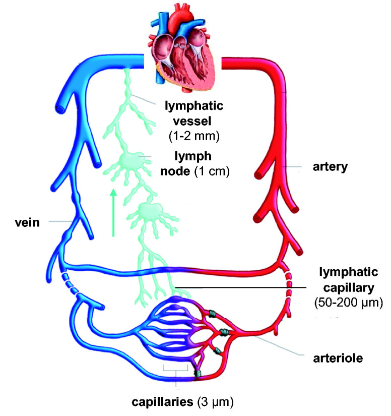
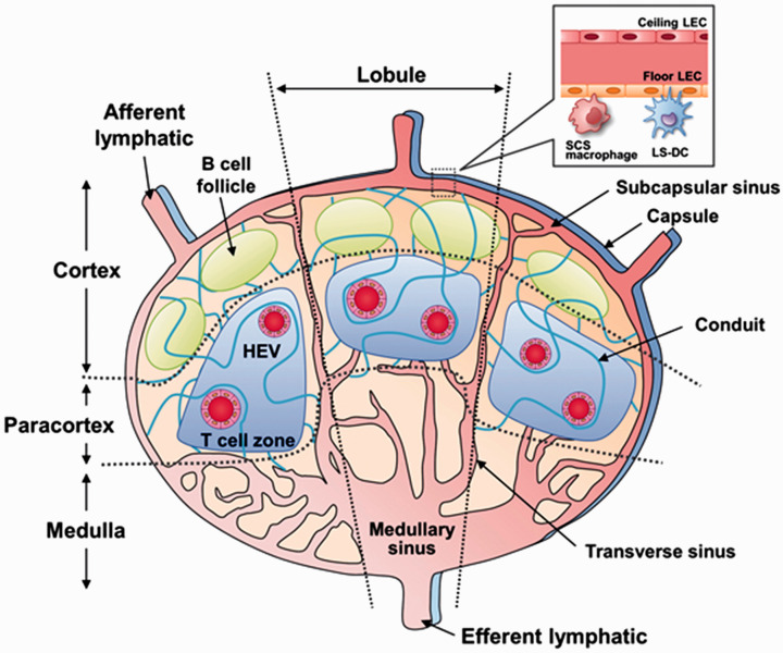
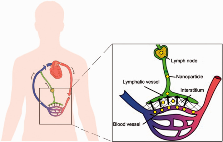
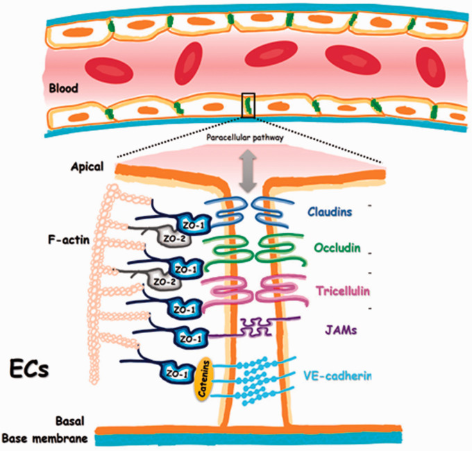
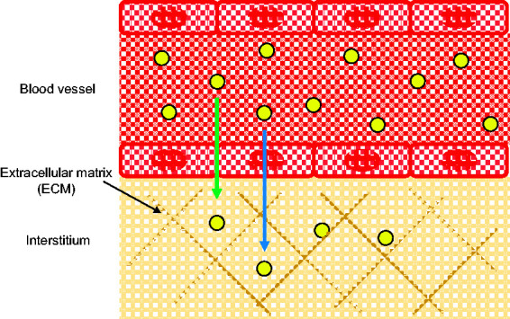
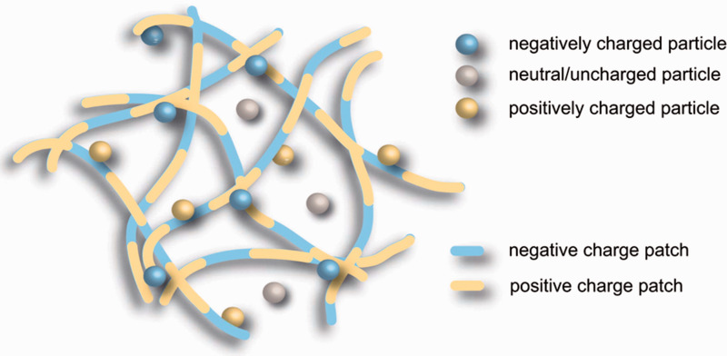
The lymph nodes are major sites of cancer metastasis and immune activity, and thus represent important clinical targets. Although not as well-studied compared to subcutaneous administration, intravenous drug delivery is advantageous for lymph node delivery as it is commonly practiced in the clinic and has the potential to deliver therapeutics systemically to all lymph nodes. However, rapid clearance by the mononuclear phagocyte system, tight junctions of the blood vascular endothelium, and the collagenous matrix of the interstitium can limit the efficiency of lymph node drug delivery, which has prompted research into the design of nanoparticle-based drug delivery systems. In this mini review, we describe the physiological and biological barriers to lymph node targeting, how they inform nanoparticle design, and discuss the future outlook of lymph node targeting.
Keywords: Nanoparticles; barrier; drug delivery; hitchhiking; lymph node; targeting.
Conflict of interest statement
Figures
Similar articles
-
Convective diffusion of nanoparticles from the epithelial barrier toward regional lymph nodes.Adv Colloid Interface Sci. 2013 Nov;199-200:23-43. doi: 10.1016/j.cis.2013.06.002. Epub 2013 Jun 10. Adv Colloid Interface Sci. 2013. PMID: 23859221 Free PMC article. Review.
-
Intraductal Drug Delivery to the Breast: Effect of Particle Size and Formulation on Breast Duct and Lymph Node Retention.Mol Pharm. 2020 Feb 3;17(2):441-452. doi: 10.1021/acs.molpharmaceut.9b00879. Epub 2020 Jan 10. Mol Pharm. 2020. PMID: 31886676
-
Impact of nanoparticle properties on immune cell interactions in the lymph node.Acta Biomater. 2025 Jan 24;193:65-82. doi: 10.1016/j.actbio.2024.12.039. Epub 2024 Dec 17. Acta Biomater. 2025. PMID: 39701340 Review.
-
Vaccine delivery systems toward lymph nodes.Adv Drug Deliv Rev. 2021 Dec;179:113914. doi: 10.1016/j.addr.2021.113914. Epub 2021 Aug 4. Adv Drug Deliv Rev. 2021. PMID: 34363861 Free PMC article. Review.
-
Multifunctional nanocarriers for targeted drug delivery and diagnostic applications of lymph nodes metastasis: a review of recent trends and future perspectives.J Nanobiotechnology. 2023 Aug 2;21(1):247. doi: 10.1186/s12951-023-01990-4. J Nanobiotechnology. 2023. PMID: 37528366 Free PMC article. Review.
Cited by
-
Targeting lymph nodes for enhanced cancer vaccination: From nanotechnology to tissue engineering.Mater Today Bio. 2024 Apr 26;26:101068. doi: 10.1016/j.mtbio.2024.101068. eCollection 2024 Jun. Mater Today Bio. 2024. PMID: 38711936 Free PMC article. Review.
-
Polymeric Nanoparticles in Cancer Chemotherapy: A Narrative Review.Iran J Public Health. 2022 Feb;51(2):226-239. doi: 10.18502/ijph.v51i2.8677. Iran J Public Health. 2022. PMID: 35866132 Free PMC article. Review.
-
Intervaginal space injection of photothermal chemotherapy nanoparticles for facilitating tumor targeting and improving outcomes in mice.Heliyon. 2024 Feb 29;10(5):e27408. doi: 10.1016/j.heliyon.2024.e27408. eCollection 2024 Mar 15. Heliyon. 2024. PMID: 38468940 Free PMC article.
-
Kinetically inert manganese (II)-based hybrid micellar complexes for magnetic resonance imaging of lymph node metastasis.Regen Biomater. 2023 May 25;10:rbad053. doi: 10.1093/rb/rbad053. eCollection 2023. Regen Biomater. 2023. PMID: 37293571 Free PMC article.
-
Locoregional Lymphatic Delivery Systems Using Nanoparticles and Hydrogels for Anticancer Immunotherapy.Pharmaceutics. 2022 Dec 8;14(12):2752. doi: 10.3390/pharmaceutics14122752. Pharmaceutics. 2022. PMID: 36559246 Free PMC article. Review.
References
Publication types
MeSH terms
Grants and funding
LinkOut - more resources
Full Text Sources
Other Literature Sources