

ASCs derived from burn patients are more prone to increased oxidative metabolism and reactive oxygen species upon passaging
- PMID: 33957963
- PMCID: PMC8100366
- DOI: 10.1186/s13287-021-02327-4
ASCs derived from burn patients are more prone to increased oxidative metabolism and reactive oxygen species upon passaging
Abstract
Background: Patients with severe burn injury (over 20% of the total body surface area) experience profound hypermetabolism which significantly prolongs wound healing. Adipose-derived stem cells (ASCs) have been proposed as an attractive solution for treating burn wounds, including the potential for autologous ASC expansion. While subcutaneous adipocytes display an altered metabolic profile post-burn, it is not known if this is the case with the stem cells associated with the adipose tissue.
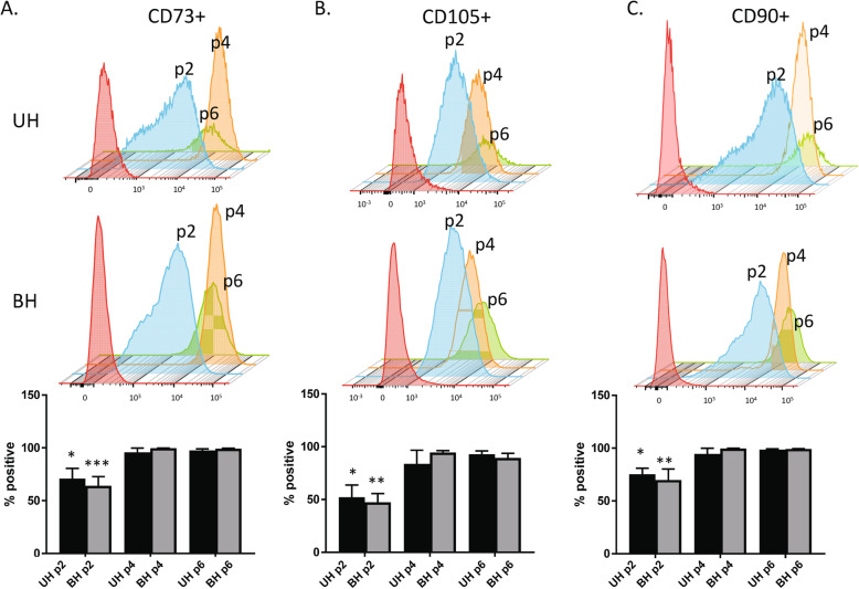
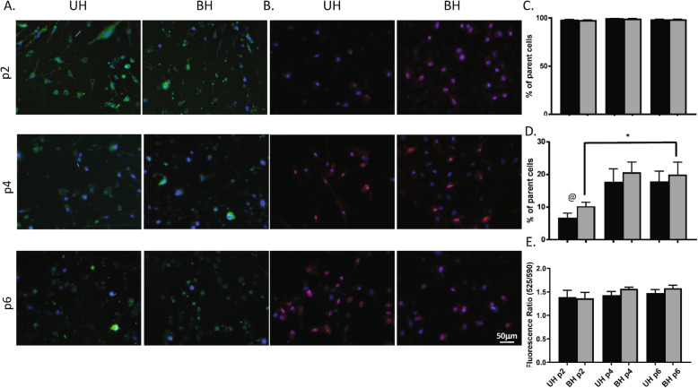
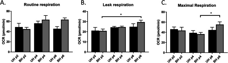
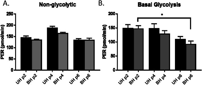
Methods: ASCs were isolated from discarded burn skin of severely injured human subjects (BH, n = 6) and unburned subcutaneous adipose tissue of patients undergoing elective abdominoplasty (UH, n = 6) and were analyzed at passages 2, 4, and 6. Flow cytometry was used to quantify ASC cell surface markers CD90, CD105, and CD73. Mitochondrial abundance and reactive oxygen species (ROS) production were determined with MitoTracker Green and MitoSOX Red, respectively, while JC-10 Mitochondrial Membrane Potential Assays were also performed. Mitochondrial respiration and glycolysis were analyzed with a high-resolution respirometer (Seahorse XFe24 Analyzer).
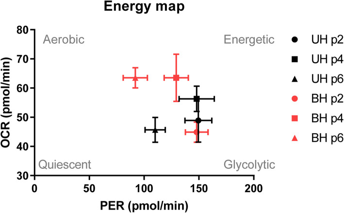
Results: There was no difference in age between BH and UH (34 ± 6 and 41 ± 4 years, respectively, P = 0.49). While passage 2 ASCs had lower ASC marker expression than subsequent passages, there were no significant differences in the expression between BH and UH ASCs. Similarly, no differences in mitochondrial abundance or membrane potential were found amongst passages or groups. Two-way ANOVA showed a significant effect (P < 0.01) of passaging on mitochondrial ROS production, with increased ROS in BH ASCs at later passages. Oxidative phosphorylation capacities (leak and maximal respiration) increased significantly in BH ASCs (P = 0.035) but not UH ASCs. On the contrary, basal glycolysis significantly decreased in BH ASCs (P = 0.011) with subsequent passaging, but not UH ASCs.
Conclusions: In conclusion, ASCs from burned individuals become increasingly oxidative and less glycolytic upon passaging when compared to ASCs from unburned patients. This increase in oxidative capacities was associated with ROS production in later passages. While the autologous expansion of ASCs holds great promise for treating burned patients with limited donor sites, the potential negative consequences of using them require further investigation.
Keywords: Adipose stem cells; Burn; Glycolysis; Mitochondria; Oxidative phosphorylation; ROS; Respirometry.
Conflict of interest statement
The authors declare that they have no competing interests.
Figures
Similar articles
-
Characterization of Adipose-Derived Stem Cells Following Burn Injury.Stem Cell Rev Rep. 2017 Dec;13(6):781-792. doi: 10.1007/s12015-017-9721-9. Stem Cell Rev Rep. 2017. PMID: 28646271 Free PMC article.
-
Characterization of human adipose-derived stem cells cultured in autologous serum after subsequent passaging and long term cryopreservation.J Stem Cells. 2014;9(3):135-48. J Stem Cells. 2014. PMID: 25157448
-
Adipose-derived human stem/stromal cells: comparative organ specific mitochondrial bioenergy profiles.Springerplus. 2016 Dec 1;5(1):2057. doi: 10.1186/s40064-016-3712-1. eCollection 2016. Springerplus. 2016. PMID: 27995034 Free PMC article.
-
Generation of reactive oxygen species in adipose-derived stem cells: friend or foe?Expert Opin Ther Targets. 2011 Nov;15(11):1297-306. doi: 10.1517/14728222.2011.628315. Epub 2011 Oct 10. Expert Opin Ther Targets. 2011. PMID: 21981031 Free PMC article. Review.
-
Cellular and molecular stimulation of adipose-derived stem cells under hypoxia.Cell Biol Int. 2014 May;38(5):553-62. doi: 10.1002/cbin.10246. Epub 2014 Feb 5. Cell Biol Int. 2014. PMID: 24446066 Review.
Cited by
-
Emerging Effects of Resveratrol on Wound Healing: A Comprehensive Review.Molecules. 2022 Oct 9;27(19):6736. doi: 10.3390/molecules27196736. Molecules. 2022. PMID: 36235270 Free PMC article. Review.
-
The total extract of Abelmoschus manihot (L.) medic flowers (TEA) mediated Nrf2-TFAM signalling to regulate mitochondrial antioxidant mechanism.Sci Rep. 2025 Jan 10;15(1):1614. doi: 10.1038/s41598-024-84022-x. Sci Rep. 2025. PMID: 39794424 Free PMC article.
-
TiO2 Nanoparticles Loaded with Polygonum cuspidatum Extract for Wound Healing Applications: Exploring Their Hemolytic, Antioxidant, Cytotoxic, and Antimicrobial Properties.Nanomaterials (Basel). 2025 Jun 14;15(12):926. doi: 10.3390/nano15120926. Nanomaterials (Basel). 2025. PMID: 40559290 Free PMC article.
-
3D culture inhibits replicative senescence of SCAPs via UQCRC2-mediated mitochondrial oxidative phosphorylation.J Transl Med. 2024 Dec 20;22(1):1129. doi: 10.1186/s12967-024-05953-7. J Transl Med. 2024. PMID: 39707408 Free PMC article.
References
-
- Hart DW, Wolf SE, Chinkes DL, Beauford RB, Mlcak RP, Heggers JP, Wolfe RR, Herndon DN. Effects of early excision and aggressive enteral feeding on hypermetabolism, catabolism, and sepsis after severe burn. J Trauma-Injury Infect Crit Care. 2003;54(4):755–761. doi: 10.1097/01.TA.0000060260.61478.A7. - DOI - PubMed
Publication types
MeSH terms
Substances
LinkOut - more resources
Full Text Sources
Other Literature Sources
Research Materials
Miscellaneous
