

Hypotony maculopathy and photoreceptor folds with disruptions after vitrectomy for epiretinal membrane removal: two case reports
- PMID: 33957968
- PMCID: PMC8103759
- DOI: 10.1186/s13256-021-02824-3
Hypotony maculopathy and photoreceptor folds with disruptions after vitrectomy for epiretinal membrane removal: two case reports
Abstract
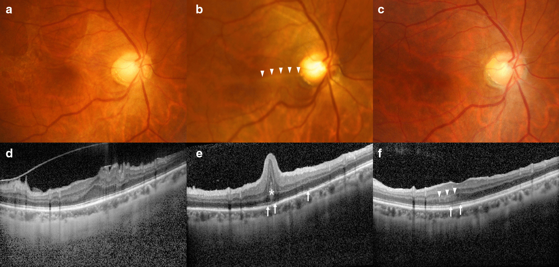
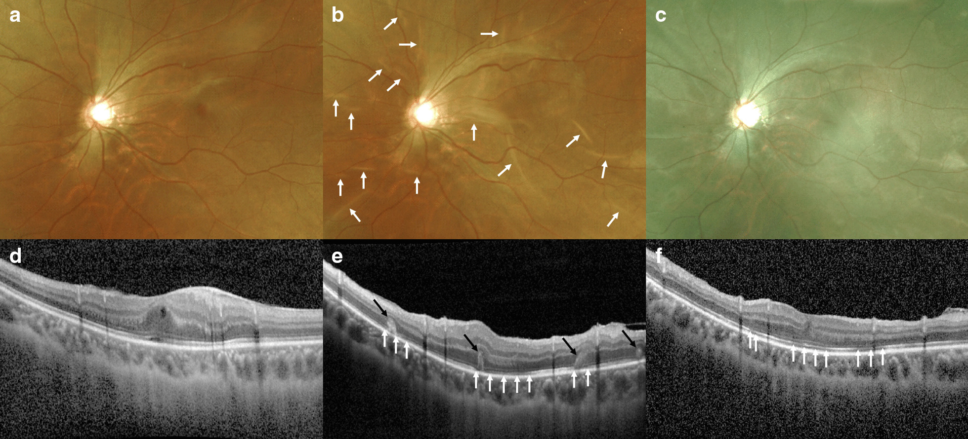
Background: Hypotony maculopathy has been classically reported as a complication of glaucoma surgery or ocular trauma. There have been only a few reports of hypotony maculopathy following pars plana vitrectomy (PPV). Here, we report two cases of hypotony maculopathy occurring after PPV for epiretinal membrane (ERM) removal and characteristic photoreceptor folds observed on optical coherence tomography (OCT).
Case presentation: A 53-year-old Korean woman (case 1) underwent phacoemulsification and posterior chamber lens implantation combined with 25-gauge PPV for ERM removal in the right eye. On the following day, she had severe ocular hypotony, with an intraocular pressure (IOP) that was unmeasurable using a pneumatic tonometer. Despite normalization of IOP, macular retinal and photoreceptor folds with photoreceptor disruptions developed, and Henle's fiber layer hyperreflectivity was identified. Thereafter, retinal and photoreceptor folds gradually disappeared but photoreceptor disruption and Henle's fiber layer hyperreflectivity did not improve until 1 year postoperatively, with persistent central visual field distortion and visual acuity worse than that at the preoperative state. A 20-year-old Korean man (case 2) underwent an additional 25-gauge PPV for ERM removal in the left eye. Examination on the following day showed ocular hypotony and retinal folds with peripheral choroidal detachment. Although IOP was normalized, further OCT revealed photoreceptor folds and photoreceptor disruptions. Since then, the photoreceptor folds resolved; however, the photoreceptor disruption remained in the macula at the 1-year follow up, with persistent distorted vision and visual acuity worse than that at the preoperative state.
Conclusions: Early hypotony after vitrectomy for ERM could result in maculopathy leading to irreversible visual decline and metamorphopsia. Photoreceptor folds on OCT are characteristic features and the predominant mechanism of central visual loss in cases of hypotony maculopathy.
Keywords: Epiretinal membrane; Hypotony maculopathy; Pars plana vitrectomy.
Conflict of interest statement
The authors declare that they have no competing interests.
Figures
Similar articles
-
Management of hypotony-related maculopathy after combined phacoemulsification and trabeculectomy: January consultation #1.J Cataract Refract Surg. 2021 Jan 1;47(1):130. doi: 10.1097/j.jcrs.0000000000000524. J Cataract Refract Surg. 2021. PMID: 33901082
-
Vitrectomy for a persisting macular fold in a case of resolved hypotony maculopathy.Am J Ophthalmol. 2004 Sep;138(3):487-9. doi: 10.1016/j.ajo.2004.03.024. Am J Ophthalmol. 2004. PMID: 15364240
-
Recalcitrant Fovea-Involving Macular Fold After Uneventful Epiretinal Membrane Surgery.Rom J Ophthalmol. 2025 Jan-Mar;69(1):124-128. doi: 10.22336/rjo.2025.20. Rom J Ophthalmol. 2025. PMID: 40330969 Free PMC article.
-
Hypotony maculopathy.Acta Ophthalmol Scand. 2007 Sep;85(6):586-97. doi: 10.1111/j.1600-0420.2007.00910.x. Epub 2007 Jun 2. Acta Ophthalmol Scand. 2007. PMID: 17542978 Review.
-
THE EFFECT OF INTERNAL LIMITING MEMBRANE PEELING ON IDIOPATHIC EPIRETINAL MEMBRANE SURGERY, WITH A REVIEW OF THE LITERATURE.Retina. 2017 May;37(5):873-880. doi: 10.1097/IAE.0000000000001263. Retina. 2017. PMID: 27617536 Review.
Cited by
-
Risk factors for hypotony after transconjunctival sutureless vitrectomy.PLoS One. 2025 Apr 28;20(4):e0321135. doi: 10.1371/journal.pone.0321135. eCollection 2025. PLoS One. 2025. PMID: 40293984 Free PMC article.
References
-
- Dellaporta A. Creasing of retina in hypotonia. Klin Monbl Augenheilkd Augenarztl Fortbild. 1954;125(6):672–678. - PubMed
-
- Gass JDM. Hypotony maculopathy. In: Bellows JG, editor. Contemporary ophthalmology, honoring sir Stewart duke-elder. Baltimore: Williams and Wilkins; 1972. pp. 343–366.
-
- Costa VP, Arcieri ES. Hypotony maculopathy. Acta Ophthalmol Scand. 2007;85(6):586–597. - PubMed
-
- Fannin LA, Schiffman JC, Budenz DL. Risk factors for hypotony maculopathy. Ophthalmology. 2003;110(6):1185–1191. - PubMed
Publication types
MeSH terms
Grants and funding
LinkOut - more resources
Full Text Sources
Other Literature Sources
Medical
Miscellaneous
