. 2021 Jun 2;7(23):eabf5632.
doi: 10.1126/sciadv.abf5632. Print 2021 Jun.

Structural insights into the cross-neutralization of SARS-CoV and SARS-CoV-2 by the human monoclonal antibody 47D11

Affiliations

Structural insights into the cross-neutralization of SARS-CoV and SARS-CoV-2 by the human monoclonal antibody 47D11

Juliette Fedry et al. Sci Adv. .

Abstract

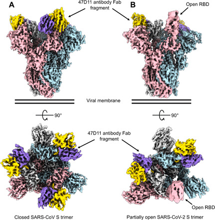
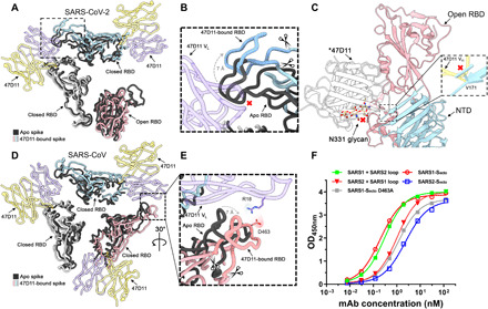
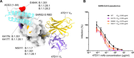
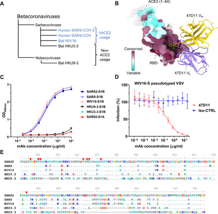
The emergence of SARS-CoV-2 antibody escape mutations highlights the urgent need for broadly neutralizing therapeutics. We previously identified a human monoclonal antibody, 47D11, capable of cross-neutralizing SARS-CoV-2 and SARS-CoV and protecting against the associated respiratory disease in an animal model. Here, we report cryo-EM structures of both trimeric spike ectodomains in complex with the 47D11 Fab. 47D11 binds to the closed receptor-binding domain, distal to the ACE2 binding site. The CDRL3 stabilizes the N343 glycan in an upright conformation, exposing a mutationally constrained hydrophobic pocket, into which the CDRH3 loop inserts two aromatic residues. 47D11 stabilizes a partially open conformation of the SARS-CoV-2 spike, suggesting that it could be used effectively in combination with other antibodies targeting the exposed receptor-binding motif. Together, these results reveal a cross-protective epitope on the SARS-CoV-2 spike and provide a structural roadmap for the development of 47D11 as a prophylactic or postexposure therapy for COVID-19.


Figures

Fig. 1
Fig. 1. 47D11 has differing conformational selectivity for the SARS-CoV and SARS-CoV-2 spike.
(A) Surface rendering of the fully closed SARS spike bound to three 47D11 antibody Fab fragments, shown as two orthogonal views. (B) Surface rendering of the partially open SARS2 spike in complex with two 47D11 antibody Fab fragments, shown as two orthogonal views. The spike protomers are colored pink, blue, and gray, and the 47D11 HC and LC are colored yellow and purple, respectively. For clarity, only the Fab variable region is shown.
Fig. 2
Fig. 2. 47D11 binds specifically to the RBD in down conformation and prevents their full compaction.
(A) Top view of the 47D11-bound SARS2 spike, shown as a ribbon diagram. The 47D11-bound spike protomers are colored pink, blue, and gray, and the 47D11 HC and LC are shown semitransparently and colored yellow and purple, respectively. Glycans and the NTD are omitted for clarity, and only the Fab variable region is shown. The superposed structure of the partially open apo SARS2 spike (PDB ID: 6ZGG) is colored black. (B) Zoomed-in view of the boxed region in (A). The region encompassing residues 470 to 490, used for the loop swap experiments, is indicated with scissors. (C) Zoomed-in view of the SARS2 up RBD and adjacent NTD, shown in cartoon representation. The overlaid 47D11 Fab is shown as a silhouette, and the N331 glycan is shown in ball-and-stick representation and colored tan. The inset shows a zoomed-in view of the clash between the NTD residue V171 and the 47D11 HC. (D) Top view of the 47D11-bound SARS spike colored as shown in (A). The superposed structure of the closed apo SARS spike (PDB ID: 5XLR) is colored black. (E) Zoomed-in view of the boxed region in (D), showing a putative salt bridge between the 47D11 variable LC and the RBD loop. The region encompassing residues 457 to 477, used for the loop swap experiments, is indicated with scissors. (F) ELISA binding curves of 47D11 binding to wild-type, loop-swapped, and D463A spike ectodomains. VH, variable region of immunoglobulin heavy chain; VL, variable region of immunoglobulin light chain; OD450nm, optical density at 450 nm.
Fig. 3
Fig. 3. The 47D11 epitope comprises a mutationally constrained hydrophobic pocket that is normally shielded by glycan N343.
(A) Ribbon diagram of the SARS2-S RBD in complex with the 47D11 antibody Fab fragment. For comparison, residues 1 to 84 of the RBD-bound ACE2 (PDB ID: 6M0J) are shown as a silhouette. (B) Close-up view of the 47D11 epitope with the hydrophobic pocket residues shown as sticks and colored dark blue. The N343 glycan is shown in ball-and-stick representation and colored tan. For clarity, only the core pentasaccharide is shown. (C) Slice through the surface rendered 47D11-bound SARS2-S RBD. The helix encompassing residues 365 to 370 is shown in darker blue. (D) Equivalent view as shown in (C) for the apo RBD (PDB ID: 6VYB). (E) Relative binding of 47D11, (F) ACE2, and (G) CR3022 to cell surface–expressed SARS2-S, determined by fluorescence-activated cell sorting. (H) Relative binding of an anti-FLAG antibody to permeabilized cells expressing the full-length SARS2 spike epitope mutants, determined by fluorescence-activated cell sorting. The data were analyzed by the unpaired, two-tailed Student’s t test using GraphPad Prism 7.0. P < 0.05 was considered significant (*P < 0.05, **P < 0.01, and ***P < 0.0001). (I) Antibody-mediated neutralization of infection of luciferase-encoding VSV particles pseudotyped with wild-type, V367A, or V367F SARS2-S. (J) Surface representation of the 47D11-bound SARS2-S RBD colored according to mean mutation effect on expression (red indicates more constrained) (58). The Fab is shown as a ribbon diagram. (K) As shown in (E) for the S309-bound SARS2 RBD.
Fig. 4
Fig. 4. 47D11 neutralizes SARS2-S pseudoviruses containing single mutations found in the RBM of recently emerged SARS-CoV-2 variants.
(A) Surface representation of the 47D11-bound RBD with the location of the K417N/T, E484K, and N501Y mutations from SARS-CoV-2 variants colored purple, red, and orange, respectively, and annotated (–39). The 47D11 Fab variable chains are shown as a ribbon diagram, and the HC and LC are colored yellow and purple, respectively. Residues 1 to 84 of the RBD-bound ACE2 (PDB ID: 6M0J) are shown for comparison and colored cyan. (B) 47D11-mediated neutralization of infection of luciferase-encoding VSV particles pseudotyped with SARS-CoV-2 S containing single RBM mutations. The average ± SD from two independent experiments with technical triplicates is shown.
Fig. 5
Fig. 5. The 47D11 epitope is conserved in SARS-like viruses.
(A) Phylogenetic tree of SARS-like viruses RBD used to assess 47D11 binding (62). (B) Surface representation of the 47D11-bound RBD colored according to sequence conservation across SARS-CoV, SARS-CoV-2, and 11 SARS-like viruses (fig. S5). The 47D11 Fab variable chains are shown as a ribbon diagram and colored gray. HC residues W102 and F103 are shown as sticks. For comparison, residues 1 to 84 of the RBD-bound ACE2 (PDB ID: 6M0J) are shown as a silhouette. (C) ELISA binding curves of 47D11 to the S1B domain of SARS, SARS2, WIV16, HKU3-3, and HKU9-3. The average ± SD from two independent experiments with technical duplicates is shown. (D) 47D11-mediated neutralization of infection of luciferase-encoding VSV particles pseudotyped with WIV16-S. An anti–Strep-tag human mAb was used as an antibody isotype control. The average ± SD from two independent experiments is shown. (E) Aligned RBD sequences of SARS-CoV-2, SARS-CoV, WIV16, HKU3-3, and HKU9-3. Key residues in the 47D11 epitope are indicated by red arrowheads.

Similar articles

Cited by

References

    1. Zhou P., Yang X.-L., Wang X.-G., Hu B., Zhang L., Zhang W., Si H.-R., Zhu Y., Li B., Huang C.-L., Chen H.-D., Chen J., Luo Y., Guo H., Jiang R.-D., Liu M.-Q., Chen Y., Shen X.-R., Wang X., Zheng X.-S., Zhao K., Chen Q.-J., Deng F., Liu L.-L., Yan B., Zhan F.-X., Wang Y.-Y., Xiao G.-F., Shi Z.-L., A pneumonia outbreak associated with a new coronavirus of probable bat origin. Nature 579, 270–273 (2020). - PMC - PubMed
    1. Ksiazek T. G., Erdman D., Goldsmith C. S., Zaki S. R., Peret T., Emery S., Tong S., Urbani C., Comer J. A., Lim W., Rollin P. E., Dowell S. F., Ling A.-E., Humphrey C. D., Shieh W.-J., Guarner J., Paddock C. D., Rota P., Fields B., DeRisi J., Yang J.-Y., Cox N., Hughes J. M., LeDuc J. W., Bellini W. J., Anderson L. J.; SARS Working Group , A novel coronavirus associated with severe acute respiratory syndrome. N. Engl. J. Med. 348, 1953–1966 (2003). - PubMed
    1. Coronaviridae Study Group of the International Committee on Taxonomy of Viruses , The species Severe acute respiratory syndrome-related coronavirus: Classifying 2019-nCoV and naming it SARS-CoV-2. Nat. Microbiol. 5, 536–544 (2020). - PMC - PubMed
    1. Prabakaran P., Zhu Z., Xiao X., Biragyn A., Dimitrov A. S., Broder C. C., Dimitrov D. S., Potent human monoclonal antibodies against SARS CoV, Nipah and Hendra viruses. Expert Opin. Biol. Ther. 9, 355–368 (2009). - PMC - PubMed
    1. Saphire E. O., Schendel S. L., Gunn B. M., Milligan J. C., Alter G., Antibody-mediated protection against Ebola virus. Nat. Immunol. 19, 1169–1178 (2018). - PMC - PubMed

Publication types

MeSH terms