

Primary elbow osteoarthritis presenting as ulnar nerve palsy with claw hand
- PMID: 33958369
- PMCID: PMC8103949
- DOI: 10.1136/bcr-2021-242773
Primary elbow osteoarthritis presenting as ulnar nerve palsy with claw hand
Abstract
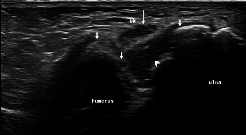
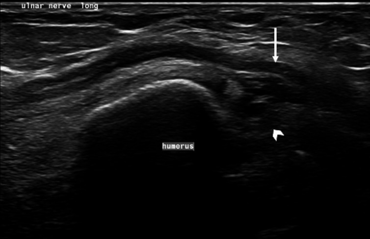
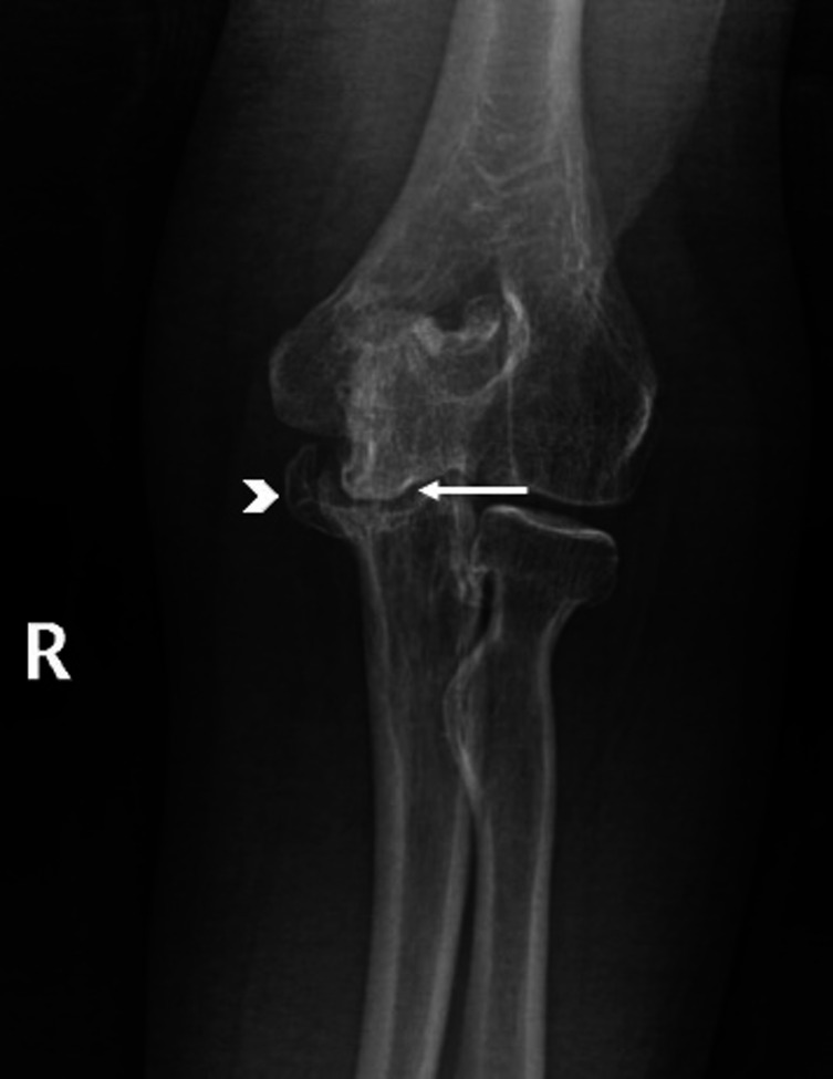
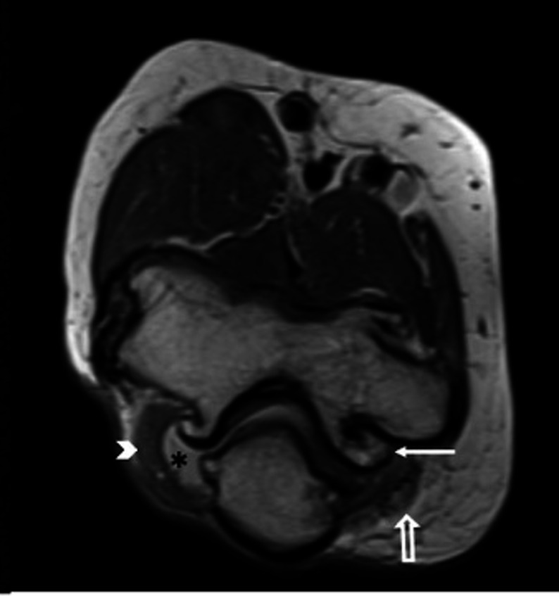
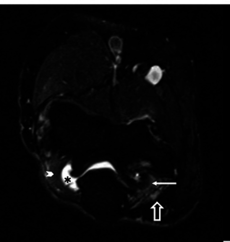
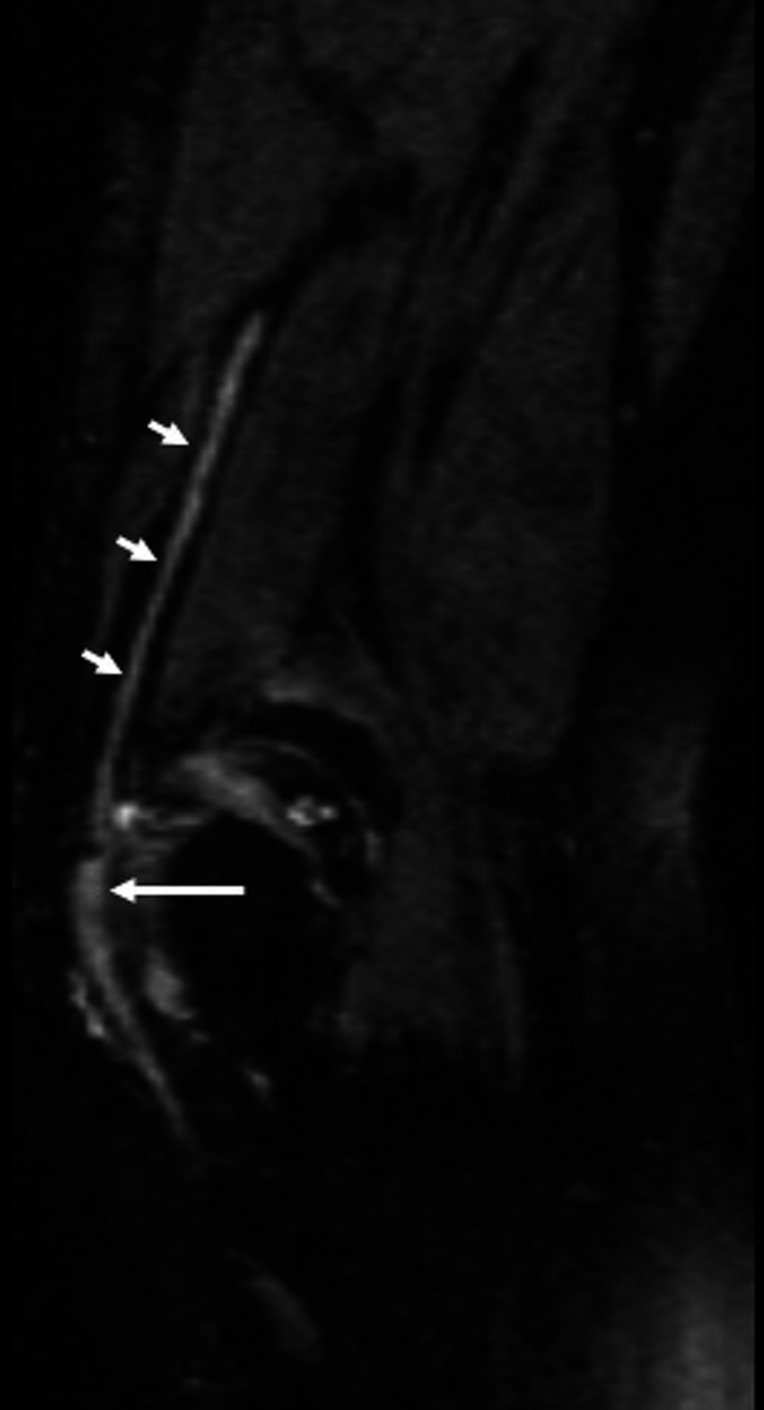
A 59-year-old woman was referred with weakness, paraesthesia, numbness and clawing of the little and ring fingers for the last 2 years. MRI of the cervical spine was normal and nerve conduction velocity revealed abnormality of the ulnar nerve. Ultrasound and MRI showed medial osteophytes and effusion of the elbow joint with stretched and thinned ulnar nerve in the cubital tunnel. The patient underwent release and anterior transposition of the ulnar nerve with significant relief of symptoms.
Keywords: musculoskeletal and joint disorders; peripheral nerve disease; plastic and reconstructive surgery.
© BMJ Publishing Group Limited 2021. No commercial re-use. See rights and permissions. Published by BMJ.
Conflict of interest statement
Competing interests: None declared.
Figures

Similar articles
-
Attrition rupture of ulnar nerve in a patient with rheumatoid elbow arthritis: A case report.Medicine (Baltimore). 2018 Apr;97(17):e0535. doi: 10.1097/MD.0000000000010535. Medicine (Baltimore). 2018. PMID: 29703029 Free PMC article.
-
Quantitative magnetic resonance imaging analysis of the cross-sectional areas of the anconeus epitrochlearis muscle, cubital tunnel, and ulnar nerve with the elbow in extension in patients with and without ulnar neuropathy.J Shoulder Elbow Surg. 2018 Jul;27(7):1306-1310. doi: 10.1016/j.jse.2018.03.021. Epub 2018 May 10. J Shoulder Elbow Surg. 2018. PMID: 29754844
-
Acute ulnar nerve palsy after Outerbridge-Kashiwagi procedure.J Hand Surg Eur Vol. 2007 Oct;32(5):596. doi: 10.1016/J.JHSE.2007.04.021. Epub 2007 Aug 3. J Hand Surg Eur Vol. 2007. PMID: 17950231 No abstract available.
-
[Progress of treatment of cubital tunnel syndrome].Zhongguo Xiu Fu Chong Jian Wai Ke Za Zhi. 2014 Aug;28(8):1043-6. Zhongguo Xiu Fu Chong Jian Wai Ke Za Zhi. 2014. PMID: 25417323 Review. Chinese.
-
Compressive neuropathies of the ulnar nerve at the elbow and wrist.Instr Course Lect. 2000;49:305-17. Instr Course Lect. 2000. PMID: 10829185 Review.
References
Publication types
MeSH terms
LinkOut - more resources
Full Text Sources
Other Literature Sources
Medical