. 2021 Jun 23;41(25):5331-5337.
doi: 10.1523/JNEUROSCI.0340-21.2021. Epub 2021 May 6.

On the Road from Phenotypic Plasticity to Stem Cell Therapy

Affiliations

On the Road from Phenotypic Plasticity to Stem Cell Therapy

Lorraine Iacovitti. J Neurosci. .

Abstract

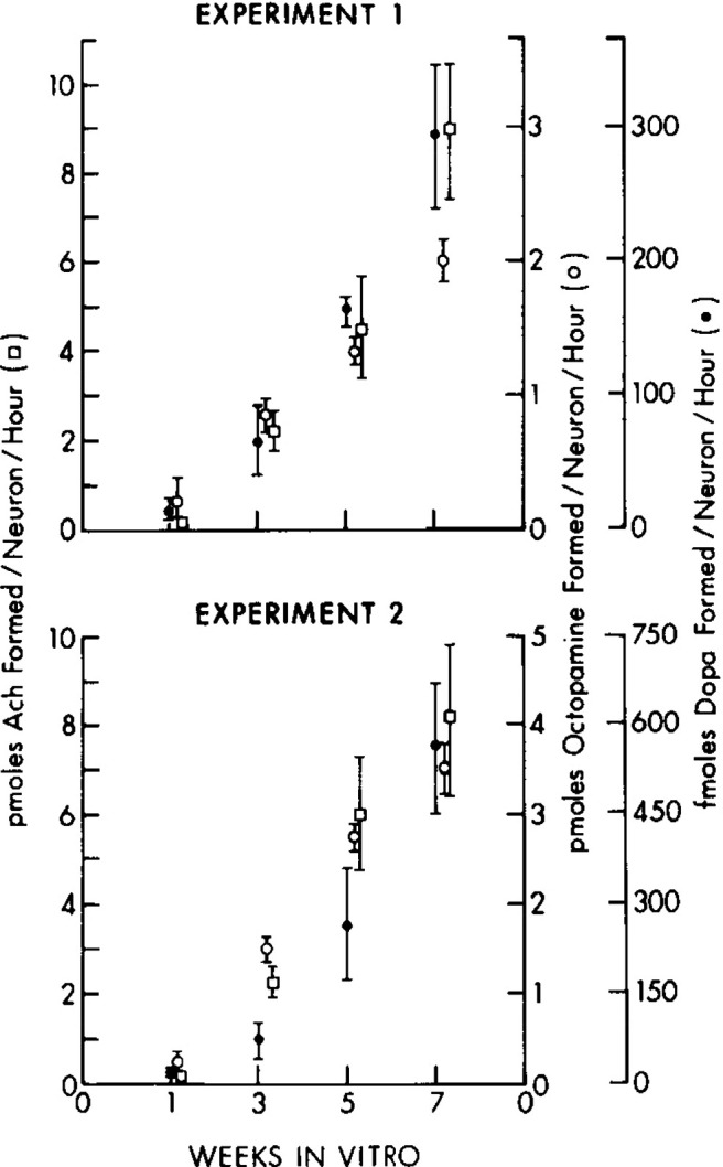
In 1981, I published a paper in the first issue of The Journal of Neuroscience with my postdoctoral mentor, Richard Bunge. At that time, the long-standing belief that each neuron expressed only one neurotransmitter, known as Dale's Principle (Dale, 1935), was being hotly debated following a report by French embryologist Nicole Le Douarin showing that neural crest cells destined for one transmitter phenotype could express characteristics of another if transplanted to alternate sites in the developing embryo (Le Douarin, 1980). In the Bunge laboratory, we were able to more directly test the question of phenotypic plasticity in the controlled environment of the tissue culture dish. Thus, in our paper, we grew autonomic catecholaminergic neurons in culture under conditions which promoted the acquisition of cholinergic traits and showed that cells did not abandon their inherited phenotype to adopt a new one but instead were capable of dual transmitter expression. In this Progressions article, I detail the path that led to these findings and how this study impacted the direction I followed for the next 40 years. This is my journey from phenotypic plasticity to the promise of a stem cell therapy.


Figures

Figure 1.
Figure 1.
Time course of changes in TH, dopamine-β-hydroxylase (DBH), and ChAT activities in cultures of dissociated perinatal rat SCG. Each experiment represents cultures derived from one group of fetuses and maintained in an identical manner. Each point represents the mean + SEM of five preparations. In Experiments 1 and 2, ChAT activity (0) increased in an identical manner, reaching levels of ∼9 pmol of acetylcholine (Ach)/neuron/hr by 7 weeks in culture. While the magnitude of the increases in the activities of TH (0) and DBH (0) remained the same for both experiments, the maximal levels of activity reached at 7 weeks in vitro differed in Experiments 1 and 2. The activity of all three enzymes in both experiments differed significantly between 3 and 7 weeks in vitro. In Experiment 1: for TH, p < 0.05; for DBH, p < 0.001; for ChAT, p < 0.05. In Experiment 2: for TH, p < 0.05; for DBH, p < 0.01; and for CAT, p < 0.001. Reprinted with permission from Iacovitti et al. (1981).
Figure 2.
Figure 2.
A model for the role of post-replicative chromatin for the association of lineage-specific TFs following induction of ESC differentiation. In uninduced ESCs, the repressive histone mark H3K27me3-containing chromatin with a high density of nucleosomes prevents binding of unwanted transcription factors (TFs) (first row). Following induction of differentiation, demethylation of H3K27me3 induced the lysine demethylase UTX leads to a decrease in nucleosome spacing (second row). This “opening” of chromatin facilitates binding of the newly induced lineage-specific TFs (third row). The return of the fast mode of accumulation of H3K27me3 correlates with condensing nucleosome spacing, preventing further association of nonspecific TFs, which may cause changes in lineage commitment (fourth row). Reprinted with permission from Petruk et al. (2017).
Figure 3.
Figure 3.
Induction of expression and association with DNA of lineage-specific transcription factors during midbrain dopamine differentiation of hESCs. A, qRT-PCR gene expression analysis of undifferentiated (0 h, control) hESCs and hESCs induced to the midbrain dopamine lineage for 6 and 12 h. B, Undifferentiated hESCs (top) and hESCs induced to the midbrain dopamine lineage for 6 or 12 h (bottom) were labeled with EdU for 15 min and then chased for 15 min. Chromatin assembly assay (CAA) is a method based on the proximity ligation assay (PLA) that allows examination of proteins associated with nascent DNA following replication in single cells. CAA was performed for LMX1A, FOXA2, and SIP1 followed by immunostaining for biotin (green). PLA only is shown in black and white. Quantification of the results of three independent CAA experiments is also shown (right): x axis, hours in midbrain dopamine differentiation cocktail; y axis, number of PLA signals per nucleus. Reprinted with permission from Petruk et al. (2017).
Figure 4.
Figure 4.
LRRK2 kinase inhibitor CZC54252 rescues LRRK2 iPS-derived dopamine neurons from neurotoxin-induced cell death. A, LRRK2 G2019S iPS-derived dopamine neurons were treated with 5 μm, 10 μm LRRK2 kinase inhibitor CZC54252 (bottom two rows), while DMSO was added to control wells (top row). On the next day, fresh medium containing neurotoxins, 200 nm rotenone (Rot, middle column) or 200 μm 1-methyl-4-phenylpyridinium (MPP+) (right column), were added together with the same doses of CZC54252. Forty-eight hours later, all wells fixed and stained with antibodies to TH (red) and bIII tubulin (green) and counterstained with DAPI nuclear stain (blue). B, In the absence of CZC5252, rotenone or MPP+ treatment caused the death of a large percentage of dopamine neurons (TH+). Both dosages of CZC5252 significantly increased the survival of TH+ cells both in control conditions (reason still unknown) and after treatment with rotenone and MPP+.

Similar articles

References

    1. Arenas E (2002) Stem cells in the treatment of Parkinson's disease. Brain Res Bull 57:795–808. 10.1016/S0361-9230(01)00772-9 - DOI - PubMed
    1. Cai J, Donaldson A, Yang M, German MS, Enikolopov G, Iacovitti L (2009) The role of Lmx1a in the differentiation of human embryonic stem cells into midbrain dopamine neurons in culture and after transplantation into a Parkinson's disease model. Stem Cells 27:220–229. 10.1634/stemcells.2008-0734 - DOI - PubMed
    1. Cai J, Yang M, Poremsky E, Kidd S, Schneider JS, Iacovitti L (2010) Dopaminergic neurons derived from human induced pluripotent stem cells survive and integrate into 6-OHDA-lesioned rats. Stem Cells Dev 19:1017–1023. 10.1089/scd.2009.0319 - DOI - PMC - PubMed
    1. Cai J, Schleidt S, Pelta-Heller J, Hutchings D, Cannarsa G, Iacovitti L (2013) BMP and TGF-β pathway mediators are critical upstream regulators of Wnt signaling during midbrain dopamine differentiation in human pluripotent stem cells. Dev Biol 376:62–73. - PMC - PubMed
    1. Cai J, Kropf E, Hou YM, Iacovitti L (2021) A stress-free strategy to correct point mutations in patient iPS cells. Stem Cell Res 53:102332. 10.1016/j.scr.2021.102332 - DOI - PMC - PubMed

Publication types

LinkOut - more resources