. 2021 May 6;11(1):9682.
doi: 10.1038/s41598-021-88809-0.

Rapid development of neutralizing and diagnostic SARS-COV-2 mouse monoclonal antibodies

Affiliations

Rapid development of neutralizing and diagnostic SARS-COV-2 mouse monoclonal antibodies

Asheley P Chapman et al. Sci Rep. .

Abstract

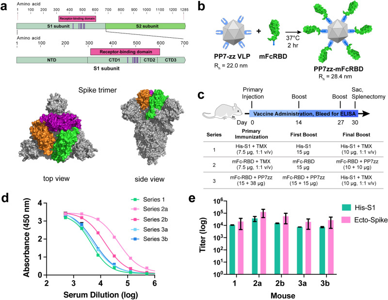
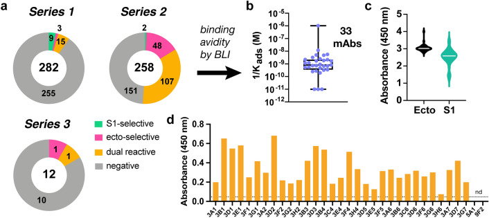
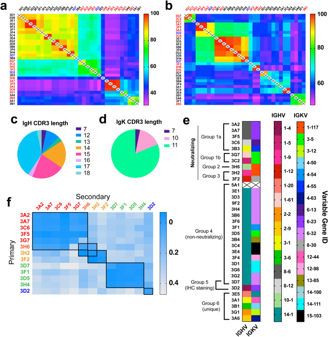
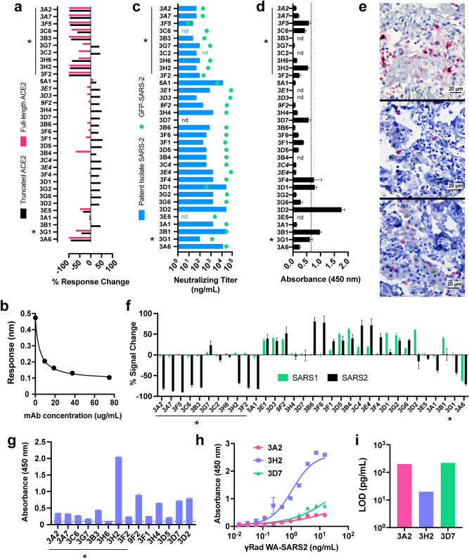
The need for high-affinity, SARS-CoV-2-specific monoclonal antibodies (mAbs) is critical in the face of the global COVID-19 pandemic, as such reagents can have important diagnostic, research, and therapeutic applications. Of greatest interest is the ~ 300 amino acid receptor binding domain (RBD) within the S1 subunit of the spike protein because of its key interaction with the human angiotensin converting enzyme 2 (hACE2) receptor present on many cell types, especially lung epithelial cells. We report here the development and functional characterization of 29 nM-affinity mouse SARS-CoV-2 mAbs created by an accelerated immunization and hybridoma screening process. Differing functions, including binding of diverse protein epitopes, viral neutralization, impact on RBD-hACE2 binding, and immunohistochemical staining of infected lung tissue, were correlated with variable gene usage and sequence.


Conflict of interest statement

The authors declare no competing interests.

Figures

Figure 1
Figure 1
SARS-CoV-2 Spike protein subunit vaccine strategy and humoral immune response in mice. (a) Recombinant spike subunit 1 protein (His-S1, residues 1–681) or S1 Receptor Binding Domain (mouse Fc-RBD, residues 319–541, ACE2 contact residues in purple; PDBID: 6vxx) antigens. (b) VLP display of Fc-tagged antigens using the PP7 particle bearing 120 ZZ-domains; a 1:1 mass ratio of mFc-RBD and VLP provides a Fc:ZZ molar ratio of approximately 0.8. Rh = hydrodynamic radius measured by dynamic light scattering in phosphate buffer. (c) Vaccine schedule and strategy. Six-week old female BALB/c mice (n = 3 per group) were immunized with primary antigen and adjuvant on day 0 followed by boosts on days 14 and 27. Blood was collected for ELISA on days 0, 14, 21 and 30. (d) ELISA responses for serum dilutions against plated His-S1 protein from the sacrificed mice at day 30. (e) Titer values from ELISA analysis as in panel (d), against plated His-S1 or spike ectodomain protein. Immunization series defined in panel (c); (a) and (b) designate different mice within that series. Experimental error represents standard deviation.
Figure 2
Figure 2
Characterization of anti-SARS-COV-2 antibodies as supernatants from hybridoma clones. (a) Antigen specificity of antibody binding from IgG-secreting hybridomas chosen from Series 1–3 immunization schedules. Numbers inside circles are total clones selected, numbers within each portion reflect binding selectivity. (b) Distribution of affinities of the 33 selected clones as measured by BLI. (c) Binding to two different Spike recombinant proteins (ecto Spike trimer and S1 polypeptide) as determined by ELISA. (d) Binding of antibodies to γ-irradiated WA-SARS-CoV-2 by ELISA (15 ng/mL virus plated, supernatant undiluted, secondary antibody = goat anti-mouse IgG HRP conjugate).
Figure 3
Figure 3
SARS-CoV-2 mAb sequences and epitope binning. (a,b) Overall similarity of (a) heavy chain and (b) light chain anti-SARS-CoV-2 antibody sequences, scored by percent of the total number of identical amino acids at each position. Antibody sequences were analyzed via MiXCR software and sequence alignment performed by Geneious. Neutralizing (red) and IHC staining mAbs (blue) determined via functional assays (Fig. 4) (c,d) Distribution of CDR3 amino acid length in heavy (c) and light (d) chains of 32 SARS-COV-2 mAbs. (e) IGHV and IGKV genes identified for mAbs from sequences in panels (a) and (b). mAb 5A1 was not sequenced. Antibody grouping includes sequence, epitope, and function-level characteristics. (f) Representative assessment of competitive mAb binding to recombinant ectodomain immobilized to streptavidin-coated BLI sensor chips via Strep-tag sequence. Binding to the “primary” antibody was validated by BLI, followed by washing and exposure to the secondary antibody to generate the binding signal of interest.
Figure 4
Figure 4
Functional utility of SARS-CoV-2 mAbs. (a) Assessment of inhibition of S1 RBD binding to human ACE2 receptor as measured by BLI, performed at a single antibody concentration. Both full-length ACE2 (residues #1-740, pink bars) and truncated ACE2 (residues #1-614, black bars) were included. Data represents percent of signal change normalized to response of ACE2/RBD binding with no mAb present, error bars represent standard deviation of technical replicates. Neutralizing antibodies in both assays are denoted by asterisks; adjacent mAbs in italics are identical. (b) Representative (mAb 3G7) dose-dependent antibody-mediated inhibition of ecto-Spike binding to full-length human ACE2 receptor; readout is the wavelength shift in nm in the BLI interferometric response,. (c) In vitro mAb neutralization of SARS-CoV-2 USA/WA1/2020 virus (blue bars) or of GFP-expressing SARS-CoV-2 (green dots). Asterisks mark particularly effective entries. (d) Binding of antibodies to biotinylated peptide (amino acids 486–501 with C488S substitution; containing four out of five reported RBD-hACE2 contacts) plated on streptavidin-coated plates as measured by ELISA. Dashed line represents the average plus twice the standard deviation of the values obtained for replicates involving three identical mAbs (3A2, 3A7, 3F5); error bars = standard deviation. (e) Immunohistochemical staining of lung tissue from an early confirmed SARS-CoV-2 fatal case by (top) known SARS polyclonal nucleocapsid antibody, (middle) mAbs 3D2, and (bottom) 3D7 (1:100 mAb dilution, antigen detection in red). (f) Specificity of mAbs determined by split nanoluciferase assay including either SARS1 or SARS2 RBD incubated with mAb followed by ACE2, normalized to ACE2/RBD signal without antibody. Negative values denote inhibition, positive indicate binding enhancement observed only with truncated ACE2. (g) ELISA detection of mAb binding to gamma-inactivated SARS-COV-2 (1 µg/mL TCID50 units plated); dotted line denotes background signal. (h) Dose-dependent gamma-inactivated SARS-COV-2 binding of select mAbs (5 µg/mL mAb) measured by ELISA, sigmoidal dose–response curve fitted by GraphPad Prism8. (i) Limit of detection quantification of mAbs in panel g. LOD calculated as the minimum virus concentration giving rise to a signal equivalent to the mean background signal plus 3 experimental standard deviations (GraphPad Prism 8).

References

    1. Shang J, et al. Structural basis of receptor recognition by SARS-CoV-2. Nature. 2020;581:221–224. doi: 10.1038/s41586-020-2179-y. - DOI - PMC - PubMed
    1. Wan Y, Shang J, Graham R, Baric RS, Li F. Receptor recognition by novel coronavirus from Wuhan: An analysis based on decade-long structural studies of SARS. J. Virol. 2020;94:e00127-00120. - PMC - PubMed
    1. Hamming I, et al. Tissue distribution of ACE2 protein, the functional receptor for SARS coronavirus. A first step in understanding SARS pathogenesis. J. Pathol. 2004;203:631–637. doi: 10.1002/path.1570. - DOI - PMC - PubMed
    1. Wu F, et al. A new coronavirus associated with human respiratory disease in China. Nature. 2020;579:265–269. doi: 10.1038/s41586-020-2008-3. - DOI - PMC - PubMed
    1. Zhao L, Kopylov M, Potter CS, Carragher B, Finn MG. Engineering the PP7 virus capsid as a peptide display platform. ACS Nano. 2019;13:4443–4454. doi: 10.1021/acsnano.8b09683. - DOI - PMC - PubMed

Publication types