. 2021 May 6;11(1):9686.
doi: 10.1038/s41598-021-88956-4.

IgE-activated mast cells enhance TLR4-mediated antigen-specific CD4+ T cell responses

Affiliations

IgE-activated mast cells enhance TLR4-mediated antigen-specific CD4+ T cell responses

Binh L Phong et al. Sci Rep. .

Abstract

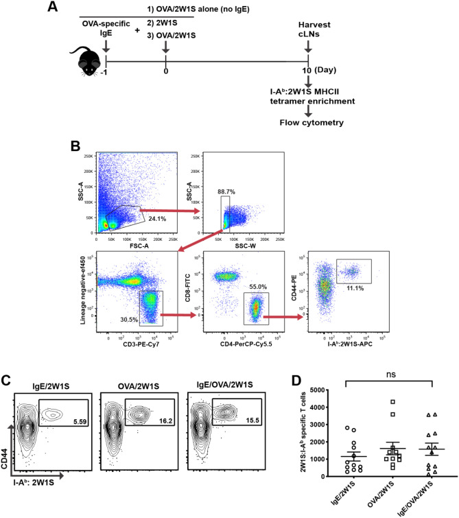
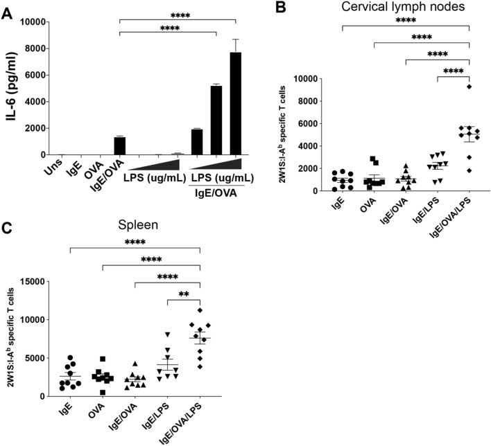
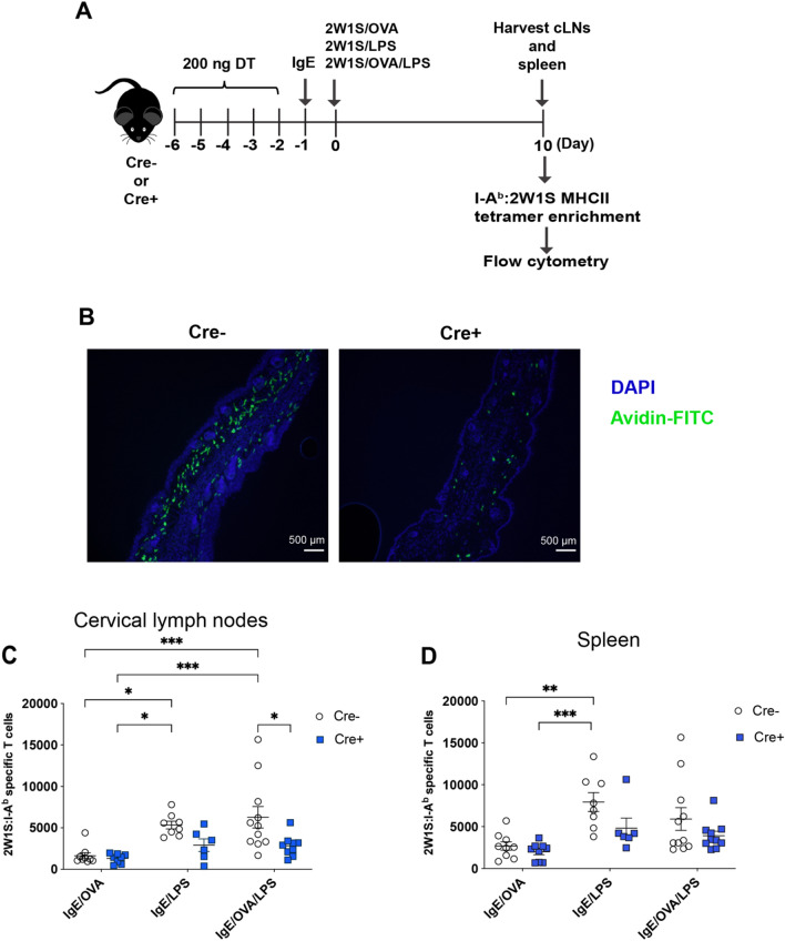
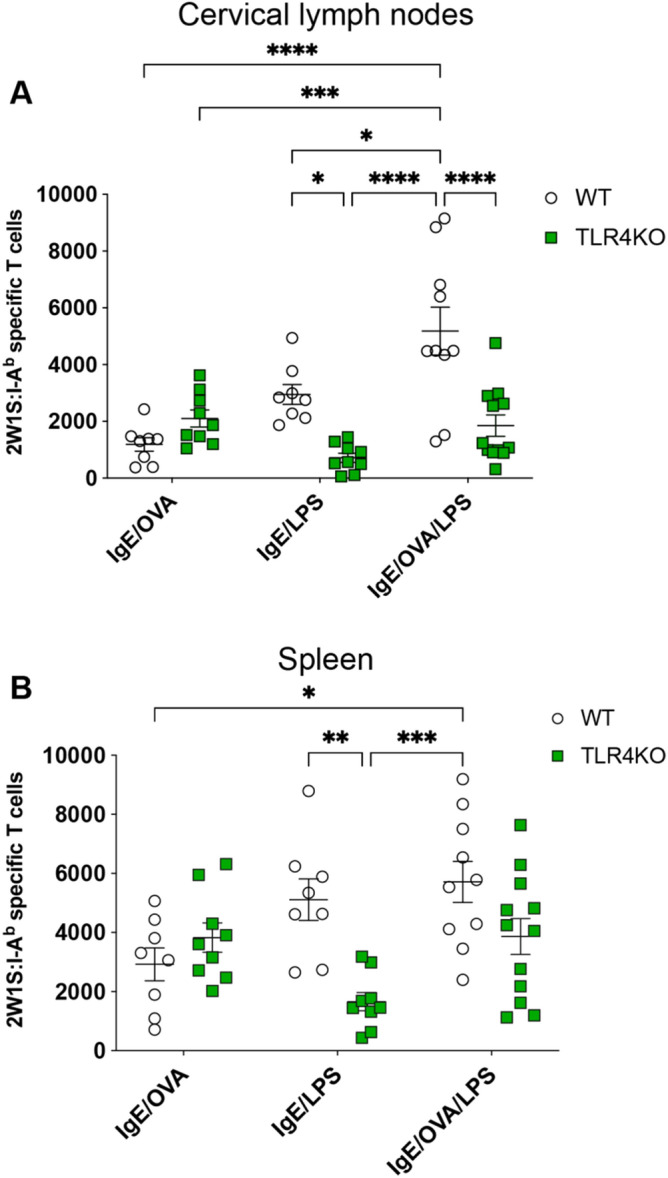
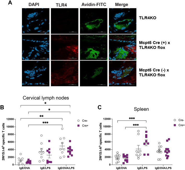
Mast cells are potent mediators of allergy and asthma, yet their role in regulating adaptive immunity remains ambiguous. On the surface of mast cells, the crosslinking of IgE bound to FcεRI by a specific antigen recognized by that IgE triggers the release of immune mediators such as histamine and cytokines capable of activating other immune cells; however, little is known about the mast cell contribution to the induction of endogenous, antigen-specific CD4+ T cells. Here we examined the effects of specific mast cell activation in vivo on the initiation of an antigen-specific CD4+ T cell response. While CD4+ T cells were not enhanced by FcεRI stimulation alone, their activation was synergistically enhanced when FcεRI activation was combined with TLR4 stimulation. This enhanced activation was dependent on global TLR4 stimulation but appeared to be less dependent on mast cell expressed TLR4. This study provides important new evidence to support the role of mast cells as mediators of the antigen-specific adaptive immune response.


Conflict of interest statement

The authors declare no competing interests.

Figures

Figure 1
Figure 1
IgE-mediated mast cell activation does not result in increased numbers of endogenous antigen-specific CD4+ T cells. (A) Strategy for intradermal ear immunization to enumerate 2W1S-specific CD4+ T cells upon IgE/OVA-induced mast cell activation. (B) Gating strategy and representative flow plots of antigen-specific CD4 + T cells in the cervical draining lymph nodes (CLNs) 7 days post immunization. 2W1S-specific CD4 + T cells post enrichment were identified as lymphocytes by forward and side scatter properties and then doublets were excluded based on side scatter width (SSC-W). Singlets were stained for T cells by eliminating T cell lineage negative cells (CD11b-, CD11c-, F4/80-, CD19-), and gating on CD3ε + cell. Helper T cells were gated as CD4 + CD8α- cells. Finally, antigen experienced 2W1S specific cells were gated as CD44hi and I-Ab :2W1S-APC + . Percentages in each gate are indicated. (C) Representative flow plot of 2W1S-specific CD4+ T cells in the cervical draining lymph nodes (CLNs) 7 days post immunization for each group is shown. Percentage of 2W1S-specific CD4+ T cells post tetramer enrichment was indicated. (C) The total number of 2W1S-specific CD4+ T cells in the CLNs 7 days post immunization. Results are shown as mean ± SEM from 4 independent experiments n = 11–12 mice per group. Statistical analysis was performed using a Kruskal–Wallis test with Dunn’s correction.
Figure 2
Figure 2
IgE/OVA and LPS co-exposure leads to expanded antigen-specific CD4+ T cell population. (A) Bone marrow-derived mast cells (BMMCs) from C57BL/6 J mice sensitized overnight with 1 μg/mL anti-OVA IgE were stimulated with 10 μg/ml OVA alone or in combination with 0.1, 1, 10 μg/ml LPS. Unsensitized BMMCs were stimulated with 0.1, 1, 10 μg/ml LPS alone. IL-6 cytokine release from supernatants was assayed by ELISA 6 h post stimulation. (B,C) C57BL/6 J mice received intradermal ear injection of 2W1S peptide and either LPS alone or IgE/OVA. The numbers of 2W1S-specific CD4+ T cells in the cervical draining lymph nodes (B) and spleens (C) were quantified 10 days post immunization. Results are representative of 2 independent experiments (A) and cumulative data from 3 independent experiments with n = 9 mice total (B,C) and shown as mean ± SEM. *P < 0.05 **P < 0.01, **P < 0.001, **** P < 0.0001. Statistical analysis was performed using one-way ANOVA with Tukey’s multiple comparisons test.
Figure 3
Figure 3
Mast cells are required for expansion of antigen-specific CD4+ T cells upon IgE mediated activation and LPS exposure. (A) Mast cell depletion and intradermal ear immunization strategy to quantify the number of 2W1S-specific CD4+ T cells in Mcpt5-Cre + (Cre +) mice and littermates (Cre −) upon 2W1S peptide, IgE/OVA, and LPS stimulation. (B) Immunofluorescent images of mast cells visualized by Avidin-FITC in ear tissue sections from Cre + and Cre − mice to show mast cell reduction in Cre + mice one day post last DT injection taken at 10 × objective. The numbers of 2W1S-specific CD4+ T cells within the cervical draining lymph nodes (C) and spleen (D) of mast cell-depleted (Cre +) mice and littermate controls (Cre −) 10 days post intradermal ear immunization. Results are cumulative data from 3 independent experiments n = 6–11 mice total and shown as mean ± SEM. *P < 0.05 **P < 0.01, **P < 0.001, **** P < 0.0001. Statistical analysis was performed using one-way ANOVA with Tukey’s multiple comparisons test (C) or a Kruskal–Wallis test with Dunn’s correction (D). Scale bar, 500 µm.
Figure 4
Figure 4
TLR4 activation by LPS is required for enhanced antigen-specific CD4+ T cell response upon IgE/OVA-induced mast cell activation. (A,B) WT or TLR4-deficient mice were immunized with 2W1S peptide, IgE/OVA, LPS, or a combination of IgE/OVA and LPS. Cervical draining lymph nodes (A) and spleens (B) were analyzed 10 days post immunization for the numbers of 2W1S-specific CD4+ T cells. Results shown are cumulative data from 3 independent experiments n = 8–12 mice and shown as mean ± SEM. *P < 0.05 **P < 0.01, **P < 0.001, **** P < 0.0001. Statistical analysis was performed using one-way ANOVA with Tukey’s multiple comparisons test.
Figure 5
Figure 5
TLR4 expression on mast cells is not entirely responsible for the IgE/OVA- and LPS-mediated expansion of antigen-specific CD4 + T cells. (A) Tissue ear sections from TLR4-deficient mice (top), Mcpt5 Cre ( −) x Tlr4KO flox (middle), and Mcpt Cre ( +) x Tlr4KO flox (bottom) were imaged using confocal microscopy (see “Materials and methods” section). Red (PE) indicates TLR4 stain, green (FITC) indicates MC-granule-specific avidin stain, and DAPI (blue) indicates cell nuclei. Image shows that Cre ( +) MCs lack TLR4 expression in comparison to Cre ( −) MCs. (B,C) Mast cell-specific TLR4-deficient mice (Cre +) and littermate controls (Cre −) were immunized intradermally in the ears with 2W1S peptide and the indicated combination of IgE, OVA, and LPS. Cervical draining lymph nodes (B) and spleens (C) were analyzed 10 days later for the numbers of 2W1S-specific CD4+ T cells. Results are cumulative data from 3 independent experiments with n = 7–10 mice total and shown as mean ± SEM. Scale bar, 25 µm. *P < 0.05 **P < 0.01, ***P < 0.001, **** P < 0.0001. Statistical analysis was performed using one-way ANOVA with Tukey’s multiple comparisons test.

Similar articles

Cited by

References

    1. Méndez-Enríquez E, Hallgren J. Mast cells and their progenitors in allergic asthma. Front. Immunol. 2019;10:821. doi: 10.3389/fimmu.2019.00821. - DOI - PMC - PubMed
    1. Theoharides TC, Kalogeromitros D. The critical role of mast cells in allergy and inflammation. Ann. Ny. Acad. Sci. 2006;1088:78–99. doi: 10.1196/annals.1366.025. - DOI - PubMed
    1. Agier J, Pastwińska J, Brzezińska-Błaszczyk E. An overview of mast cell pattern recognition receptors. Inflamm. Res. 2018;67:737–746. doi: 10.1007/s00011-018-1164-5. - DOI - PMC - PubMed
    1. Marshall JS. Mast-cell responses to pathogens. Nat. Rev. Immunol. 2004;4:787–799. doi: 10.1038/nri1460. - DOI - PubMed
    1. Malaviya R, Ikeda T, Ross E, Abraham SN. Mast cell modulation of neutrophil influx and bacterial clearance at sites of infection through TNF-alpha. Nature. 1996;381:77–80. doi: 10.1038/381077a0. - DOI - PubMed

Publication types

MeSH terms

LinkOut - more resources