. 2021 May 6;11(1):9666.
doi: 10.1038/s41598-021-88508-w.

Steroid hormone bioavailability is controlled by the lymphatic system

Affiliations

Steroid hormone bioavailability is controlled by the lymphatic system

Rahel Klossner et al. Sci Rep. .

Abstract

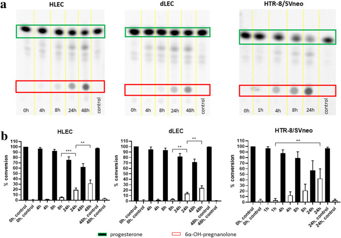
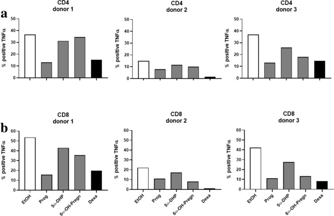
The steroid hormone progesterone accounts for immune tolerance in pregnancy. Enhanced progesterone metabolism to 6α-OH-pregnanolone occurs in complicated pregnancies such as in preeclampsia with preterm delivery or intrauterine growth restriction, and in cancer. As lymphatic endothelial cells (LECs) promote tumor immunity, we hypothesized that human LECs modify progesterone bioavailability. Primary human LECs and mice lymph nodes were incubated with progesterone and progesterone metabolism was analyzed by thin layer chromatography and liquid chromatography-mass spectrometry. Expression of steroidogenic enzymes, down-stream signal and steroid hormone receptors was assessed by Real-time PCR. The placental cell line HTR-8/SV neo was used as reference. The impact of the progesterone metabolites of interest was investigated on the immune system by fluorescence-activated cell sorting analysis. LECs metabolize progesterone to 6α-OH-pregnanolone and reactivate progesterone from a precursor. LECs highly express 17β-hydroxysteroid dehydrogenase 2 and are therefore antiandrogenic and antiestrogenic. LECs express several steroid hormone receptors and PIBF1. Progesterone and its metabolites reduced TNF-α and IFN-γ production in CD4+ and CD8+ T cells. LECs modify progesterone bioavailability and are a target of steroid hormones. Given the global area represented by LECs, they might have a critical immunomodulatory control in pregnancy and cancer.


Conflict of interest statement

The authors declare no competing interests.

Figures

Figure 1
Figure 1
Characterization of progesterone metabolism in HLEC, dLEC and HTR8/SV neo by TLC. Metabolism of progesterone in HLEC, dLEC and HTR-8/SV neo. (a) Characteristic phosphorimager pictures of time-dependent conversion of progesterone to a main metabolite, 6α-OH-pregnanolone. Cell-free controls were run for time point 0 h (HLEC, dLEC and HTR-8/SV neo), for time point 24 h (HTR-8/SVneo) and for time point 48 h (HLEC and dLEC). (b) Densitometric quantification of all performed experiments. Progesterone was time-dependently and significantly converted to 6α-OH-pregnanolone in all three cell lines. Data were normalized to condition 0 h = 100% progesterone. One-way ANOVA, Dunnett’s multiple comparisons test, n = 5. Production of 6α-OH-pregnanolone: HLEC: 8 h-24 h ***p < 0.0001, 24 h-48 h **p = 0.0002. dLEC: 8 h-24 h ** p = 0.0026, 24 h-48 h ** p = 0.0020. HTR-8/SV neo: 1 h-24 h ** p = 0.0002. Green rectangle/black column: substrate (progesterone). Red rectangle/white column: product (6α-OH-pregnanolone). * p < 0.05, ** p < 0.01, *** p < 0.0001, ns = not significant.
Figure 2
Figure 2
Identification of 6α-OH-pregnanolone by LC–MS/MS in HTR-8/SV neo cells: LC–MS trace of m/z 317.2469 corresponding to a sum formula of C21H34O3 in an authentic standard (a) and in HTR-8/SV neo cells (b). MS/MS fragment spectra recorded at LC–MS peak maximum for an authentic standard (c) and in HTR-8/SVneo cells (d).
Figure 3
Figure 3
Identification of 6α-OH-pregnanolone by LC–MS/MS in HLEC and dLEC cells. LC–MS trace of m/z 317.2469 corresponding to a sum formula of C21H34O3 in an authentic standard (a), in HLEC cells (b) and in dLEC cells (c). MS/MS fragment spectra recorded at LC–MS peak maximum in an authentic standard (d), in HLEC cells (e) and in dLEC cells (f).
Figure 4
Figure 4
Putative progesterone metabolism pathway in HLECs, dLECs and HTR-8/SV neo. Based on our mRNA, proteomics, TLC and LC–MS data, we propose the following pathway in green to take place. The pathway with the bulky arrows down to 6α-OH-pregnanolone is favored over the pathway to the 20α-hydroxyprogesterone in HLECs, dLECs and HTR-8/SV neo. HLEC and dLEC highly express HSD17B2 and efficiently back-convert 20α-hydroxyprogesterone to progesterone.
Figure 5
Figure 5
Inhibitory effect of dutasteride on 5α-reductase (SRD5A1-3) in HLECs, dLECs and HTR-8/SV neo. Characteristic phorphorimager pictures of (a) HLEC, (b) dLEC, (c) HTR-8/SV neo and their densitometry (d) of three independent experiments. Cells were cultured without (lane 1) and with (lane 2 and 3) the indicated concentration of dutasteride and 14C-progesterone. Lane 4 is the cell-free control, where 14C-progesterone was incubated for 24 h without cells. HLEC and HTR-8/SV neo: (1) no dutasteride = DMSO control, (2) dutasteride 10−5 M, (3) dutasteride 10−6 M, (4) no cells; dLEC: (1) no dutasteride, (2) dutasteride 10−6 M, (3) dutasteride 10−8 M, (4) no cells. Progesterone was significantly metabolized to 6α-OH-pregnanolone in the DMSO control, while dutasteride significantly inhibited the conversion of progesterone to 6α-OH-pregnanolone in all three cell lines. Data were normalized to condition 0 h = 100% progesterone. One-way ANOVA, Dunnett’s multiple comparisons test, n = 3. HLEC: DMSO control *** p < 0.0001; dutasteride 10−5 M *** p < 0.0001; dutasteride 10−6 M *** p < 0.0001. dLEC: DMSO control *** p < 0.0001; dutasteride 10−6 M ** p = 0.0001; dutasteride 10−8 M ** p = 0.0006. HTR-8/SV neo: DMSO control *** p < 0.0001; dutasteride 10−5 M ** p = 0.0005; dutasteride 10−6 M ** p = 0.0006. Green rectangle/black column: substrate (progesterone). Red rectangle/white column: product (6α-OH-pregnanolone). * p < 0.05, ** p < 0.01, *** p < 0.0001, ns = not significant.
Figure 6
Figure 6
Activity of HSD17B2 in HLECs. Time-dependent conversion of testosterone to androstenedione, 17β-estradiol to estrone, androstenediol to DHEA-S and DHEA as well as 20α-hydroxyprogesterone to progesterone was assessed by LC–MS. Data were normalized to condition at 0 h = 1000 nM = 10−6 M for each compound. Y-axis shows steroid hormone concentrations in nM. Testosterone was significantly converted to androstenedione (testosterone 0 h/4 h/24 h: *** p < 0.0001/** p = 0.0019; androstenedione 0 h/4 h/24 h: *** p < 0.0001/ns). 17β-estradiol was significantly converted to estrone (17β-estradiol 0 h/4 h/24 h: *** p < 0.0001/ns; estrone 0 h/4 h/24 h: *** p < 0.0001/* p = 0.01). Androstenediol was significantly converted to DHEA (androstenediol 0 h/4 h/24 h: ns/*** p < 0.0001; DHEA 0 h/4 h/24 h: ** p = 0.0087/** p = 0.0002). 20α-OHP was significantly converted to progesterone (20α-OHP 0 h/4 h/24 h: *** p < 0.0001/*** p < 0.0001; progesterone 0 h/4 h/24 h: *** p < 0.0001/*** p < 0.0001). One-way ANOVA, Dunnett’s multiple comparisons test, n = 3. Black rectangle/column: substrate. White rectangle/column: product. * p < 0.05, ** p < 0.01, *** p < 0.0001, ns = not significant.
Figure 7
Figure 7
Progesterone metabolism in male mice lymph nodes and adrenal gland. Phosphorimager picture of TLC showing time-dependent conversion of progesterone to down-stream metabolites in lymph nodes. For positive control, adrenal glands are used under similar experimental conditions. Tissue-free controls were run for both time points. Line 1 at timepoint 24 h and line 2 at 48 h were entire lymph nodes, while all other lymph nodes were cut in half. The figure shows results of 7 isolated lymph nodes. Each lymph node was from a separate mouse. * p < 0.05, ** p < 0.01, *** p < 0.0001, ns = not significant.
Figure 8
Figure 8
TNF-α production in CD4+ and CD8+ T cells upon stimulation with progesterone metabolites. Progesterone (Prog), 5α-dihydroprogesterone (5α-DHP), 6α-hydroxypregnanolone (6α-OH-Pregn) and dexamethasone (Dexa), were added at a concentration of 10−3 M to the PBMCs obtained from 3 different female donors for 24 h. 6 h after first steroid hormone contact, PBMCs were activated with PMA/Ionomycin. TNF-α positive CD4+ and CD8+ T cells were counted by FACS analysis. The y-axis shows the % of CD4+ (a) and CD8+ (b) T cells positively staining for TNF-α. * p < 0.05, ** p < 0.01, *** p < 0.0001, ns = not significant.
Figure 9
Figure 9
IFN-γ production in CD4+ and CD8+ T cells upon stimulation with progesterone metabolites. Progesterone (Prog), 5α-dihydroprogesterone (5α-DHP), 6α-hydroxypregnanolone (6α-OH-Pregn) and dexamethasone (Dexa), were added at a concentration of 10−3 M to the PBMCs of 3 different female donors for 24 h. 6 h after first steroid hormone contact, PBMCs were activated with PMA/Ionomycin. IFN-γ positive CD4+ and CD8+ T cells were counted by FACS analysis. The y-axis shows the % of CD4+ (a) and CD8+ (b) T cells positively staining for IFN-γ. * p < 0.05, ** p < 0.01, *** p < 0.0001, ns = not significant.

Similar articles

Cited by

References

    1. Swartz MA, Lund AW. Lymphatic and interstitial flow in the tumour microenvironment: linking mechanobiology with immunity. Nat. Rev. Cancer. 2012;12(3):210–219. doi: 10.1038/nrc3186. - DOI - PubMed
    1. Lund AW, Duraes FV, Hirosue S, et al. VEGF-C promotes immune tolerance in B16 melanomas and cross-presentation of tumor antigen by lymph node lymphatics. Cell Rep. 2012;1(3):191–199. doi: 10.1016/j.celrep.2012.01.005. - DOI - PubMed
    1. Cohen JN, Guidi CJ, Tewalt EF, et al. Lymph node-resident lymphatic endothelial cells mediate peripheral tolerance via Aire-independent direct antigen presentation. J. Exp. Med. 2010;207(4):681–688. doi: 10.1084/jem.20092465. - DOI - PMC - PubMed
    1. Tewalt EF, Cohen JN, Rouhani SJ, et al. Lymphatic endothelial cells induce tolerance via PD-L1 and lack of costimulation leading to high-level PD-1 expression on CD8 T cells. Blood. 2012;120(24):4772–4782. doi: 10.1182/blood-2012-04-427013. - DOI - PMC - PubMed
    1. Hirosue S, Vokali E, Raghavan VR, et al. Steady-state antigen scavenging, cross-presentation, and CD8+ T cell priming: a new role for lymphatic endothelial cells. J. Immunol. 2014;192(11):5002–5011. doi: 10.4049/jimmunol.1302492. - DOI - PMC - PubMed

Publication types

MeSH terms