. 2021 May 6;11(1):9738.
doi: 10.1038/s41598-021-89121-7.

Repetitive mild head trauma induces activity mediated lifelong brain deficits in a novel Drosophila model

Affiliations

Repetitive mild head trauma induces activity mediated lifelong brain deficits in a novel Drosophila model

Joseph A Behnke et al. Sci Rep. .

Abstract

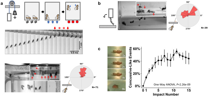
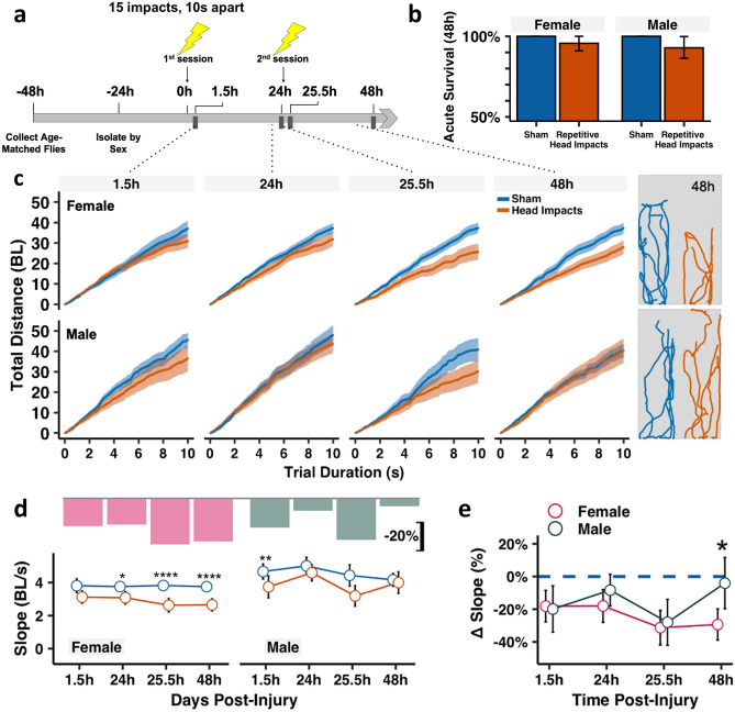
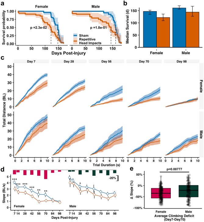
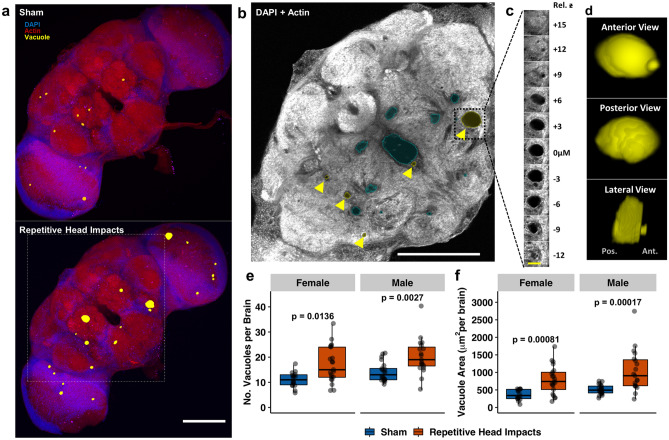
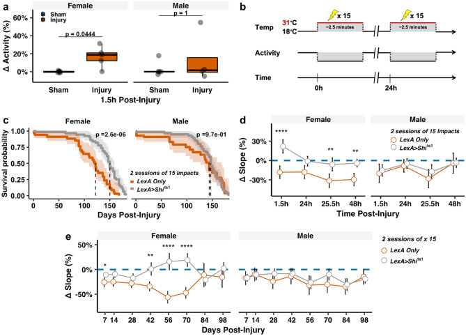
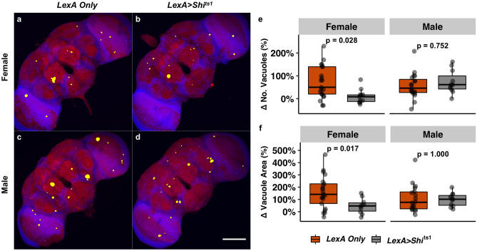
Mild head trauma, including concussion, can lead to chronic brain dysfunction and degeneration but the underlying mechanisms remain poorly understood. Here, we developed a novel head impact system to investigate the long-term effects of mild head trauma on brain structure and function, as well as the underlying mechanisms in Drosophila melanogaster. We find that Drosophila subjected to repetitive head impacts develop long-term deficits, including impaired startle-induced climbing, progressive brain degeneration, and shortened lifespan, all of which are substantially exacerbated in female flies. Interestingly, head impacts elicit an elevation in neuronal activity and its acute suppression abrogates the detrimental effects in female flies. Together, our findings validate Drosophila as a suitable model system for investigating the long-term effects of mild head trauma, suggest an increased vulnerability to brain injury in female flies, and indicate that early altered neuronal excitability may be a key mechanism linking mild brain trauma to chronic degeneration.


Conflict of interest statement

The authors declare no competing interests.

Figures

Figure 1
Figure 1
Development of a novel repetitive head impact Drosophila model. (a) (Top) Diagram of our novel headfirst impact model that utilizes a counterweight pulley system to accelerate a vial of flies upward ultimately resulting in acceleration-deceleration headfirst impacts. To prime proper body orientation, the vial is lightly tamped down, resulting in the natural negative geotactic upwards orientation, after which the vial is release, causing the counterweight to accelerate the vial unidirectionally. The unidirectional movement upwards, together with the priming sequences facilitates headfirst orientation at impact once the vial reaches its maximum height. (Middle) High-speed image sequence showing headfirst movement through impact with the surface of the injury vial (arrows indicate frames immediately before and after contact, which is denoted by red stars, which lasts ~ 1 ms long). (Bottom) Angular histogram comparisons demonstrate more reliable and consistent headfirst impacts and improves upon (b) previously existing strategies which encourage rotational body movement that results in inconsistent head orientation at impact. (c) Immediately following headfirst impacts, flies sustain acute signs of neurological injury, such as temporary loss of consciousness that becomes more prevalent with increasing number of repetitive head impacts. Impact number increases concussive-like events (one-way ANOVA, F (1,89) = 44.26645, P = 2.26e−09). Data represented as mean ± SD, n = 4–8 vials (10 flies per vial) of both male and female flies.
Figure 2
Figure 2
Acute recovery of climbing deficits following minimally lethal repetitive head impacts is sexually dimorphic. (a) Injury timeline schematic showing that flies are subjected to two sessions of repetitive head impacts (24 h apart). Each session consists of 15 iterative impacts spaced 10 s apart. Behavior and mortality are longitudinally monitored throughout life. (b) Bar graph showing acute survival following repetitive impacts with 95% confidence intervals. n = 56–69 flies per injured group/sex and n = 42–50 flies per sham group/sex. (c) Total cumulative distance traversed during the negative geotaxis assay. Plotted values are median with 95% confidence intervals as shaded regions, (Right) Representative movement tracings from 5 representative flies/group of sham (blue) and injured (orange) flies during the 10 s trial duration at 48 h. Distance units are in body lengths (BL). (d,e) Plotted climbing slope showing that injured females exhibit a progressive decrease that worsens after the second impact session, while injured male flies show active acute recovery 24 h after each session of impacts. Bar plots in (d) correspond to the median relative decrease in climbing performance between injured and sham groups (∆ slope = (Injured Slope-Median Sham Slope)/Median Sham Slope). Plotted values are median with 95% confidence interval error bars in (d,e). Mann–Whitney U test between (d) injured and non-injured groups, with Holm correction and (e) injured female and male performance relative to non-injured, with Bonferroni correction. *p < 0.05, **p < 0.01, ***p < 0.001, ****p < 0.0001, n = 25–35 flies per sex/time/injury group.
Figure 3
Figure 3
Repetitive head impacts result in a shortened lifespan and long-term behavioral climbing deficits. (a,b) Repetitive head impacts result in a shortened overall lifespan that is significantly different in injured female flies compared to sham injured flies. Kaplan–Meier p-values were determined using the Mantel-Cox log rank test with Bonferroni correction. (c–e) Repetitive head impacts exacerbate age-related climbing deficits that are more pronounced in female flies. Plotted values are median with 95% confidence intervals as shaded regions in (c) or error bars in (b,d) Bar plots in (d) indicate median relative decrease in climbing performance between injured and sham groups (∆ slope = (Injured Slope-Median Sham Slope)/Median Sham Slope). Boxplots in (e) contain individually plotted ∆ slope values with whiskers corresponding to the maximum 1.5 interquartile range. Mann–Whitney U test between (d,e) injured and non-injured groups, with Holm correction: *p < 0.05, **p < 0.01, ***p < 0.001, n ≥ 22 flies per sex/time/injury group except day 56 (n ≥ 11 flies).
Figure 4
Figure 4
Repetitive head impacts exacerbate age-related neurodegeneration five weeks after injury. (a) Representative max projection of whole-brain slices (1 µm thick) imaged using two-photon microscopy with brain vacuoles (neurodegeneration) depicted as yellow overlays which correspond to regions devoid of DAPI (blue) and phalloidin (red) signal from a (Top) sham injury brain and (Bottom) a repetitively injured brain, captured five weeks following repetitive head impact exposure (2 sessions of 15 impacts). (b) Representative two-photon microscopy slice from white square outline of injured brain in (a) where pathological vacuoles of varying size (enclosed within yellow outline with yellow arrow) are found throughout the midbrain while blue outlines designate physiologically normal holes. (c,d) Representative vacuole outlined in (b) is depicted in (c) as a series of z-stack images and (d) 3-D reconstruction of the vacuole. (e,f) Quantification of (e) vacuole number and (f) vacuole area per brain from sham and repetitively injured brains. Boxplots contain individually plotted values with whiskers corresponding to the maximum 1.5 interquartile range. Within sex differences between sham and repetitive head impact conditions were analyzed with the Mann–Whitney U test with Bonferroni correction. White scale bar = 100 µm, yellow scale bar = 20 µm.
Figure 5
Figure 5
Long-term benefit of suppressing acute injury-induced neuronal activity following repetitive head impacts preferentially affects females. (a) Quantification of neuronal activity using mushroom body in vivo calcium monitoring (OK107 > CaLexA-LUC) revealed an acute increase in neuronal activity 1.5 h following repetitive head impact exposure in lysates collected from injured female brains, but not males. Within sex differences between injury and sham were analyzed using the Mann–Whitney U test with Bonferroni correction, n = 4–7 lysates per group (2–4 fly heads per lysate). ∆ activity = ( (Sample luminescence)- (Median Sham Luminescence))/(Median Sham Luminescence). Boxplots contain individually plotted values with whiskers corresponding to the maximum 1.5 interquartile range. (b) Schematic overview of strategy to conditionally silence neuronal activity using the pan-neuronally expressed temperature-sensitive hypomorphic null allele, Shibire ts1 during the repetitive injury induction period. 18 °C → 31 °C shift conditionally suppresses neuronal activity for 2.5 min during delivery of repetitive head impacts in pan-neuronally shits1-expressing flies (LexA > shits1). (c) Kaplan–Meier survival curves showing that blocking neuronal activity at the time of injury protects against shortened lifespan following injury exposure which preferentially benefits female flies. p-values were calculated using the Mantel-Cox log rank test with Bonferroni correction and correspond to within sex differences between injured LexA only and Shibirets1-containing flies (LexA > shits1). (d,e) Blocking activity protects against (d) acute and (e) chronic climbing deficits related to repetitive head impact exposure that are preferentially found in females. Plotted values in (d,e) are relative median slopes (relative to respective uninjured sham) with 95% confidence interval error bars. Differences in climbing behavior were analyzed using the Mann–Whitney U test with Holm correction, between injured LexA Only and Shits1-containing flies (LexA > shits1). *p < 0.05, **p < 0.01, ***p < 0.001, ****p < 0.0001.
Figure 6
Figure 6
Acute suppression of neuronal activity mitigates injury-induced chronic neurodegeneration in a sex-dependent manner. (a–d) Representative max projection of whole-brain slices (1 µm thick) imaged using two-photon microscopy with brain vacuoles (neurodegeneration) depicted as yellow overlays which correspond to regions devoid of DAPI (blue) and phalloidin (red) signal from injured (a) female nSyb-LexA brain, (b) female nSyb > Shits1 brain, (c) male nSyb-LexA brain, and (d) male nSyb > Shits1 brain. White scale bar = 100 µm. (e,f) Boxplot of relative quantification of (e) vacuole number and (f) vacuole area (relative to respective uninjured sham) with whiskers corresponding to the maximum 1.5 interquartile range. Within sex differences between injured nSyb-LexA (LexA Only) and nSyb > Shits1 (LexA > Shits1) were analyzed with the Mann–Whitney U test with Bonferroni correction. White scale bar = 100 µm.

Similar articles

Cited by

References

    1. Katsumoto A, Takeuchi H, Tanaka F. Tau pathology in chronic traumatic encephalopathy and Alzheimer's disease: Similarities and differences. Front. Neurol. 2019;10:980. - PMC - PubMed
    1. Barnes DE, Byers AL, Gardner RC, Seal KH, Boscardin WJ, Yaffe K. Association of mild traumatic brain injury with and without loss of consciousness with dementia in US Military Veterans. JAMA Neurol. 2018;75(9):1055–1061. - PMC - PubMed
    1. DeKosky ST, Blennow K, Ikonomovic MD, Gandy S. Acute and chronic traumatic encephalopathies: Pathogenesis and biomarkers. Nat Rev Neurol. 2013;9(4):192–200. - PMC - PubMed
    1. Little DM, Geary EK, Moynihan M, Alexander A, Pennington M, Glang P, Schulze ET, Dretsch M, Pacifico A, Davis ML, Stevens AB, Huang JH. Imaging chronic traumatic brain injury as a risk factor for neurodegeneration. Alzheimers Dement. 2014;10(3 Suppl):S188–S195. - PubMed
    1. Mackay DF, Russell ER, Stewart K, MacLean JA, Pell JP, Stewart W. Neurodegenerative disease mortality among former professional soccer players. N. Engl. J. Med. 2019;381(19):1801–1808. - PMC - PubMed

Publication types

LinkOut - more resources